. 2021 Feb 26;21(1):78.
doi: 10.1186/s12906-021-03218-0.

Taohuajing reduces oxidative stress and inflammation in diabetic cardiomyopathy through the sirtuin 1/nucleotide-binding oligomerization domain-like receptor protein 3 pathway

Affiliations

Taohuajing reduces oxidative stress and inflammation in diabetic cardiomyopathy through the sirtuin 1/nucleotide-binding oligomerization domain-like receptor protein 3 pathway

Rui Yao et al. BMC Complement Med Ther. .

Abstract

Background: Oxidative stress and inflammation promote the development of diabetic cardiomyopathy (DCM). Therefore, inhibiting these processes may show beneficial effects in the treatment of patients with DCM. Taohuajing (THJ) is prepared using Persicae semen (Taoren), Polygonatum sibiricum (Huangjing), and Carthami flos (Honghua) and may have applications in the treatment of DCM. However, the protective effects of THJ have not been thoroughly assessed. Accordingly, in this study, we aimed to investigate the protective effects of THJ in a model of DCM and further clarify the potential mechanisms.

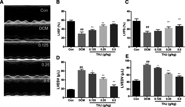
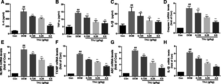
Methods: A type 2 diabetes mellitus model was generated using male C57BL/6 mice. Echocardiography and histopathology were used to evaluate cardiac function. The expression levels of cytokines were measured using enzyme-linked immunosorbent assays. Western blotting and small interfering RNA were used to evaluate the targets of THJ.

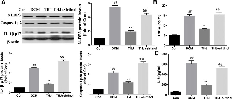
Results: Compared with the control group, DCM mice showed cardiac dysfunction, metabolic disorder, fibrosis, and disorganized ultrastructure, and THJ treatment significantly inhibited these changes significantly. THJ treatment also inhibited the production of reactive oxygen species (ROS) and malondialdehyde (MDA), induced the production of glutathione peroxidase (GSH-Px) and superoxide dismutase (SOD), decreased the levels of pro-inflammatory cytokines, and suppressed the activation of the nucleotide-binding oligomerization domain-like receptor protein 3 (NLRP3) inflammasome. These protective effects were abolished by sirtinol, an inhibitor of sirtuin1 (SIRT1).

Conclusions: Overall, THJ protected the heart from hyperglycemia-induced oxidative stress and inflammation in DCM mice via a mechanism involving SIRT1-mediated antioxidant proteins and suppression of the NLRP3 inflammasome.

Keywords: Diabetic cardiomyopathy; Inflammation; NLRP3; Oxidative stress; Sirtuin 1; Taohuajing.


Conflict of interest statement

The authors declare no conflicts of interest.

Figures

Fig. 1
Fig. 1
Effects of THJ on cardiac hypertrophy and dysfunction in DCM model mice. a. M-mode echocardiography. Representative images were selected from three measurements. Effects of THJ treatment on the cardiac function indicators LVEF (b), LVFS (c), LVESV (d), and LVEDV (e). Values are shown as means ± standard deviations (n = 6–8). ##P < 0.01 versus the Con group; *P < 0.05, **P < 0.01 versus the DCM group
Fig. 2
Fig. 2
Effects of THJ on pathological changes in myocardial tissues in DCM model mice. a. Representative images of Masson’s trichrome staining of interstitial and perivascular tissues. b. Representative images of HE staining of longitudinal and transverse sections of heart tissues. c. Representative images of TEM
Fig. 3
Fig. 3
Effects of THJ on oxidative stress in DCM model mice. a. DHE staining images from three measurements are shown. Statistical results of DHE intensity are given in the lower panel. b. Effects of THJ on MDA levels in myocardial tissues. c. Effects of THJ on SOD activities in myocardial tissues. d. Effects of THJ on GSH-Px activities in myocardial tissues. Values are showed as means ± standard deviations (n = 6–8). ##P < 0.01 versus the Con group, **P < 0.01 versus the DCM group
Fig. 4
Fig. 4
Effects of THJ on the expression levels of inflammatory factors and NLRP3. a. Effects of THJ on IL-6 levels in the serum. b. Effects of THJ on TNF-α levels in the serum. c. Effects of THJ on IL-1β levels in the serum. Effects of THJ on the relative mRNA expression levels of caspase 1 (d), NLRP3 (e), TXNIP (f), ASC (g), and IL-1β (h) in myocardial tissues. n = 6–8. ##P < 0.01 versus the Con group, **P < 0.01 versus the DCM group
Fig. 5
Fig. 5
Effects of THJ on the expression levels of NLRP3-related proteins. Representative blots of NLRP3, TXNIP, caspase 1 p20, IL-1β p17, and ASC are shown on the left. Histograms show quantitative expression levels of these targets on the right. Expression levels were normalized to the expression of β-actin. Values are shown as means ± standard deviations from three different experiments. ##P < 0.01 versus the Con group, **P < 0.01 versus the DCM group
Fig. 6
Fig. 6
Effects of THJ on the SIRT1 pathway. a. Effects of THJ on the NAD+/NADH ratio in myocardial tissues. b. Detection of protein expression levels of Ac-SOD2, SIRT1, and Ac- FOXO3a using western blotting. c. Mice were treated with the SIRT1 inhibitor sirtinol for 12 weeks and treated with THJ. The effects of THJ on SIRT1 expression levels were assessed. d. DHE intensity levels after treatment with the SIRT1 inhibitor sirtinol. e. Effects of sirtinol on MDA levels. F. Effects of sirtinol on SOD levels. Values are shown as means ± standard deviations from three different experiments. ##P < 0.01 versus the Con group, **P < 0.01 versus the DCM group, &&P < 0.01 versus the THJ group
Fig. 7
Fig. 7
Pharmacological modulation of SIRT1-regulated THJ-induced anti-inflammatory activity. a. Mice were treated with the SIRT1 inhibitor sirtinol for 12 weeks and then treated with THJ. Protein expression levels of NLRP3, caspase-1 p20, and IL-1β p17 were evaluated after different treatments. b. TNF-α levels in DCM mice. c. IL-6 levels in DCM mice. Values are shown as means ± standard deviations from three different experiments. ##P < 0.01 versus the Con group, **P < 0.01 versus the DCM group, &&P < 0.01 versus the THJ group

Similar articles

Cited by

References

    1. Shaw JE, Sicree RA, Zimmet PZ. Global estimates of the prevalence of diabetes for 2010 and. Diab Res Clin Pract. 2030;87(1):4–14. doi: 10.1016/j.diabres.2009.10.007. - DOI - PubMed
    1. Fox CS, Sean C, Sorlie PD, D'Agostino RB, Pencina MJ, Vasan RS, Meigs JB, Daniel L, Savage PJ. Increasing cardiovascular disease burden due to diabetes mellitus: the Framingham heart study. Circulation. 2007;115(12):1544. doi: 10.1161/CIRCULATIONAHA.106.658948. - DOI - PubMed
    1. Cai L, Kang YJ. Oxidative stress and diabetic cardiomyopathy. Cardiovasc Toxicol. 2001;1(3):181–193. doi: 10.1385/CT:1:3:181. - DOI - PubMed
    1. Bugger H, Abel ED. Molecular mechanisms of diabetic cardiomyopathy. Diabetologia. 2014;57(4):660–671. doi: 10.1007/s00125-014-3171-6. - DOI - PMC - PubMed
    1. Jia G, Demarco VG, Sowers JR. Insulin resistance and hyperinsulinaemia in diabetic cardiomyopathy. Nat Rev Endocrinol. 2016;12(3):144–153. doi: 10.1038/nrendo.2015.216. - DOI - PMC - PubMed

MeSH terms