. 2021 Feb 26;21(1):137.
doi: 10.1186/s12935-021-01824-z.

Immune subtyping for pancreatic cancer with implication in clinical outcomes and improving immunotherapy

Affiliations

Immune subtyping for pancreatic cancer with implication in clinical outcomes and improving immunotherapy

Jingkai Liu et al. Cancer Cell Int. .

Abstract

Background: Emerging evidence has shown that intra-tumor immune features are associated with response to immune checkpoint blockade (ICB) therapy. Accordingly, patient stratification is needed for identifying target patients and designing strategies to improve the efficacy of ICB therapy. We aimed to depict the specific immune features of patients with pancreatic cancer and explore the implication of immune diversity in prognostic prediction and individualized immunotherapy.

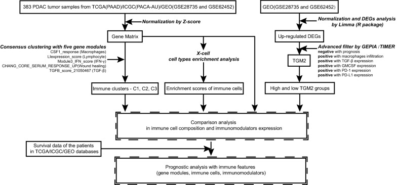
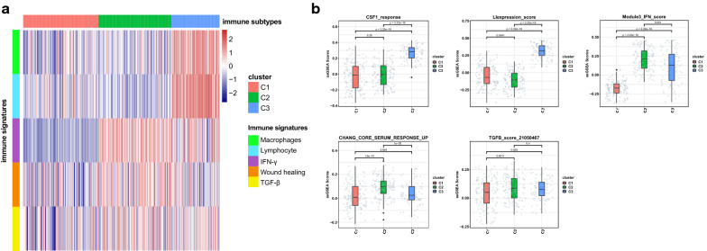
Methods: From transcriptional profiles of 383 tumor samples in TCGA, ICGC, and GEO database, robust immune subtypes which had different response immunotherapy, including ICB therapy, were identified by consensus clustering with five gene modules. DEGs analysis and tumor microarray were used to screen and demonstrate potential targets for improving ICB therapy.

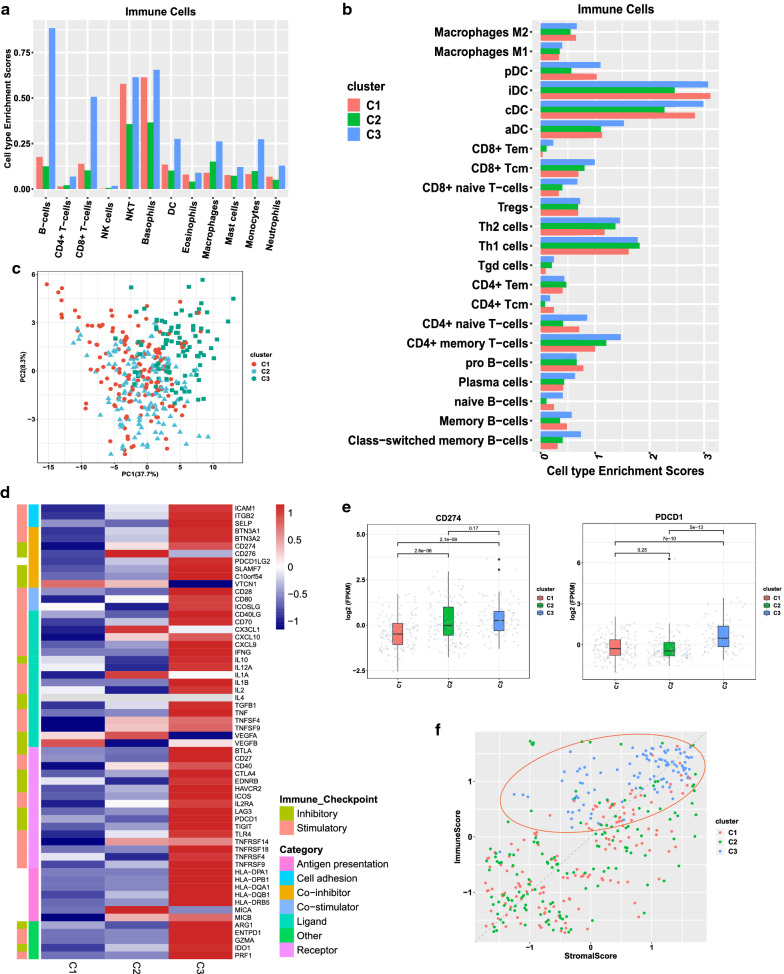
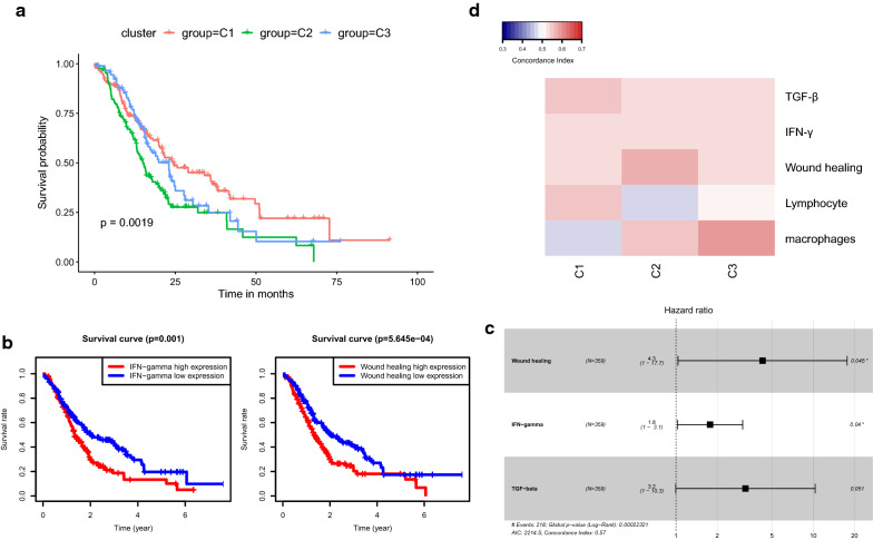
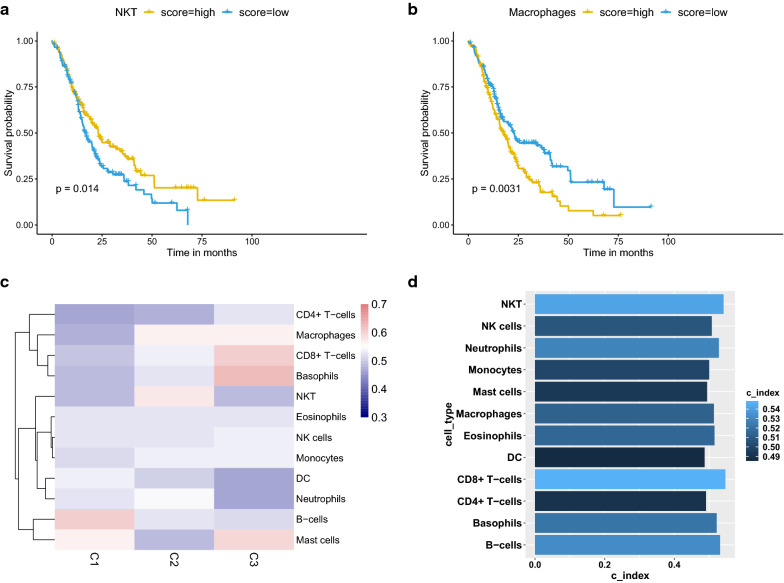
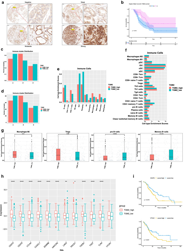
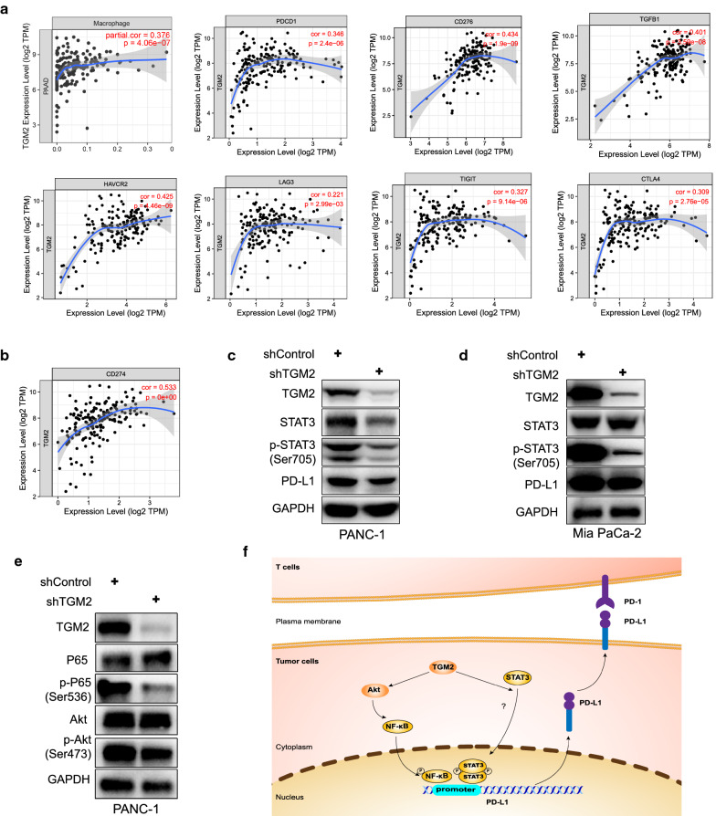
Results: Three subtypes of pancreatic cancer, namely cluster 1-3 (C1-C3), characterized with distinct immune features and prognosis, were generated. Of that, subtype C1 was an immune-cold type in lack of immune regulators, subtype C2, with an immunosuppression-dominated phenotype characterized by robust TGFβ signaling and stromal reaction, showed the worst prognosis, subtype C3 was an immune-hot type, with massive immune cell infiltration and in abundance of immune regulators. The disparity of immune features uncovered the discrepant applicability of anti-PD-1/PD-L1 therapy and potential sensitivity to other alternative immunotherapy for each subtype. Patients in C3 were more suitable for anti-PD-1/PD-L1 therapy, while patients in the other two clusters may need combined strategies targeted on other immune checkpoints or oncogenic pathways. A promising target for improving anti-PD-1/PD-L1 treatment, TGM2, was screened out and its role in the regulation of PD-L1 was investigated for the first time.

Conclusion: Collectively, immune features of pancreatic cancer contribute to distinct immunosuppressive mechanisms that are responsible for individualized immunotherapy. Despite pancreatic cancer being considered as a poor immunogenic cancer type, the derived immune subtypes may have implications in tailored designing of immunotherapy for the patients. TGM2 has potential synergistic roles with ICB therapy.

Keywords: Heterogeneity; Immune cell; Immune checkpoints; Immunotherapy; Pancreatic cancer; Transglutaminase 2.


Conflict of interest statement

The authors declare that they have no competing interests.

Figures

Fig. 1
Fig. 1
Flowchart of data collection and analysis in present study. Transcriptional profiles were collected from TCGA (PAAD)/ICGC (PACA-AU)/GEO (GSE28735 and GSE62452). Three immune clusters were derived by consensus clustering with five gene modules. The enrichment scores of cell types were calculated by X-cell. Then, comparison analysis in immune cell composition and immunomodulators expression were performed in groups and whole cohort. Combined with survival data of the patients TCGA/ICGC/GEO cohort, the prognostic analysis with immune features (gene modules, immune cells, and immunomodulators) were performed. To find promising targets for anti-PD-1/PD-L1 treatment, DEGs analysis and correlation analysis were performed in GEO (GSE28735 and GSE62452) database. The results led to gene TGM2, an oncogenic target for PDAC. The comparison analysis and prognostic analysis were performed in high-/low- TGM2 groups
Fig. 2
Fig. 2
Immune subtypes of PDAC and immune signatures in TCGA and ICGC and GEO mix. a Immune subtypes of PDAC: column was for tumor samples and row was for five immune signatures. b Enrichment comparison of the five immune gene modules across three immune subtypes. Gene modules include CSF1_response (correspond to Macrophages), LIexpression_score (correspond to Lymphocyte), Module3_IFN_score (correspond to IFN-γ), CHANG_CORE_SERUM_RESPONSE_UP (correspond to Wound healing), and TGFB_score_21050467 (correspond to TGF-β)
Fig. 3
Fig. 3
Immune cell composition and expression patterns of IMs in three subtypes. a, b Immune cell composition of three immune subtypes. c PCA analysis of IMs genes. d Expression patterns of IMs in three subtypes. e Enrichment disparity of CD274 and PDCD1 across the three subtypes. f Analysis of stromal and immune scores across the three subtypes: the distribution of samples in C3 was indicated by the red circle
Fig. 4
Fig. 4
Prognostic impact of immune subtypes and immune signatures. a Overall survival of three immune subtypes. b Correlation of immune signatures with overall survival. The Left was for IFN-γ module. The Right was for Wound healing module. c Multivariate COX analysis of three immune signatures. Wound healing module (coef = 1.45, HR = 4.28, 95%CI 1.03–17.67, p = 0.045), IFN-γ module (coef = 0.57, HR = 1.77, 95%CI 1.03–3.06, p = 0.04), TGF-β module (coef = 1.16, HR = 3.21, 95%CI 0.99–10.34, p = 0.051). d Concordance index (CI) of five immune signature expression with the overall survival. Columns and rows represented immune subtypes and immune signatures respectively. Red indicated higher prediction accuracy while blue indicated lower prediction accuracy, along with the increasing expression of immune signature enrichment scores
Fig. 5
Fig. 5
Prognostic impact of the immune cells. a, b Survival analysis of NKT cells (p = 0.014) and macrophages (p = 0.0031). c CI analysis with immune cells across the three subtypes: red indicated higher prediction accuracy while blue indicated lower prediction accuracy, along with the increasing expression of immune cells enrichment scores. d CI analysis with immune cells in the whole cohort: the lighter blue indicated greater prognostic impact while the darker blue indicated weaker prognostic impact
Fig. 6
Fig. 6
Relation between TGM2 and tumor immunosuppression in PDAC. a IHC analysis of TGM2 with our tissue microarray (n = 97). The scale bars were shown as indicated: 100 μm and 20 μm. b Survival analysis of TGM2 with tissue microarray data (p = 0.015). c, d Distribution of high- and low-TGM2 group across the three immune subtypes showed as percentage and number. e, f Immune cell composition of high- and low-TGM2 groups. g Enrichment comparison of M2 type macrophages (p = 0.014), Tregs (p = 0.068), pro-B cells (p = 0.00052) and memory B cells (p = 3.7e−06) between high- and low-TGM2 groups. h Comparison of IMs expression between high- and low-TGM2 groups: row is for the immunomodulators and column is for the gene expression value. (CD274, p = 1.0e−14; CD276, p = 1.1e−10; CTLA4, p = 5.6e−07; CX3CL1, p = 1.6e−06; EDNRB, p = 3.8e−06; HAVCR2, p = 4.3e−10; LAG3, p = 0.0046; PDCD1, p = 0.0121; TGFB1, p = 1.2e−12; TIGIT, p = 6.5e−06; TLR4, p = 3.8e−08; VTCN1, p = 0.0585). i Survival analysis of CD276 expression in high-TGM2 group (top) and VTCN1 expression in low-TGM2 group (bottom)
Fig. 7
Fig. 7
Proposed mechanism that TGM2 may regulate PD-L1 expression via STAT3/NF-κB signaling pathways in PDAC. a Correlation analysis of TGM2 and immunosuppressive factors with TIMER. b Correlation analysis of TGM2 and CD274 (PD-L1) by TIMER. c, d Western blot results showed that TGM2 knocking down resulted in a decreased expression of PD-L1 and p-STAT3 in PANC-1 and Mia PaCa-2 cells. e TGM2 knocking down in PANC-1 cells led to a decreased expression of p-Akt (Ser473) and p-P65 (Ser536) which was consistent with previous studies. f TGM2 may regulate PD-L1 via NF-κB/STAT3 signaling pathways: a TGM2 activated AKT pathway and then promotes the activation of downstream transcription factor NF-κB which has been reported to be able to directly bind with the promoter of PD-L1 and stimulate its transcription. b TGM2 may promote the phosphorylation of STAT3 despite the underlying pathways remains unclear, and then p-STAT3 binds to the promoter of PD-L1 and stimulate its transcription

Similar articles

Cited by

References

    1. Siegel RL, Miller KD, Jemal A. Cancer statistics, 2019. CA A Cancer J Clin. 2019;69(1):7–34. doi: 10.3322/caac.21551. - DOI - PubMed
    1. Neoptolemos JP, Kleeff J, Michl P, Costello E, Greenhalf W, Palmer DH. Therapeutic developments in pancreatic cancer: current and future perspectives. Nat Rev Gastroenterol Hepatol. 2018;15(6):333–348. doi: 10.1038/s41575-018-0005-x. - DOI - PubMed
    1. Antoni R, Jedd DW. Cancer immunotherapy using checkpoint blockade. Science. 2018;359:1350–1355. doi: 10.1126/science.aar4060. - DOI - PMC - PubMed
    1. Macherla S, Laks S, Naqash A, Bulumulle A, Zervos E, Muzaffar M. Emerging role of immune checkpoint blockade in pancreatic cancer. Int J Mol Sci. 2018;19(11):3505. doi: 10.3390/ijms19113505. - DOI - PMC - PubMed
    1. Mahoney KM, Rennert PD, Freeman GJ. Combination cancer immunotherapy and new immunomodulatory targets. Nat Rev Drug Discov. 2015;14(8):561–584. doi: 10.1038/nrd4591. - DOI - PubMed