. 2021 Mar 12;8(2):ENEURO.0565-20.2021.
doi: 10.1523/ENEURO.0565-20.2021. Print 2021 Mar-Apr.

A Role for STOML3 in Olfactory Sensory Transduction

Affiliations

A Role for STOML3 in Olfactory Sensory Transduction

Emilio Agostinelli et al. eNeuro. .

Abstract

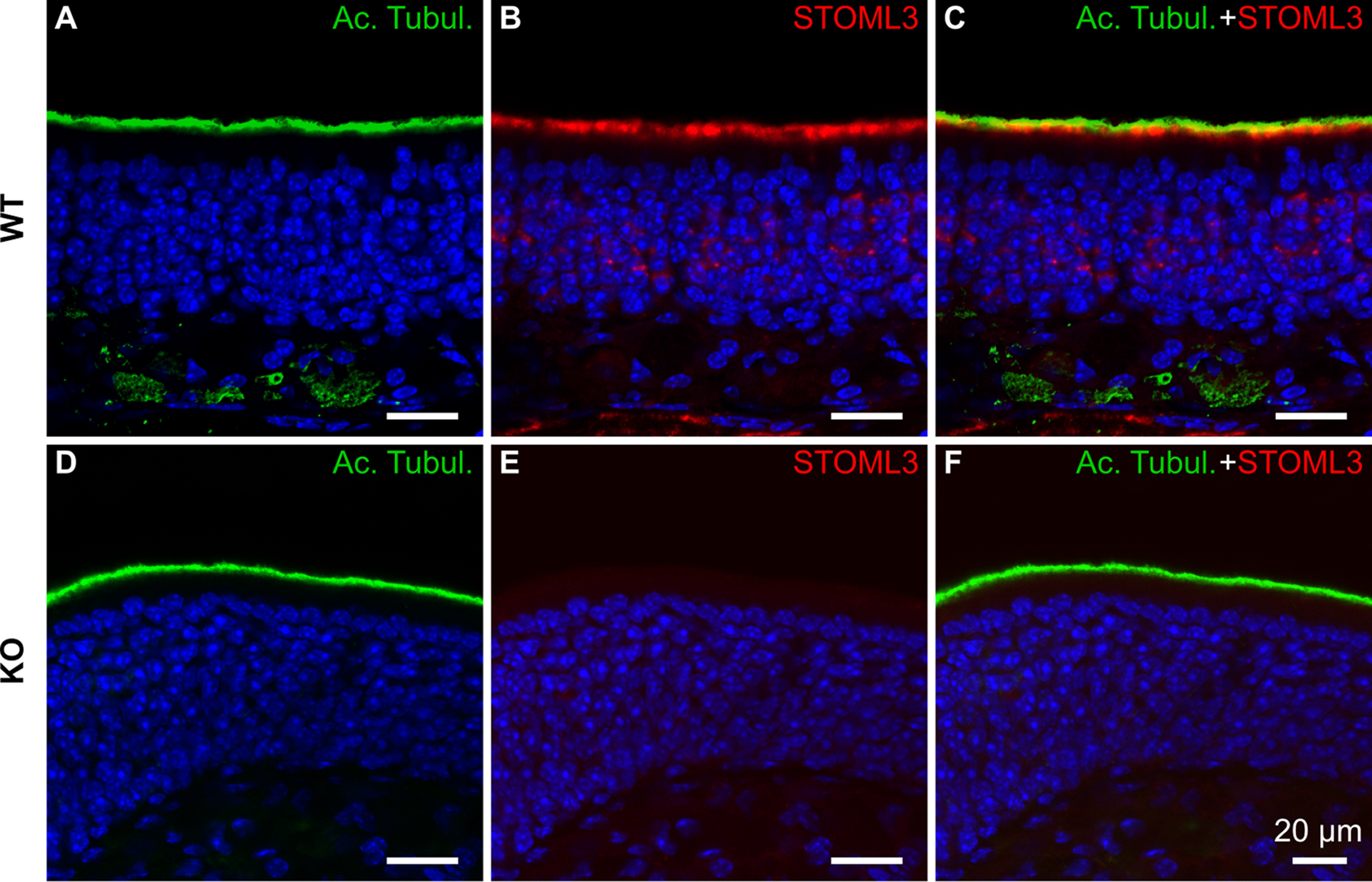
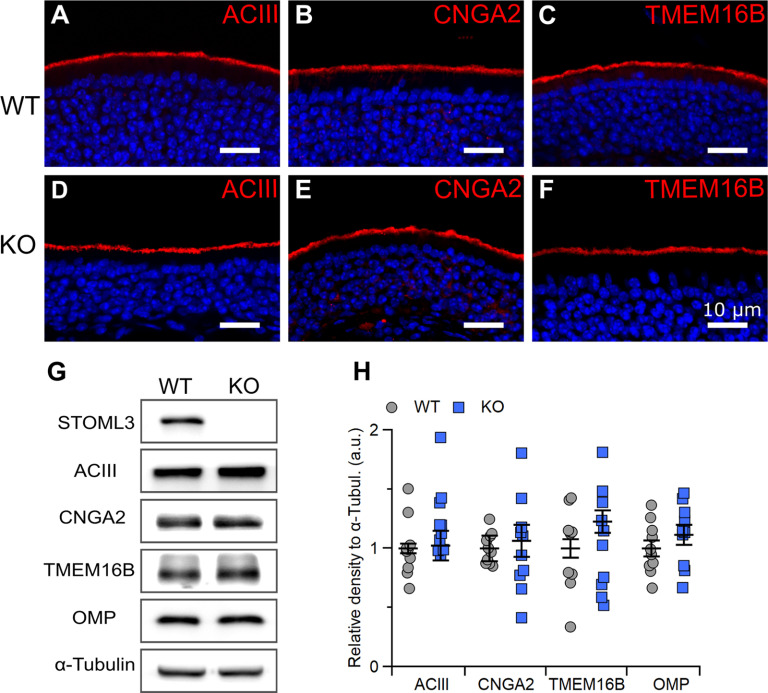
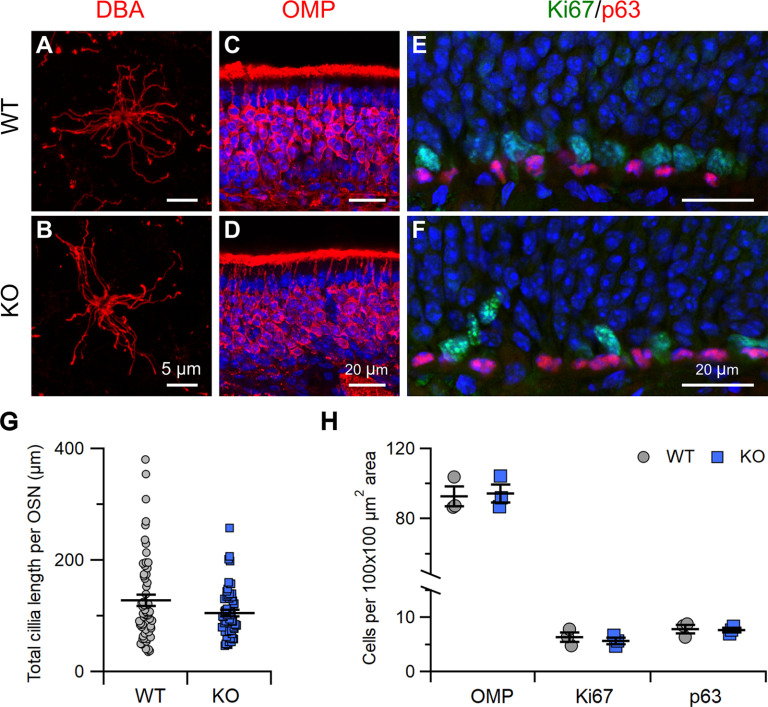
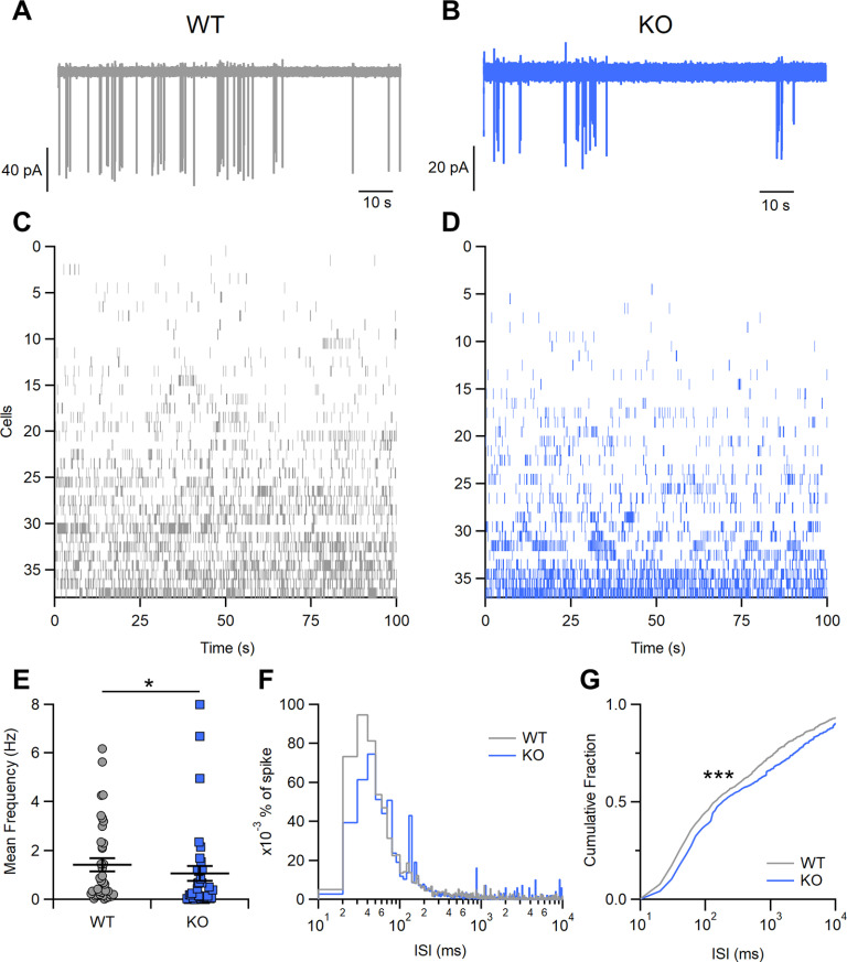
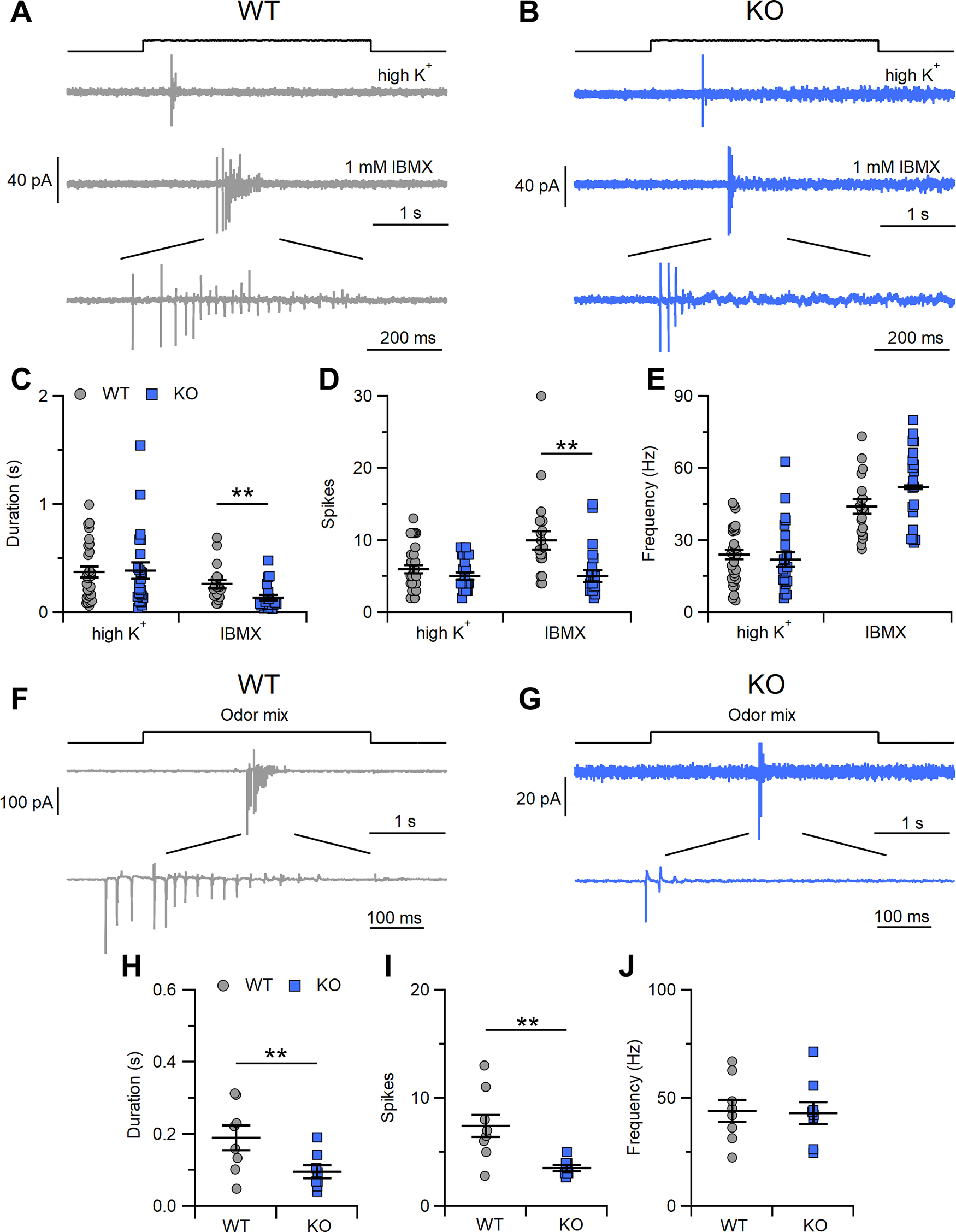
Stomatin-like protein-3 (STOML3) is an integral membrane protein expressed in the cilia of olfactory sensory neurons (OSNs), but its functional role in this cell type has never been addressed. STOML3 is also expressed in dorsal root ganglia neurons, where it has been shown to be required for normal touch sensation. Here, we extended previous results indicating that STOML3 is mainly expressed in the knob and proximal cilia of OSNs. We additionally showed that mice lacking STOML3 have a morphologically normal olfactory epithelium. Because of its presence in the cilia, together with known olfactory transduction components, we hypothesized that STOML3 could be involved in modulating odorant responses in OSNs. To investigate the functional role of STOML3, we performed loose patch recordings from wild-type (WT) and Stoml3 knock-out (KO) OSNs. We found that spontaneous mean firing activity was lower with additional shift in interspike intervals (ISIs) distributions in Stoml3 KOs compared with WT neurons. Moreover, the firing activity in response to stimuli was reduced both in spike number and duration in neurons lacking STOML3 compared with WT neurons. Control experiments suggested that the primary deficit in neurons lacking STOML3 was at the level of transduction and not at the level of action potential generation. We conclude that STOML3 has a physiological role in olfaction, being required for normal sensory encoding by OSNs.

Keywords: ion channel; olfactory; transduction.


Figures

Figure 1.
Figure 1.
STOML3 is expressed in knob/proximal cilia of OSNs. A, D, Confocal micrographs of coronal sections of the olfactory epithelium from WT and KO mice immunostained with antibody against the ciliary marker acetylated tubulin (Ac. Tubul.). B, E, Immunostaining for STOML3 from WT and KO mice. C, Merging of the signals shows that STOML3 has diffuse staining that colocalizes with acetylated tubulin (yellow regions in C) and more defined staining of the layer below the Ac. Tubul. in the region occupied by the OSN knobs. The signal disappeared in the olfactory epithelium of the Stoml3 KO mice (E, F). Nuclei were stained with DAPI (blue).
Figure 2.
Figure 2.
Stoml3 KO does not alter the localization or expression level of molecular components of olfactory transduction. A–F, Confocal micrographs of coronal sections of the olfactory epithelium from WT and KO mice immunostained for ACIII, CNGA2, or TMEM16B. Nuclei were stained with DAPI (blue). G, Western blot analysis of olfactory epithelium proteins for STOML3, ACIII, CNGA2, TMEM16B, OMP, and α-tubulin. The specific 32-kDa band for STOML3 is missing in proteins from KO mice. H, Expression levels relative to α-tubulin for the indicated proteins in olfactory epithelium from 10 WT and KO mice.
Figure 3.
Figure 3.
Stoml3 KO mice have grossly normal olfactory epithelium. A, B, Enface view of a whole-mount preparation of olfactory epithelium with OSN cilia labeled by biotinylated DBA detected by streptavidin-Alexa Fluor 594. C–F, Confocal micrographs of coronal sections of the olfactory epithelium from WT and KO mice immunostained for OMP (C, D) or Ki67 and p63 (E, F). Nuclei were stained with DAPI (blue). G, Quantification of the total cilia length per OSN from WT and KO mice (OSNs: n = 61 from 4 WT mice, n = 57 from 4 KO mice). H, Quantification of OMP-immunopositive, Ki67-immunopositive, and p63-immunopositive cells in olfactory epithelium from WT and KO mice.
Figure 4.
Figure 4.
Spontaneous firing activity in OSNs in WT and Stoml3 KO mice A, B, Representative loose-patch recordings showing the spontaneous activity of OSNs from acute slices of olfactory epithelium from WT and KO mice. C, D, Raster plots of recordings of spontaneous activity from several OSNs from WT and KO mice. Each row represents spike activity from a different OSN. E, Mean frequency of spontaneous activity in OSNs from WT and KO mice (n = 38 for WT and n = 37 for KO, *p < 0.05 U test). F, ISI distributions of spontaneous firing from cells shown in C, D (bin = 5 ms). Values were normalized to the area under each curve to show the spike percentages for WT or KO mice. G, Cumulative fraction of ISI distributions (***p < 0.001, Kolmogorov–Smirnov test).
Figure 5.
Figure 5.
Evoked activity in OSNs in WT and Stoml3 KO mice. A, B, Representative loose-patch recordings of OSNs from WT and KO mice stimulated with high K+ (upper trace) or with 1 mm IBMX (middle and lower traces). Comparison of response duration (C), number of evoked spikes (D), and mean frequency of the response (E) to high K+ or 1 mm IBMX in OSNs from WT and KO mice (n = 19 for WT, n = 20 for KO; **p < 0.01 U test). F, G, Representative loose-patch recordings of OSNs from WT and KO mice stimulated with odor mixture. Comparison of response duration (H), number of evoked spikes (I), and mean frequency of the response (J) to odor mixture in OSNs from WT and KO mice (n = 8 for WT and KO; **p < 0.01 U test).

Similar articles

Cited by

References

    1. Arnson HA, Holy TE (2011) Chemosensory burst coding by mouse vomeronasal sensory neurons. J Neurophysiol 106:409–420. 10.1152/jn.00108.2011 - DOI - PMC - PubMed
    1. Baumgart S, Jansen F, Bintig W, Kalbe B, Herrmann C, Klumpers F, Köster SD, Scholz P, Rasche S, Dooley R, Metzler-Nolte N, Spehr M, Hatt H, Neuhaus EM (2014) The scaffold protein MUPP1 regulates odorant-mediated signaling in olfactory sensory neurons. J Cell Sci 127:2518–2527. 10.1242/jcs.144220 - DOI - PubMed
    1. Billig GM, Pál B, Fidzinski P, Jentsch TJ (2011) Ca2+-activated Cl− currents are dispensable for olfaction. Nat Neurosci 14:763–769. 10.1038/nn.2821 - DOI - PubMed
    1. Brand J, Smith ESJ, Schwefel D, Lapatsina L, Poole K, Omerbašić D, Kozlenkov A, Behlke J, Lewin GR, Daumke O (2012) A stomatin dimer modulates the activity of acid-sensing ion channels. EMBO J 31:3635–3646. 10.1038/emboj.2012.203 - DOI - PMC - PubMed
    1. Browman DT, Hoegg MB, Robbins SM (2007) The SPFH domain-containing proteins: more than lipid raft markers. Trends Cell Biol 17:394–402. 10.1016/j.tcb.2007.06.005 - DOI - PubMed

Publication types