. 2021 Apr 7;41(14):3094-3104.
doi: 10.1523/JNEUROSCI.2609-20.2021. Epub 2021 Feb 26.

The GARP Domain of the Rod CNG Channel's β1-Subunit Contains Distinct Sites for Outer Segment Targeting and Connecting to the Photoreceptor Disk Rim

Affiliations

The GARP Domain of the Rod CNG Channel's β1-Subunit Contains Distinct Sites for Outer Segment Targeting and Connecting to the Photoreceptor Disk Rim

Jillian N Pearring et al. J Neurosci. .

Abstract

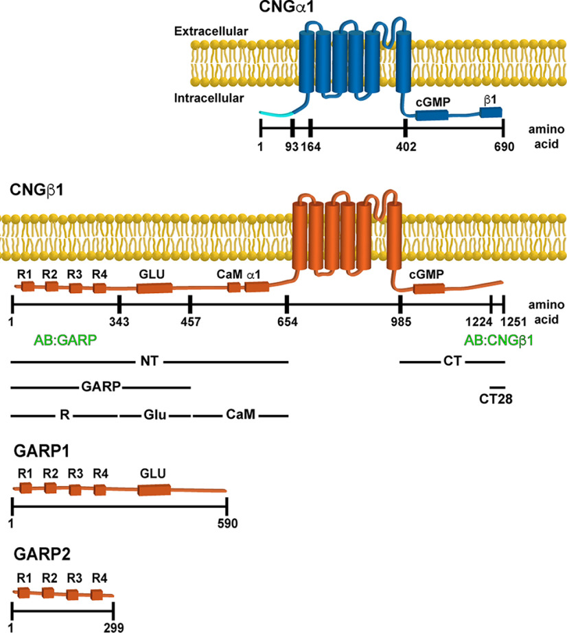
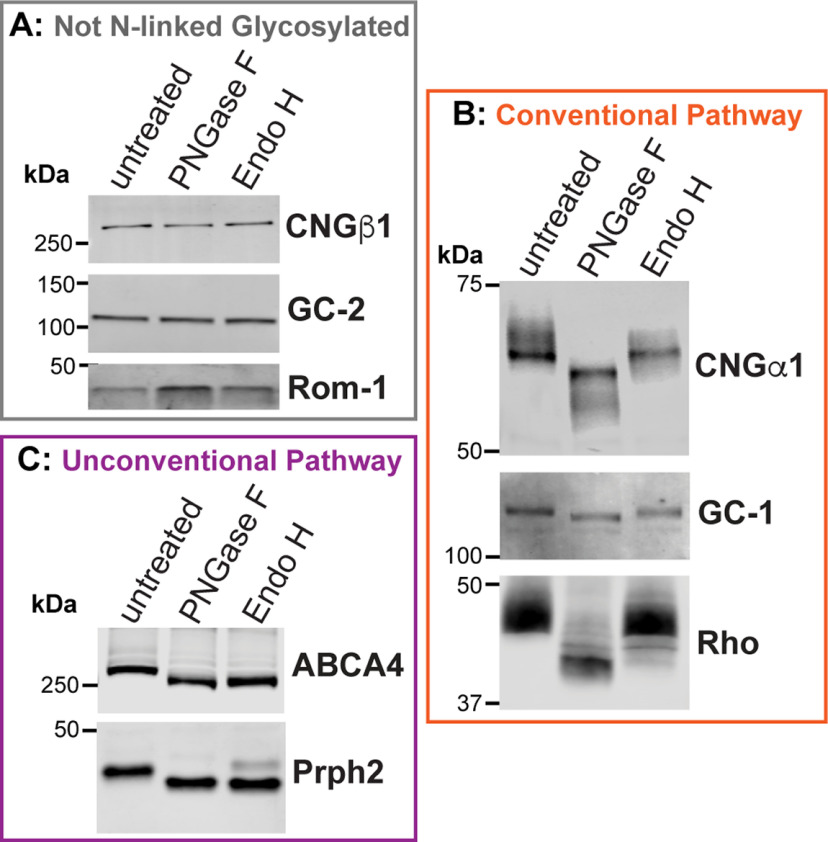
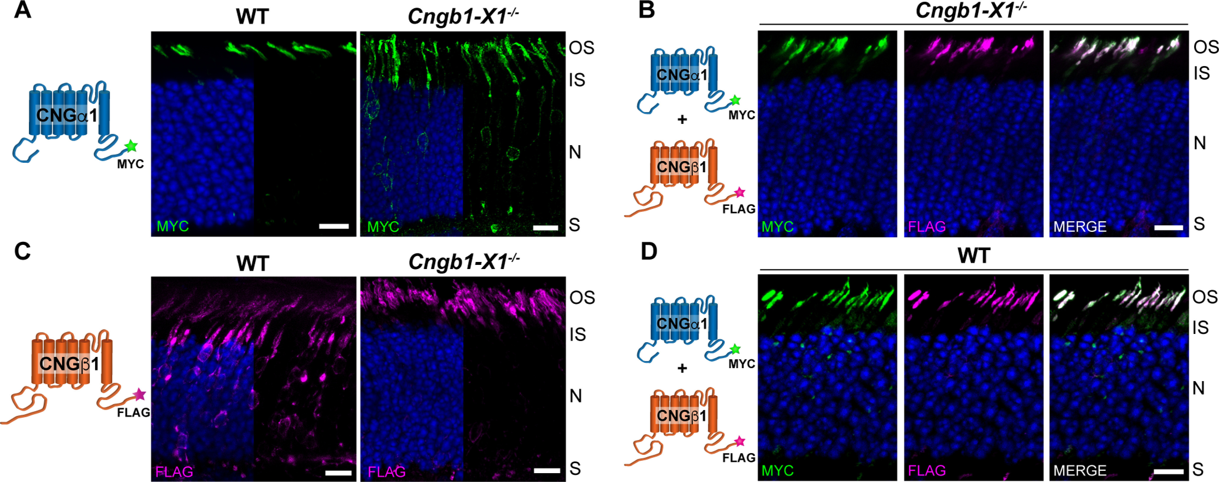
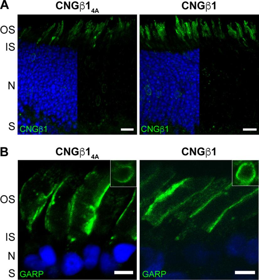
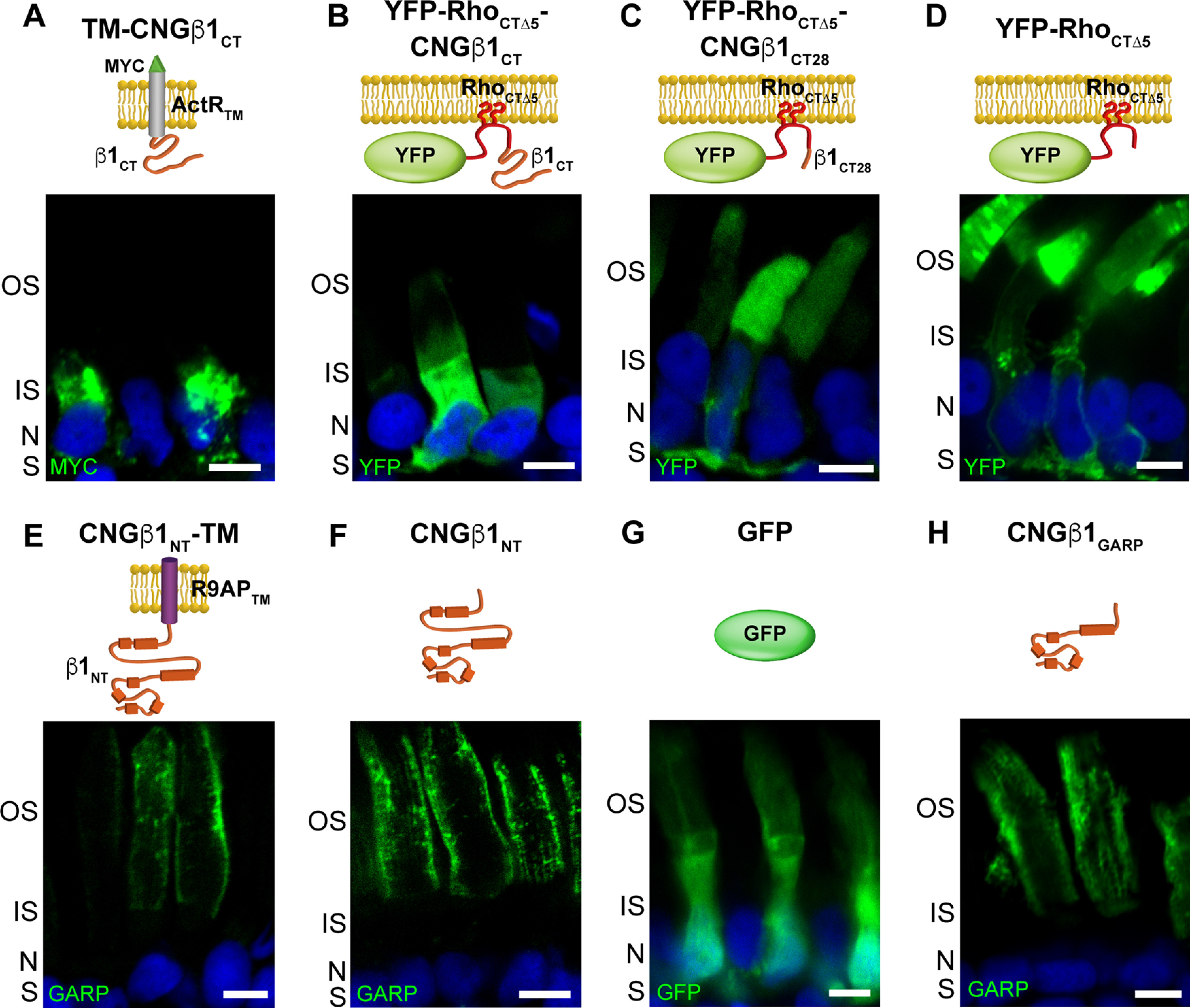
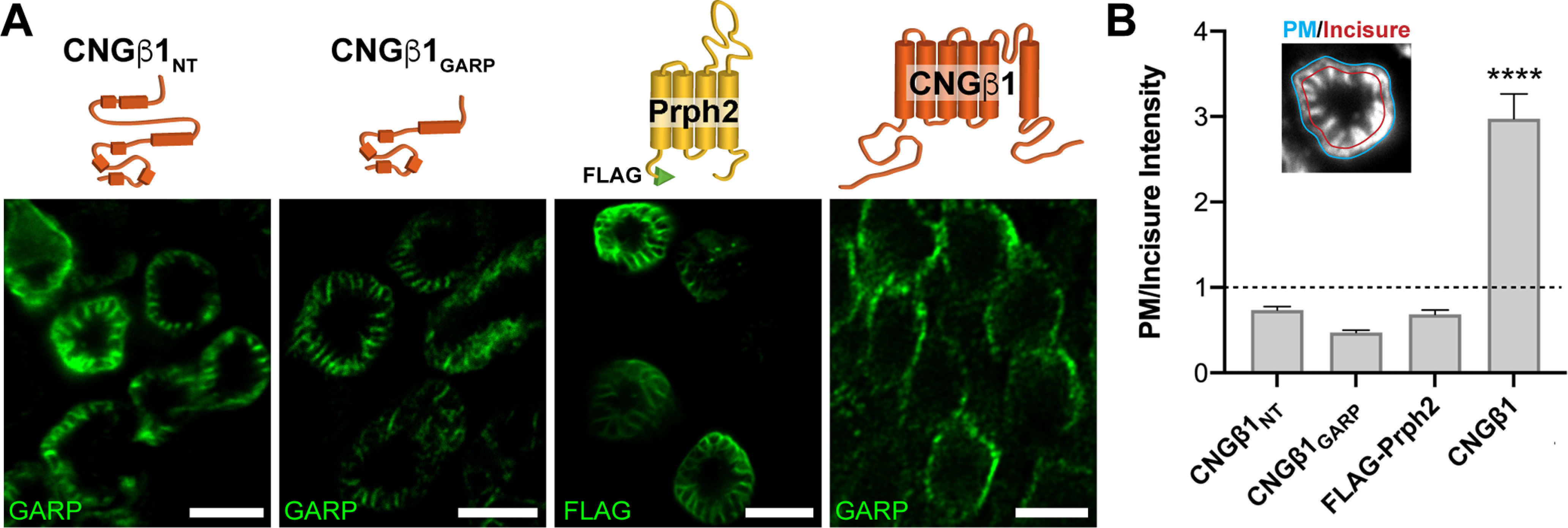
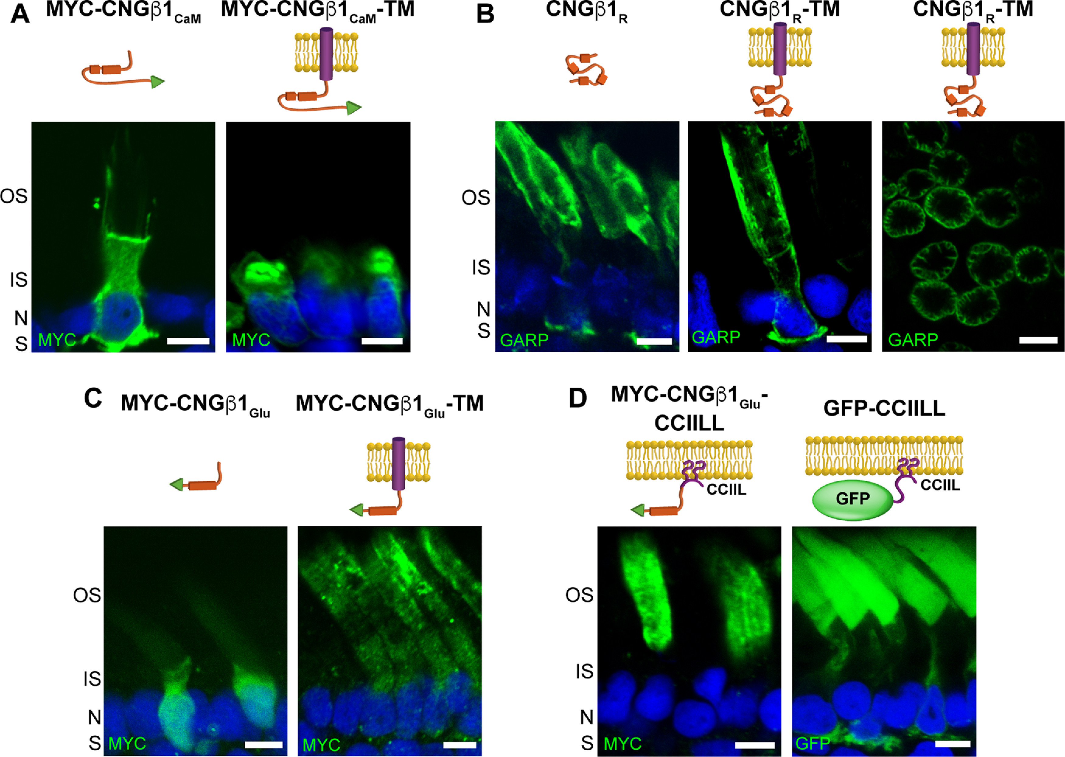
Vision begins when light is captured by the outer segment organelle of photoreceptor cells in the retina. Outer segments are modified cilia filled with hundreds of flattened disk-shaped membranes. Disk membranes are separated from the surrounding plasma membrane, and each membrane type has unique protein components. The mechanisms underlying this protein sorting remain entirely unknown. In this study, we investigated the outer segment delivery of the rod cyclic nucleotide-gated (CNG) channel, which is located in the outer segment plasma membrane, where it mediates the electrical response to light. Using Xenopus and mouse models of both sexes, we now show that the targeted delivery of the CNG channel to the outer segment uses the conventional secretory pathway, including protein processing in both ER and Golgi, and requires preassembly of its constituent α1 and β1 subunits. We further demonstrate that the N-terminal glutamic acid-rich protein (GARP) domain of CNGβ1 contains two distinct functional regions. The glutamic acid-rich region encodes specific information targeting the channel to rod outer segments. The adjacent proline-enriched region connects the CNG channel to photoreceptor disk rims, likely through an interaction with peripherin-2. These data reveal fine functional specializations within the structural domains of the CNG channel and suggest that its sequestration to the outer segment plasma membrane requires an interaction with peripherin-2.SIGNIFICANCE STATEMENT Neurons and other differentiated cells have a remarkable ability to deliver and organize signaling proteins at precise subcellular locations. We now report that the CNG channel, mediating the electrical response to light in rod photoreceptors, contains two specialized regions within the N terminus of its β-subunit: one responsible for delivery of this channel to the ciliary outer segment organelle and another for subsequent channel sequestration into the outer segment plasma membrane. These findings expand our understanding of the molecular specializations used by neurons to populate their critical functional compartments.

Keywords: CNG channel; cilium; membrane trafficking; outer segment; photoreceptor.


Figures

Figure 1.
Figure 1.
Domain composition of CNGα1, CNGβ1, GARP1 and GARP2. Cytosolic domains include cGMP binding, calmodulin (CaM) binding, CNGα1 (α1) and CNGβ1 (β1) binding, Glu, and four proline-rich repeats (R1–R4). Notably, the first 92 amino acids of CNGα1 (marked in light blue) are cleaved during intracellular CNG channel processing (Molday et al., 1991). The epitope location for anti-GARP and anti-CNGβ1 antibodies as well as boundaries of CNGβ1-derived constructs expressed in this study are shown below the diagram. The numbering of amino acids corresponds to human protein sequences. For mouse variants, the corresponding amino acids for CNGα1 would be 1, 93, 156, 394, and 684, and for CNGβ1 would be 1, 358, 512, 731, 935, 1278, and 1305.
Figure 2.
Figure 2.
The rod CNG channel is trafficked using the conventional secretory pathway. Mouse retinal lysates (untreated) were incubated with PNGase F or Endo H and analyzed by Western blotting. A, Electrophoretic mobility of nonglycosylated proteins CNGβ1, guanylate cyclase-2 (GC-2) and Rom-1 is unaffected by enzymatic treatments. B, CNGα1, GC-1, and rhodopsin are sensitive to PNGase F and resistant to Endo H treatment, indicating their processing through the conventional secretory pathway. C, ABCA4 and peripherin-2 are sensitive to both PNGase F and Endo H treatments, indicating their processing through the unconventional secretory pathway.
Figure 3.
Figure 3.
Outer segment localization of the CNG channel requires expression of the CNGβ1 subunit. A, Retinal cross sections from WT and Cngb1-X1−/− rods transfected with MYC-tagged, full-length mouse CNGα1. B, Cngb1-X1−/− rods were cotransfected with CNGα1-MYC and CNGβ1-FLAG. The merged image is shown on the far right. C, WT or Cngb1-X1−/− rods transfected with FLAG-tagged, full-length mouse CNGβ1. D, WT rods were cotransfected with CNGα1-MYC and CNGβ1-FLAG. In all panels, retinal sections were stained for each construct using an anti-MYC (green) and/or anti-FLAG (magenta) antibodies as indicated in the panel, and nuclei are stained by Hoechst (blue). Cartoon of transfected constructs are depicted on the left. Scale bar, 10 µm. Here and in the following figures, photoreceptor cell layer abbreviations are outer segment (OS), inner segment (IS), nucleus (N), and synapse (S). All mice are analyzed at P21.
Figure 4.
Figure 4.
The C-terminus RVxP motif is not involved in CNG channel localization to the plasma membrane of the outer segment. A, Cngb1-X1−/− rods were transfected with untagged mouse CNGβ14A mutant or CNGβ1 control constructs. Retinal cross-sections were stained for each construct using an anti-CNGβ1 (green) antibody. Scale bar: 10 µm. B, Transgenic Xenopus rods expressing full-length human CNGβ14A mutant or CNGβ1 control. Retinal cross-sections were stained for each construct using an anti-GARP (green) antibody. Scale bar, 5 µm. Nuclei are counterstained with Hoechst (blue).
Figure 5.
Figure 5.
CNGβ1 N-terminal GARP domain is confined to the rod outer segment. A–H, Retinal cross-sections from transgenic Xenopus rods expressing membrane-anchored TM-CNGβ1CT (A), lipidated YFP-RhoCTΔ5-CNGβ1CT (B), lipidated YFP-RhoCTΔ5-CNGβ1CT28 (C), lipidated YFP-RhoCTΔ5 (D), membrane-anchored CNGβ1NT-TM (E), soluble CNGβ1NT (F), soluble GFP (G), and soluble CNGβ1GARP constructs (H). Diagram of transfected constructs are shown above their corresponding panel. Constructs that include N terminal of human CNGβ1 were detected using an anti-GARP antibody. Nuclei are counterstained with Hoechst (blue). Scale bar: 5 µm in all panels. All Xenopus tadpoles were analyzed at stage 43–45.
Figure 6.
Figure 6.
CNGβ1's N-terminal GARP domain localizes to disk incisures within rod outer segments. A, Tangential sections through Xenopus rod outer segments expressing soluble CNGβ1NT and CNGβ1GARP; FLAG-tagged full-length bovine peripherin-2 or untagged full-length CNGβ1. Diagram of transfected constructs are shown above their corresponding panel. Constructs that include N terminal of human CNGβ1 were detected using an anti-GARP antibody. Scale bar: 5 µm in all panels. B, Quantification of plasma membrane intensity (blue line, inset) normalized to incisure intensity (red line, inset) was performed for all the constructs shown in panel A. Protein residing in the plasma membrane yield a value >1, whereas proteins residing in disk incisures yield a value <1. Unpaired Student's t test was performed for full-length CNGβ1 compared with the other three constructs, p < 0.0001.
Figure 7.
Figure 7.
CNGβ1's GARP domain contains separate sites for peripherin-2 binding and outer segment targeting. A–D, Retinal cross-sections from transgenic Xenopus rods comparing localization of soluble and membrane-anchored CNGβ1CaM (A), soluble and membrane-anchored CNGβ1R and a tangential image of outer segment's expressing CNGβ1R-TM (B), soluble and membrane-anchored CNGβ1Glu (C), and CNGβ1Glu or GFP fused to a C-terminal CCIIL double lipidation anchor (D). Cartoon of transgenic constructs are shown above their corresponding panel. Constructs that include the N terminal of human CNGβ1 were detected using an anti-GARP antibody. All other constructs were N-terminally tagged with the MYC epitope and detected using an anti-MYC antibody. Scale bar, 5 µm. Nuclei are counterstained with Hoechst (blue).

Similar articles

Cited by

References

    1. Amaya E, Kroll KL (1999) A method for generating transgenic frog embryos. Methods Mol Biol 97:393–414. 10.1385/1-59259-270-8:393 - DOI - PubMed
    1. Ardell MD, Makhija AK, Oliveira L, Miniou P, Viegas-Péquignot E, Pittler SJ (1995) cDNA, gene structure, and chromosomal localization of human GAR1 (CNCG3L), a homolog of the third subunit of bovine photoreceptor cGMP-gated channel. Genomics 28:32–38. 10.1006/geno.1995.1102 - DOI - PubMed
    1. Ardell MD, Aragon I, Oliveira L, Porche GE, Burke E, Pittler SJ (1996) The β subunit of human rod photoreceptor cGMP-gated cation channel is generated from a complex transcription unit. FEBS Lett 389:213–218. 10.1016/0014-5793(96)00588-1 - DOI - PubMed
    1. Ardell MD, Bedsole DL, Schoborg RV, Pittler SJ (2000) Genomic organization of the human rod photoreceptor cGMP-gated cation channel β-subunit gene. Gene 245:311–318. 10.1016/S0378-1119(00)00023-8 - DOI - PubMed
    1. Baker SA, Haeri M, Yoo P, Gospe SM, Skiba NP, Knox BE, Arshavsky VY (2008) The outer segment serves as a default destination for the trafficking of membrane proteins in photoreceptors. J Cell Biol 183:485–498. 10.1083/jcb.200806009 - DOI - PMC - PubMed

Publication types

MeSH terms