Site-Specific and Residualizing Linker for 18F Labeling with Enhanced Renal Clearance: Application to an Anti-HER2 Single-Domain Antibody Fragment
- PMID: 33637584
- PMCID: PMC8612331
- DOI: 10.2967/jnumed.120.261446
Site-Specific and Residualizing Linker for 18F Labeling with Enhanced Renal Clearance: Application to an Anti-HER2 Single-Domain Antibody Fragment
Abstract
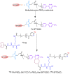
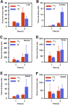
Single-domain antibody fragments (sdAbs) are promising vectors for immuno-PET; however, better methods for labeling sdAbs with 18F are needed. Herein, we evaluate a site-specific strategy using an 18F residualizing motif and the anti-epidermal growth factor receptor 2 (HER2) sdAb 5F7 bearing an engineered C-terminal GGC tail (5F7GGC). Methods: 5F7GGC was site-specifically attached with a tetrazine-bearing agent via thiol-maleimide reaction. The resultant conjugate was labeled with 18F by inverse electron demand Diels-Alder cycloaddition with a trans-cyclooctene attached to 6-18F-fluoronicotinoyl moiety via a renal brush border enzyme-cleavable linker and a PEG4 chain (18F-5F7GGC). For comparisons, 5F7 sdAb was labeled using the prototypical residualizing agent, N-succinimidyl 3-(guanidinomethyl)-5-125I-iodobenzoate (iso-125I-SGMIB). The 2 labeled sdAbs were compared in paired-label studies performed in the HER2-expressing BT474M1 breast carcinoma cell line and athymic mice bearing BT474M1 subcutaneous xenografts. Small-animal PET/CT imaging after administration of 18F-5F7GGC in the above mouse model was also performed. Results:18F-5F7GGC was synthesized in an overall radiochemical yield of 8.9% ± 3.2% with retention of HER2 binding affinity and immunoreactivity. The total cell-associated and intracellular activity for 18F-5F7GGC was similar to that for coincubated iso-125I-SGMIB-5F7. Likewise, the uptake of 18F-5F7GGC in BT474M1 xenografts in mice was similar to that for iso-125I-SGMIB-5F7; however, 18F-5F7GGC exhibited significantly more rapid clearance from the kidney. Small-animal PET/CT imaging confirmed high uptake and retention in the tumor with very little background activity at 3 h except in the bladder. Conclusion: This site-specific and residualizing 18F-labeling strategy could facilitate clinical translation of 5F7 anti-HER2 sdAb as well as other sdAbs for immuno-PET.
Keywords: HER2; click chemistry; immuno-PET; single-domain antibody fragment; site-specific labeling.
© 2021 by the Society of Nuclear Medicine and Molecular Imaging.
Figures


Similar articles
-
Labeling a TCO-functionalized single domain antibody fragment with 18F via inverse electron demand Diels Alder cycloaddition using a fluoronicotinyl moiety-bearing tetrazine derivative.Bioorg Med Chem. 2020 Sep 1;28(17):115634. doi: 10.1016/j.bmc.2020.115634. Epub 2020 Jul 9. Bioorg Med Chem. 2020. PMID: 32773089 Free PMC article.
-
Site-specific radioiodination of an anti-HER2 single domain antibody fragment with a residualizing prosthetic agent.Nucl Med Biol. 2021 Jan;92:171-183. doi: 10.1016/j.nucmedbio.2020.05.002. Epub 2020 May 12. Nucl Med Biol. 2021. PMID: 32448731 Free PMC article.
-
Labeling single domain antibody fragments with 18F using a novel residualizing prosthetic agent - N-succinimidyl 3-(1-(2-(2-(2-(2-[18F]fluoroethoxy)ethoxy)ethoxy)ethyl)-1H-1,2,3-triazol-4-yl)-5-(guanidinomethyl)benzoate.Nucl Med Biol. 2021 Sep-Oct;100-101:24-35. doi: 10.1016/j.nucmedbio.2021.06.002. Epub 2021 Jun 11. Nucl Med Biol. 2021. PMID: 34146837 Free PMC article.
-
An Efficient Method for Labeling Single Domain Antibody Fragments with 18F Using Tetrazine- Trans-Cyclooctene Ligation and a Renal Brush Border Enzyme-Cleavable Linker.Bioconjug Chem. 2018 Dec 19;29(12):4090-4103. doi: 10.1021/acs.bioconjchem.8b00699. Epub 2018 Nov 14. Bioconjug Chem. 2018. PMID: 30384599 Free PMC article.
-
Fluorine-18 labeling of an anti-HER2 VHH using a residualizing prosthetic group via a strain-promoted click reaction: Chemistry and preliminary evaluation.Bioorg Med Chem. 2018 May 1;26(8):1939-1949. doi: 10.1016/j.bmc.2018.02.040. Epub 2018 Mar 15. Bioorg Med Chem. 2018. PMID: 29534937 Free PMC article.
Cited by
-
Rapid cleavage of 6-[18F]fluoronicotinic acid prosthetic group governs BT12 glioblastoma xenograft uptake: implications for radiolabeling design of biomolecules.EJNMMI Radiopharm Chem. 2025 Jul 8;10(1):40. doi: 10.1186/s41181-025-00368-1. EJNMMI Radiopharm Chem. 2025. PMID: 40629197 Free PMC article.
-
Molecular probes targeting HER2 PET/CT and their application in advanced breast cancer.J Cancer Res Clin Oncol. 2024 Mar 11;150(3):118. doi: 10.1007/s00432-023-05519-y. J Cancer Res Clin Oncol. 2024. PMID: 38466436 Free PMC article. Review.
-
Radiosynthesis and preclinical evaluations of [18F]AlF-RESCA-5F7 as a novel molecular probe for HER2 tumor imaging.Am J Nucl Med Mol Imaging. 2024 Jun 15;14(3):175-181. doi: 10.62347/BVPK1360. eCollection 2024. Am J Nucl Med Mol Imaging. 2024. PMID: 39027646 Free PMC article.
-
Effective Treatment of Human Breast Carcinoma Xenografts with Single-Dose 211At-Labeled Anti-HER2 Single-Domain Antibody Fragment.J Nucl Med. 2023 Jan;64(1):124-130. doi: 10.2967/jnumed.122.264071. Epub 2022 May 26. J Nucl Med. 2023. PMID: 35618478 Free PMC article.
-
Trends in nanobody radiotheranostics.Eur J Nucl Med Mol Imaging. 2025 May;52(6):2225-2238. doi: 10.1007/s00259-025-07077-6. Epub 2025 Jan 13. Eur J Nucl Med Mol Imaging. 2025. PMID: 39800806 Review.
References
-
- Martin V, Cappuzzo F, Mazzucchelli L, Frattini M. HER2 in solid tumors: more than 10 years under the microscope—where are we now? Future Oncol. 2014;10:1469–1486. - PubMed
-
- Wolff AC, Hammond MEH, Allison KH, Harvey BE, McShane LM, Dowsett M. HER2 testing in breast cancer: American Society of Clinical Oncology/College of AMERICAN Pathologists clinical practice guideline focused update summary. J Oncol Pract. 2018;14:437–441. - PubMed
-
- Xavier C, Vaneycken I, D’Huyvetter M, et al. . Synthesis, preclinical validation, dosimetry, and toxicity of 68Ga-NOTA-anti-HER2 nanobodies for iPET imaging of HER2 receptor expression in cancer. J Nucl Med. 2013;54:776–784. - PubMed
-
- Morris O, Fairclough M, Grigg J, Prenant C, McMahon A. A review of approaches to 18F radiolabelling affinity peptides and proteins. J Labelled Comp Radiopharm. 2019;62:4–23. - PubMed
MeSH terms
Substances
LinkOut - more resources
Full Text Sources
Other Literature Sources
Research Materials
Miscellaneous
