

Head-to-Head Comparison of 68Ga-NOTA (68Ga-NGUL) and 68Ga-PSMA-11 in Patients with Metastatic Prostate Cancer: A Prospective Study
- PMID: 33637585
- PMCID: PMC8724887
- DOI: 10.2967/jnumed.120.258434
Head-to-Head Comparison of 68Ga-NOTA (68Ga-NGUL) and 68Ga-PSMA-11 in Patients with Metastatic Prostate Cancer: A Prospective Study
Abstract
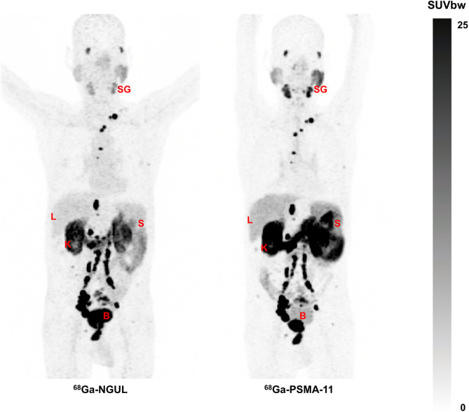
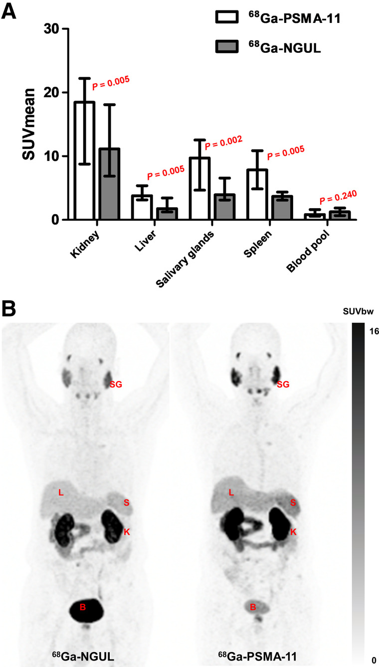
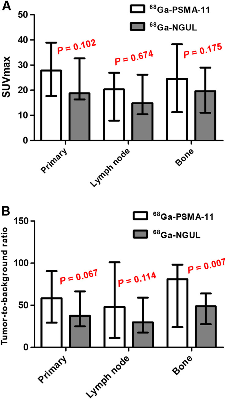
68Ga-NOTA Glu-Urea-Lys (NGUL) is a novel prostate-specific membrane antigen (PSMA)-targeting tracer used for PET/CT imaging. This study aimed to compare performance in the detection of primary and metastatic lesions and to compare biodistribution between 68Ga-NGUL and 68Ga-PSMA-11 in the same patients with prostate cancer. Methods: Eleven patients with metastatic prostate cancer were prospectively recruited. The quantitative tracer uptake was determined in normal organs and in primary and metastatic lesions. Results:68Ga-NGUL showed significantly lower normal-organ uptake and rapid urinary clearance. The number and sites of detected PSMA-positive primary and metastatic lesions were identical, and no significant quantitative uptake difference was observed. 68Ga-NGUL showed a relatively lower tumor-to-background ratio than 68Ga-PSMA-11. Conclusion: In a head-to-head comparison with 68Ga-PSMA-11, 68Ga-NGUL showed lower uptake in normal organs and similar performance in detecting PSMA-avid primary and metastatic lesions. 68Ga-NGUL could be a valuable option for PSMA imaging.
Keywords: 68Ga-NGUL; 68Ga-PSMA-11; biodistribution; prostate-specific membrane antigen.
© 2021 by the Society of Nuclear Medicine and Molecular Imaging.
Figures

References
-
- Iravani A, Violet J, Azad A, Hofman MS. Lutetium-177 prostate-specific membrane antigen (PSMA) theranostics: practical nuances and intricacies. Prostate Cancer Prostatic Dis. 2020;23:38–52. - PubMed
-
- Siva S, Udovicich C, Tran B, Zargar H, Murphy DG, Hofman MS. Expanding the role of small-molecule PSMA ligands beyond PET staging of prostate cancer. Nat Rev Urol. 2020;17:107–118. - PubMed
-
- Eder M, Schafer M, Bauder-Wust U, et al. 68Ga-complex lipophilicity and the targeting property of a urea-based PSMA inhibitor for PET imaging. Bioconjug Chem. 2012;23:688–697. - PubMed
-
- Ceci F, Uprimny C, Nilica B, et al. 68Ga-PSMA PET/CT for restaging recurrent prostate cancer: which factors are associated with PET/CT detection rate? Eur J Nucl Med Mol Imaging. 2015;42:1284–1294. - PubMed
-
- Fendler WP, Eiber M, Beheshti M, et al. 68Ga-PSMA PET/CT: joint EANM and SNMMI procedure guideline for prostate cancer imaging: version 1.0. Eur J Nucl Med Mol Imaging. 2017;44:1014–1024. - PubMed
Publication types
MeSH terms
Substances
LinkOut - more resources
Full Text Sources
Other Literature Sources
Medical
Miscellaneous