Comparative Study
. 2021 Feb;9(2):e001798.
doi: 10.1136/jitc-2020-001798.

TGFβ1 neutralization displays therapeutic efficacy through both an immunomodulatory and a non-immune tumor-intrinsic mechanism

Affiliations
Comparative Study

TGFβ1 neutralization displays therapeutic efficacy through both an immunomodulatory and a non-immune tumor-intrinsic mechanism

Stefania Canè et al. J Immunother Cancer. 2021 Feb.

Abstract

Background: Transforming growth factor-β (TGFβ) is emerging as a promising target for cancer therapy, given its ability to promote progression of advanced tumors and to suppress anti-tumor immune responses. However, TGFβ also plays multiple roles in normal tissues, particularly during organogenesis, raising toxicity concerns about TGFβ blockade. Dose-limiting cardiovascular toxicity was observed, possibly due to the blockade of all three TGFβ isoforms. The dominant isoform in tumors is TGFβ1, while TGFβ2 and TGFβ3 seem to be more involved in cardiovascular development. Recent data indicated that selective targeting of TGFβ1 promoted the efficacy of checkpoint inhibitor anti-PD1 in transplanted preclinical tumor models, without cardiovascular toxicity.

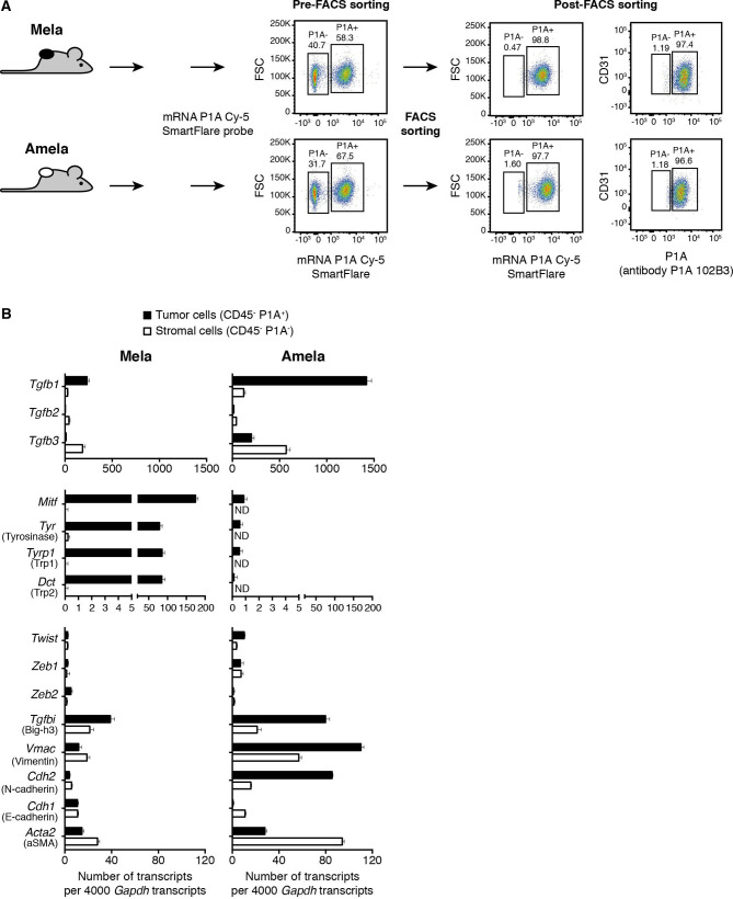
Methods: To further explore the therapeutic potential of isoform-specific TGFβ blockade, we developed neutralizing mAbs targeting mature TGFβ1 or TGFβ3, and tested them, in parallel with anti-panTGFβ mAb 1D11, in two preclinical models: the transplanted colon cancer model CT26, and the autochthonous melanoma model TiRP.

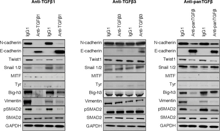
Results: We observed that the blockade of TGFβ1, but not that of TGFβ3, increased the efficacy of a prophylactic cellular vaccine against colon cancer CT26. This effect was similar to pan-TGFβ blockade, and was associated with increased infiltration of activated CD8 T cells in the tumor, and reduced levels of regulatory T cells and myeloid-derived suppressor cells. In contrast, in the autochthonous TiRP melanoma model, we observed therapeutic efficacy of the TGFβ1-specific mAb as a single agent, while the TGFβ3 mAb was inactive. In this model, the anti-tumor effect of TGFβ1 blockade was tumor intrinsic rather than immune mediated, as it was also observed in T-cell depleted mice. Mechanistically, TGFβ1 blockade increased mouse survival by delaying the phenotype switch, akin to epithelial-to-mesenchymal transition (EMT), which transforms initially pigmented tumors into highly aggressive unpigmented tumors.

Conclusions: Our results confirm TGFβ1 as the relevant isoform to target for cancer therapy, not only in combination with checkpoint inhibitors, but also with other immunotherapies such as cancer vaccines. Moreover, TGFβ1 blockade can also act as a monotherapy, through a tumor-intrinsic effect blocking the EMT-like transition. Because human melanomas that resist therapy often express a gene signature that links TGFβ1 with EMT-related genes, these results support the clinical development of TGFβ1-specific mAbs in melanoma.

Keywords: cytokines; drug evaluation; immunomodulation; immunotherapy; melanoma; preclinical.


Conflict of interest statement

Competing interests: BJVdE is co-founder of, has ownership interest in, and is SAB member of iTeos Therapeutics.

Figures

Figure 1
Figure 1
Specificity and inhibitory capacity of anti-TGFβ1 mAb 13A1 and anti-TGFβ3 mAb 1901. (A) ELISA plates coated with human TGFβ1, TGFβ2 or TGFβ3 (100 ng/mL) were incubated with serial dilutions of anti-TGFβ1 mAb 13A1 (IgG1), anti-TGFβ3 mAb 1901 (IgG1), or anti-panTGFβ mAb 1D11 (IgG1), and revealed with HRP-labeled goat anti-mouse Ab. Similar results were obtained when using mouse TGFβ1 or TGFβ3. One representative experiment (±SEM) of at least three performed is shown. The EC50 (±SEM) of the different batches used throughout the work are indicated. (B) Inhibition of the biological activity of the different TGFβ isoforms was evaluated in a TMLEC-TGFβ reporter assay. One representative experiment of at least three is illustrated graphically, and the IC50±SEM of the different batches used throughout the work are indicated. (C) Mature human TGFβ1 (300 ng/mL) was coated on ELISA plates and incubated with 100 ng/mL (0.67 nM) anti-TGFβ1 mAb 13A1 preincubated for 1 hour at room temperature with increasing concentrations of recombinant mature or latent human TGFß1 (R&D systems). Binding of mAb 13A1 to coated TGFβ1 was revealed as in (A). One representative experiment (±SEM) out of two performed. HRP, horseradish peroxidase; TGFβ, transforming growth factor-β; TMLEC, transformed mink lung epithelial cell.
Figure 2
Figure 2
Synergistic effect of anti-TGFβ1 with a prophylactic vaccine against colon carcinoma CT26. (A) Schedule for tumor induction and immunotherapy. BALB/c mice received a prophylactic vaccination with 1×10⁵ irradiated (250 Gy) CT26CL1 tumor cells, injected subcutaneously (s.c.) in the right flank. The same day they received 200 µg intraperitoneally (i.p.) of either anti-TGFβ1 13A1, anti-TGFβ3 1901 or anti-panTGFβ 1D11, or isotype control antibody. Anti-TGFβ antibodies were then administered three times a week at the dose of 100 µg. Three weeks after vaccination, mice received 1×10⁶ CT26CL1 s.c. in the left flank together with 200 µg of anti-TGFβ antibodies. The treatment with anti-TGFβ antibodies then continued three times a week, 100 µg i.p. until the end of the experiment. (B) Tumor growth (left panels) and mouse survival (right panels) were monitored. The figures represent the cumulative data of 6 independent experiments totaling 193 mice (experiment 1, cohort n=44 (groups: no vaccine=4; vaccine=5; vaccine+IgG1=4; vaccine+anti-TGFβ1=6; vaccine+anti-panTGFβ=6; vaccine+anti-TGFβ3=5; anti-TGFβ1=5; anti-panTGFβ=5; anti-TGFβ3=4); experiment 2, cohort n=52 (groups: no vaccine=4; vaccine=4; vaccine+IgG1=4; vaccine+anti-TGFβ1=7; vaccine+anti-panTGFβ=7; vaccine+anti-TGFβ3=7; anti-TGFβ1=6; anti-panTGFβ=7; anti-TGFβ3=6); experiment 3, cohort n=39 (groups: no vaccine=4; vaccine=4; vaccine+IgG1=4; vaccine+anti-TGFβ1=5; vaccine+anti-panTGFβ=5; vaccine+anti-TGFβ3=5; anti-TGFβ1=4; anti-panTGFβ=4; anti-TGFβ3=4); experiment 4, cohort n=20 (no vaccine=5; vaccine=5; vaccine+anti-TGFβ1=5; vaccine+anti-panTGFβ=5); experiment 5, cohort n=20 (no vaccine=5; vaccine=5; vaccine+anti-TGFβ1=5; vaccine+anti-panTGFβ=5); experiment 6, cohort n=18 (no vaccine=5; vaccine=5; vaccine+anti-TGFβ1=4; vaccine+anti-panTGFβ=4)). Tumor growth is reported (left panels) as tumor volume (mm3) over time. Data are presented as mean±SEM, and were analyzed with one-way ANOVA with Tukey’s multiple comparisons correction. All groups were compared with vaccine alone: vaccine versus no vaccine, *p<0.05; vaccine versus vaccine+IgG1, NS; vaccine versus vaccine+anti-TGFβ1, *p<0.05; vaccine versus vaccine+anti-panTGFβ, *p<0.05; vaccine versus vaccine+anti-TGFβ3, NS. Survival percentages are reported (right panels), and were analyzed with Log-rank (Mantel-Cox). All groups were compared with vaccine alone: vaccine versus no vaccine, *p<0.05; vaccine versus vaccine+anti-TGFβ1, **p<0.01; vaccine versus vaccine+anti-panTGFβ, **p<0.01; vaccine versus vaccine+anti-TGFβ3, NS. (C) CT26CL1 tumors from tumor-bearing mice treated as indicated in (A) were analyzed by ex vivo FACS staining for CD8, CD69, CD25, Tim-3, PD-1, CD44 and CD62L. Cumulative data are presented from 6 experiments totaling 193 mice, as detailed in (B). Results are expressed as mean+SEM unpaired Student’s t-test (two-tailed). All groups were compared with vaccine alone: *p<0.05; **p<0.01; ***p<0.001; ****p<0.0001. ANOVA, analysis of variance; NS, not significant; TGFβ, transforming growth factor-β.
Figure 3
Figure 3
Expression of TGFβ isoforms and EMT-associated genes in TiRP melanoma cells and stromal cells. (A) Separation of tumor cells and stromal cells from Mela and Amela TiRP tumors. CD45-negative cells were sorted based on P1A expression using a fluorescently labeled P1A-mRNA SmartFlare probe. Sorted cells were verified by staining with P1A-specific antibody P1A102B3. (B) Cells sorted in (A) were tested by RT-qPCR for the indicated genes. Results are expressed as mean number of transcripts±SEM, from a total 35 Mela and 110 Amela tumors tested. EMT, epithelial-to-mesenchymal transition; TGFβ, transforming growth factor-β.
Figure 4
Figure 4
In vivo neutralization of TGFβ1 increases survival of TiRP tumor-bearing mice and delays EMT-like transition. (A) TiRP mice (B10.D2;Ink4a/ARFflox/flox;TiRP+/+) received two injections of 4OH-Tamoxifen and weekly injections of anti-TGFβ antibodies (0.5 mg) as indicated, until the end of the experiment. Mice were monitored once a week, tumor size (left panels) and survival (middle panels) were measured, and median time to transition from Mela to Amela tumor was calculated (right panels). The figures represent the cumulative data of 5 independent experiments totaling 128 mice (experiment 1, cohort n=27 (groups: IgG1=6; anti-TGFβ1=7; anti-TGFβ3=7; anti-panTGFβ=7); experiment 2, cohort n=23 (groups: IgG1=5; anti-TGFβ1=6; anti-TGFβ3=6; anti-panTGFβ=6); experiment 3, cohort n=26 (groups: IgG1=6; anti-TGFβ1=7; anti-TGFβ3=6; anti-panTGFβ=7); experiment 4, cohort n=21 (groups: IgG1=4; anti-TGFβ1=7; anti-TGFβ3=5; anti-panTGFβ=5); experiment 5, cohort n=31 (groups: IgG1=6; anti-TGFβ1=10; anti-TGFβ3=5; anti-panTGFβ=10)). Tumor growth is reported as tumor volume (mm3) over time (left panel). Data are presented as mean±SEM, and were analyzed with one-way ANOVA with Tukey’s multiple comparisons correction: anti-TGFβ1 versus IgG1, *p<0.05; anti-panTGFβ versus IgG1, *p<0.05. Survival percentages (middle panel) are reported and were analyzed with Log-rank (Mantel-Cox): anti-TGFβ1 versus IgG1, ****p<0.0001; anti-panTGFβ versus IgG1, ****p<0.0001; anti-TGFβ3 versus IgG1, NS; anti-panTGFβ versus anti-TGFβ1, NS. Means+SEM of median times to EMT-like transition in the five experiments are reported (right panel), and were analyzed with the unpaired Student’s t-test (two-tailed). All groups compared with IgG1: *p<0.05; **p<0.01. (B) TiRP mice received anti-CD4 and anti-CD8 depleting antibodies (1 mg each intraperitoneally (i.p.)) as described, starting at day 15 and continuing once a week up to the end of the experiment. Depletion was verified by FACS on PBMC every 2 weeks. Mice were then injected two times with 4 mg 4OH-Tamoxifen on days 0 and 15. At day 7, some mice received either anti-TGFβ1 or IgG1 control, 0.5 mg/mouse i.p. in PBS. Weekly injections continued as indicated until the end of the experiment. Mice were monitored and data reported as described in (A). The figure represents the cumulative data of 4 independent experiments totaling 116 mice (experiment 1, cohort n=28 (anti-CD4/anti-CD8+IgG1=7; anti-CD4/anti-CD8+anti-TGFβ1=7; IgG1=7, anti-TGFβ1=7); experiment 2, cohort n=32 (anti-CD4/anti-CD8+IgG1=10; anti-CD4/anti-CD8+anti-TGFβ1=10; IgG1=5, anti-TGFβ1=7); experiment 3, cohort n=31 (anti-CD4/anti-CD8+IgG1=8; anti-CD4/anti-CD8+anti-TGFβ1=8; IgG1=7, anti-TGFβ1=8); experiment 4, cohort n=25 (anti-CD4/anti-CD8+IgG1=5; anti-CD4/anti-CD8+anti-TGFβ1=5; IgG1=7, anti-TGFβ1=8)). Tumor growth is reported as tumor volume (mm3) over time (left panel). Data are presented as mean±SEM, and were analyzed with one-way ANOVA with Tukey’s multiple comparisons correction: anti-TGFβ1 versus IgG1, **p<0.01; anti-CD4/anti-CD8+anti-TGFβ1 versus anti-CD4/anti-CD8+IgG1, **p<0.01. Survival percentages are reported (middle panel) and were analyzed with Log-rank (Mantel-Cox): anti-TGFβ1 versus IgG1, ****p<0.0001; anti-CD4/anti-CD8+anti-TGFβ1 versus anti-CD4/anti-CD8+IgG1, ****p<0.0001. Means+SEM of median times to EMT-like transition in the four experiments are reported (right panel), and were analyzed with the unpaired Student’s t-test (two-tailed): IgG1 versus anti-TGFβ1, **p<0.01; anti-CD4/anti-CD8+IgG1 versus anti-CD4/anti-CD8+anti-TGFβ1, **p<0.01. (C) TiRP mice received a prime/boost vaccine regimen of recombinant adenovirus (Adeno.Ii.P1At, 108 PFU i.d.) and SemlikiForest virus (SFV.P1A, 107 IU i.d.) as described. Mice then received 4OH-Tamoxifen and antibody injections as indicated, and were monitored as in (A). The strong CD8+ T cell immune response induced by the vaccine is shown on online supplemental figure S4A–C. The figures represent the cumulative data of 6 independent experiments totaling 161 mice (experiment 1, cohort n=27 (groups: immunized=6; immunized+anti-TGFβ1=7; immunized+anti-TGFβ3=7; immunized+anti-panTGFβ=7); experiment 2, cohort n=23 (groups: immunized=5; immunized+anti-TGFβ1=6; immunized+anti-TGFβ3=6; immunized+anti-panTGFβ=6); experiment 3, cohort n=26 (groups: immunized=6; immunized+anti-TGFβ1=7; immunized+anti-TGFβ3=6; immunized+anti-panTGFβ=7); experiment 4, cohort n=21 (groups: immunized=4; immunized+anti-TGFβ1=7; immunized+anti-TGFβ3=5; immunized+anti-panTGFβ=5); experiment 5, cohort n=30 (groups: immunized=5; immunized+anti-TGFβ1=10; immunized+anti-TGFβ3=5; immunized+anti-panTGFβ=10); experiment 6, cohort n=34 (groups: immunized=5; immunized+anti-TGFβ1=10; immunized+anti-TGFβ3=8; immunized+anti-panTGFβ=11)). Tumor growth (left panel) is reported as tumor volume (mm3) over time. Data are presented as means±SEM and were analyzed with one-way ANOVA with Tukey’s multiple comparisons correction: immunized versus immunized+anti-TGFβ1, *p<0.05; immunized versus immunized+anti-panTGFβ, *p<0.05. Survival percentages are reported (middle panel) and were analyzed with Log-rank (Mantel-Cox): immunized versus immunized+anti-TGFβ1, ****p<0.0001; immunized versus immunized+anti-panTGFβ, ****p<0.0001. Means+SEM of median times to EMT-like transition in the six experiments are reported (right panel), and were analyzed with the unpaired Student’s t-test (two-tailed): immunized versus immunized+anti-TGFβ1, ***p<0.001; immunized versus immunized+anti-panTGFβ, **p<0.01; immunized versus immunized+anti-TGFβ3, NS. ANOVA, analysis of variance; EMT, epithelial-to-mesenchymal transition; i.d., intradermal injection; NS, not significant; PBMC, peripheral blood mononuclear cell; PBS, phosphate-buffered saline; TGFβ, transforming growth factor-β.
Figure 5
Figure 5
The effects of TGFβ1 blockade are linked to a partial silencing of the EMT-associated proteins and transcription factors. Representative Western blot of transcription factors (Snail1/2 and Twist) and other proteins involved either in EMT (N-cadherin, E-cadherin, vimentin, Big-h3) or in melanocyte differentiation (MITF, Tyr). Tumor tissue lysate was obtained from mice treated with anti-TGFβ1 13A1, anti-TGFβ3 1901, anti-panTGFβ 1D11 or isotype control IgG1. Western blot of pSMAD2 was used as control of a TGFβ-dependent effect. EMT, epithelial-to-mesenchymal transition; TGFβ, transforming growth factor-β.

Similar articles

Cited by

References

    1. Massagué J. Tgfbeta in cancer. Cell 2008;134:215–30. 10.1016/j.cell.2008.07.001 - DOI - PMC - PubMed
    1. David CJ, Massagué J. Contextual determinants of TGFβ action in development, immunity and cancer. Nat Rev Mol Cell Biol 2018;19:419–35. 10.1038/s41580-018-0007-0 - DOI - PMC - PubMed
    1. Busse A, Keilholz U. Role of TGF-β in melanoma. Curr Pharm Biotechnol 2011;12:2165–75. 10.2174/138920111798808437 - DOI - PubMed
    1. Huang JJ, Blobe GC. Dichotomous roles of TGF-β in human cancer. Biochem Soc Trans 2016;44:1441–54. 10.1042/BST20160065 - DOI - PMC - PubMed
    1. Roberts AB, Wakefield LM. The two faces of transforming growth factor beta in carcinogenesis. Proc Natl Acad Sci U S A 2003;100:8621–3. 10.1073/pnas.1633291100 - DOI - PMC - PubMed

Publication types

MeSH terms

Substances