. 2021 Feb 26;12(2):224.
doi: 10.1038/s41419-021-03461-w.

Long non-coding RNA Lnc-LALC facilitates colorectal cancer liver metastasis via epigenetically silencing LZTS1

Affiliations

Long non-coding RNA Lnc-LALC facilitates colorectal cancer liver metastasis via epigenetically silencing LZTS1

Chuan Zhang et al. Cell Death Dis. .

Abstract

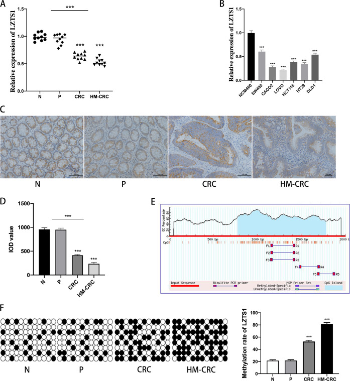
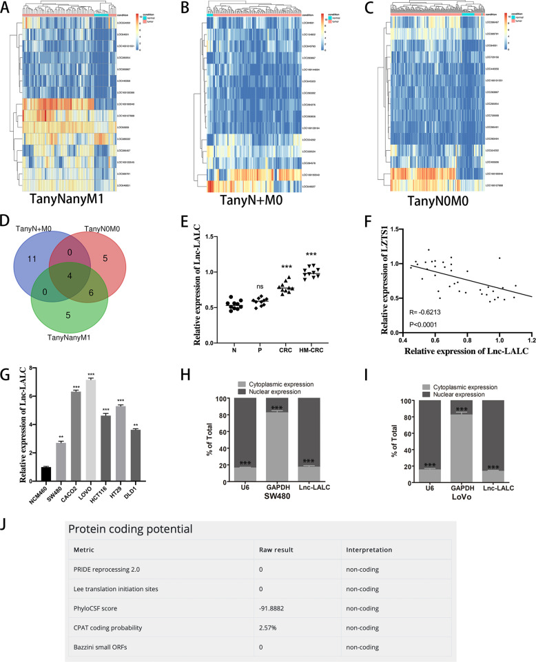
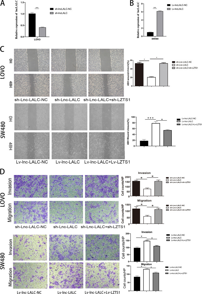
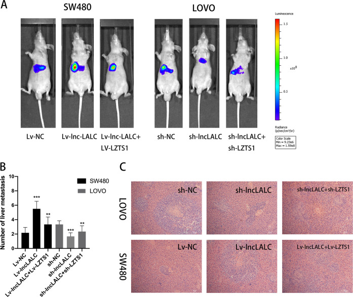
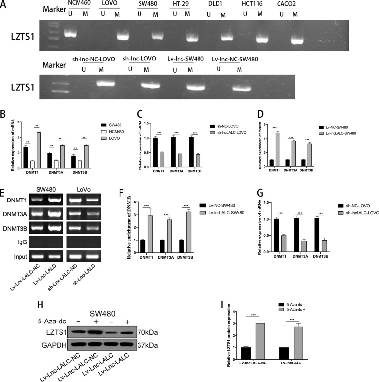
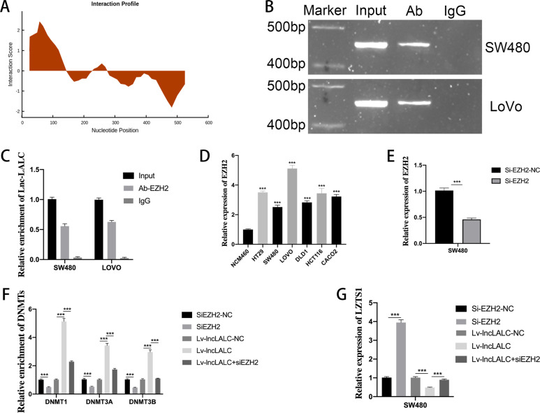
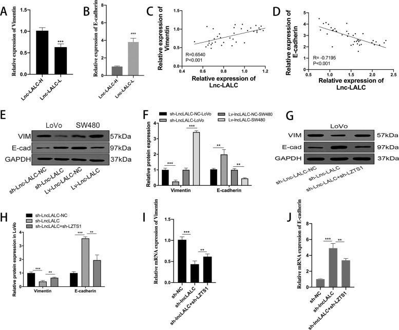
Colorectal cancer (CRC) is one of the most common cancers around the world and endangers human health seriously. Liver metastasis is an important factor affecting the long-term prognosis of CRC and the specific mechanism of CRLM (colorectal cancer with liver metastasis) is not fully understood. LZTS1 has been found dysregulated in many cancers, especially in CRC. Theories suggested that hypermethylation of the promoter regions of LZTS1 was responsible for LZTS1 abnormal expression in multiple malignant tumors. Although the role of LZTS1 in CRC cell proliferation has been reported, its role in CRLM remains unclear. Numerous studies reported Long non-coding RNA (lncRNA) could regulate the gene expression level by regulating gene methylation status in many tumors. However, whether there were lncRNAs could change the methylation status of LZTS1 or not in CRLM was unknown. In this study, we aimed to investigate whether there are lncRNAs can regulate the expression of LZTS1 through affecting DNA methylation in CRLM. We found that upregulated Lnc-LALC in CRC was negatively correlated with LZTS1 expression, and Lnc-LALC could regulate LZTS1 expression in both mRNA and protein level in our study. Functionally, Lnc-LALC enhanced the CRC cells metastasis ability in vitro and vivo through inhibiting the expression of LZTS1. Furthermore, the precise mechanisms exploration showed that lnc-LALC could recruit DNA methyltransferases (DNMTs) to the LZTS1 promoter by combining with Enhancer of zeste homolog 2(EZH2) and then altered the expression of LZTS1 via DNMTs-mediated DNA methylation. Collectively, our data demonstrated the important role of Lnc-LALC/ LZTS1 axis in CRLM development.


Conflict of interest statement

The author declares no competing interests.

Figures

Fig. 1
Fig. 1. LZTS1 expression level was negatively correlated with CRC metastasis.
A Relative expression of LZTS1 in the 40 tissues (normal intestinal epithelial tissues(N), non-metastatic paraneoplastic intestinal epithelial tissues(P), non-metastatic primary CRC tumor tissues(CRC) and primary tumor tissues with hepatic metastatic(HM-CRC),10 samples respectively) were assessed by qRT-PCR.. Statistical analysis was performed between group N and CRC,N and HM-CRC,N + P and CRC + HM-CRC groups. B The LZTS1 expression patterns in CRC cell lines and NCM460 cell lines were assessed by qRT-PCR. The LZTS1 expression level of NCM460 cells was used as a control. C Immunohistochemical assessment of LZTS1 expression in normal intestinal epithelial tissues(N), non-metastatic paraneoplastic intestinal epithelial tissues(P), non-metastatic primary tumor tissues(CRC) and primary tumor tissues with hepatic metastatic tissues(HM-CRC)(original magnification × 200). D Integrated optical density (IOD) value of the immunohistochemical assessment in samples. LZTS1 was down expressed in CRC samples and much more down expressed in HM-CRC samples compared with N samples. Statistical analysis was performed between group N and CRC, N and HM-CRC, N + P and CRC + HM-CRC groups. E The methylated CpG islands in the LZTS1 promoter region predicted by MethPrimer. F The methylation level of LZTS1 in four sample groups was detected by heavy sulfite sequencing and the methylation rate in the HM-CRC group was much higher than the other three groups. N group was used as a control. *P < 0.05; **P < 0.01; ***P < 0.001.
Fig. 2
Fig. 2. Lnc-LALC was upregulated in CRC cells and positively correlated with metastatic factors.
A The top 15 dysregulated lncRNAs in TanyNanyM1 group were shown by hierarchical clustering analysis. B The top 15 dysregulated lncRNAs in TanyN+M0 group were shown by hierarchical clustering analysis. C The top 15 dysregulated lncRNAs in TanyN0M0 group were shown by hierarchical clustering analysis. D VENNY analysis identified 4 lncRNAs which were all aberrantly regulated in the three comparisons (TanyNanyM1, TanyN+M0, and TanyN0M0). E Relative expression of LINC00922 in the above 40 tissues was assessed by qRT-PCR. N group was used as a control. F A negative correlation between expression levels of Lnc-LALC and LZTS1 in tissues was determined by Person analysis (n = 40, r = −0.6213, P < 0.0001). *** P < 0.001; **P < 0.01 and *P < 0.05. G The LINC00922 expression patterns in CRC cell lines and NCM460 cell lines were assessed by qRT-PCR. H Cellular characterization of lnc-LALC in SW480 cells, the levels of nuclear control transcript (U6), cytoplasmic control transcript (glyceraldehyde-3-phosphate dehydrogenase [GAPDH] mRNA), and lnc-LALC were assessed by qRT-PCR in nuclear and cytoplasmic fractions. Data are presented as a percentage of U6, GAPDH, and lnc-LALC levels and total levels for each were taken as 100%. I Cellular characterization of lnc-LALC in LoVo cells. J The non-coding property of Lnc-LALC was confirmed using bioinformatics software (http://cpc.cbi.pku.edu.cn/programs/run_cpc.jsp).
Fig. 3
Fig. 3. Lnc-LALC promoted CRC cells migration and invasion in vitro.
A The knockdown efficiency of lncLALC by sh-lncLALC was assessed by qRT-PCR in LoVo cells. B The overexpression efficiency of lncLALC by Lv-lncLALC was assessed by qRT-PCR in SW480 cells. C Wound-healing assay showed that knockdown of Lnc-LALC inhibited the migratory ability of LoVo cells and knockdown of LZTS1 could recover this inhibiting effect; overexpression of Lnc-LALC promoted the migratory ability of SW480 cells and overexpression of LZTS1 could recover this promoting effect. *P < 0.05(original magnification × 40). D Transwell assay showed that knockdown of Lnc-LALC inhibited the invasive ability of LoVo cells and knockdown of LZTS1 could recover this inhibiting effect; overexpression of Lnc-LALC promoted the invasive ability of SW480 cells and overexpression of LZTS1 could recover this promoting effect(original magnification × 100). *P < 0.05. The data represent the mean ± SD from three independent experiments.
Fig. 4
Fig. 4. Lnc-LALC enhanced CRC cells metastasis ability in vivo.
A Hepatic metastasis models were conducted by injecting CRC cells (2 × 107 cells suspended in 200 μL PBS) into the spleen of 48 BALB/c nude mice in 6 groups (8 in each group) and liver metastasis was investigated respectively using the IVIS Lumina II system. Representative images in each group were presented. The fluorescence intensity was highest in Lv-lnc-LALC-SW480 group and lowest in sh-lncLALC-LoVo group. B Hepatic metastatic tumors in different groups with volumes >2 mm3 were identified and compared.Lv-NC-SW480 group and sh-NC-LoVo group were used as a control in two cell lines respectively. *P < 0.05; **P < 0.01; ***P < 0.001. C H&E staining confirmed the pathological pattern of the hepatic metastatic nodules in these six groups was metastatic adenocarcinoma(original magnification × 100).
Fig. 5
Fig. 5. Lnc-LALC regulated methylation of LZTS1 promoters via binding with DNA methyltransferases.
A Methylation specific PCR (MSP) revealed that the promoter region of LZTS1 was hypermethylated in CRC cells but hypomethylated in the NCM460 cells; and also revealed that the methylation level has a positive correlation with lnc-LALC expression level. U means unmethylated;M means methylated. B The expression level of DNMTs (DNMT1, DNMT3A, DNMT3B) was detected by qRT-PCR in SW480, LoVo cells, and NCM460 cells. C qRT-PCR assay showed knockdown of lncLALC could inhibit expression of DNMTs in LoVo cells. D qRT-PCR assay showed overexpression of lncLALC could promote the expression of DNMTs in SW480 cells. EG ChIP assays revealed that the enrichment of DNMT1, DNMT3A, DNMT3B in LZTS1 promoter increased significantly after lncLALC overexpression in SW480 cells and reduced dramatically by lncLALC knockdown in LoVo cells. H, I Western blotting showed that methylation inhibition by 5-aza-2′-deoxycytidine (5-Aza-dC) could restore LncLALC-induced LZTS1 suppression. *P < 0.05; **P < 0.01; ***P < 0.001.
Fig. 6
Fig. 6. LncLALC recruited DNA methyltransferases to the LZTS1 promoter regions by combining with EZH2.
A Preliminary prediction by bioinformatics system showed that Lnc-LALC may have certain binding ability to EZH2. (http://service.tartaglialab.com/page/catrapid_group). B, C RIP test using the EZH2 antibody showed that there was a strong combining capacity between lncLALC and EZH2 in SW480 cells and LOVO cells. D qRT-PCR showed that EZH2 was elevated in CRC cells than NCM460 cells. E The silencing efficacy of EZH2 in SW480 cells was determined by qRT-PCR. F The CHIP results showed that siEZH2 could reduce the enrichment of DNMT1, DNMT3A, DNMT3B in LZTS1 promoter and could also reverse the DNMTs, high enrichment induced by Lv-lncLALC in SW480 cells. G qRT-PCR showed that siEZH2 could increase LZTS1 expression level and abolish the suppression of LZTS1 induced by Lv-lncLALC in SW480 cells. *P < 0.05; **P < 0.01; ***P < 0.001.
Fig. 7
Fig. 7. LncLALC regulated Epithelial–mesenchymal transition (EMT) in CRC cells through LZTS1.
A The qRT-PCR results showed that Vimentin was overexpressed in lncLALC High group than lncLALC Low group. B The qRT-PCR results showed that E-cadherin was lower expressed in lncLALC High group than in LncLALC low group. C The Pearson correlation analysis confirmed that lncLALC expression had a positive correlation with Vimentin expression (r = 0.6540, P < 0.001). D The Pearson correlation analysis confirmed that lncLALC expression had a negative correlation with E-cadherin expression (r = −0.7195, P < 0.001). E, F Western blot assays showed that sh-LncLALC could enhanced the expression of E-cadherin but reduced the expression of Vimentin in LoVo cells and Lv-Lnc-LALC had the opposite effects in SW480 cells. G, H Western blot assays showed that knockdown of LZTS1 could restore the function of sh-lncLALC on EMT markers in LoVo cells. I qRT-PCR assays showed that knockdown of LZTS1 could restore the function of sh-lncLALC on Vimentin in LoVo cells. J qRT-PCR assays showed that knockdown of LZTS1 could restore the function of sh-lncLALC on E-cadherin in LoVo cells.

Similar articles

Cited by

References

    1. Siegel RL, Miller KD, Jemal A. Cancer statistics, 2018. CA Cancer J. Clin. 2018;68:7–30. doi: 10.3322/caac.21442. - DOI - PubMed
    1. Lykoudis PM, O’Reilly D, Nastos K, Fusai G. Systematic review of surgical management of synchronous colorectal liver metastases. Br. J. Surg. 2014;101:605–612. doi: 10.1002/bjs.9449. - DOI - PubMed
    1. Siegel RL, et al. Colorectal cancer statistics, 2017. CA Cancer J. Clin. 2017;67:177–193. doi: 10.3322/caac.21395. - DOI - PubMed
    1. Ishii H, et al. The FEZ1 gene at chromosome 8p22 encodes a leucine-zipper protein, and its expression is altered in multiple human tumors. Proc. Natl Acad. Sci. USA. 1999;96:3928–3933. doi: 10.1073/pnas.96.7.3928. - DOI - PMC - PubMed
    1. Hou X, et al. miR-1207-5p regulates the sensitivity of triple-negative breast cancer cells to Taxol treatment via the suppression of LZTS1 expression. Oncol. Lett. 2019;17:990–998. - PMC - PubMed

Publication types

MeSH terms