Review
. 2021 Feb 26;12(2):215.
doi: 10.1038/s41419-021-03505-1.

The crosstalk between HIFs and mitochondrial dysfunctions in cancer development

Affiliations
Review

The crosstalk between HIFs and mitochondrial dysfunctions in cancer development

Xingting Bao et al. Cell Death Dis. .

Abstract

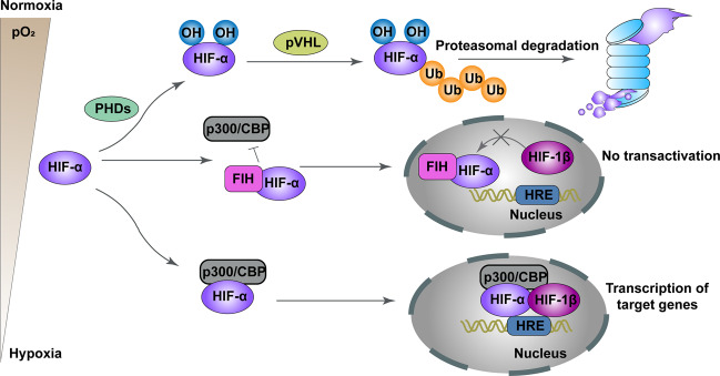
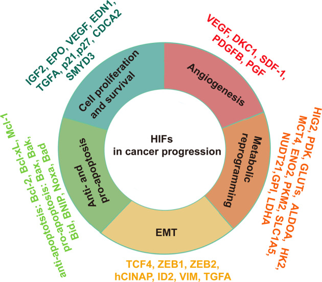
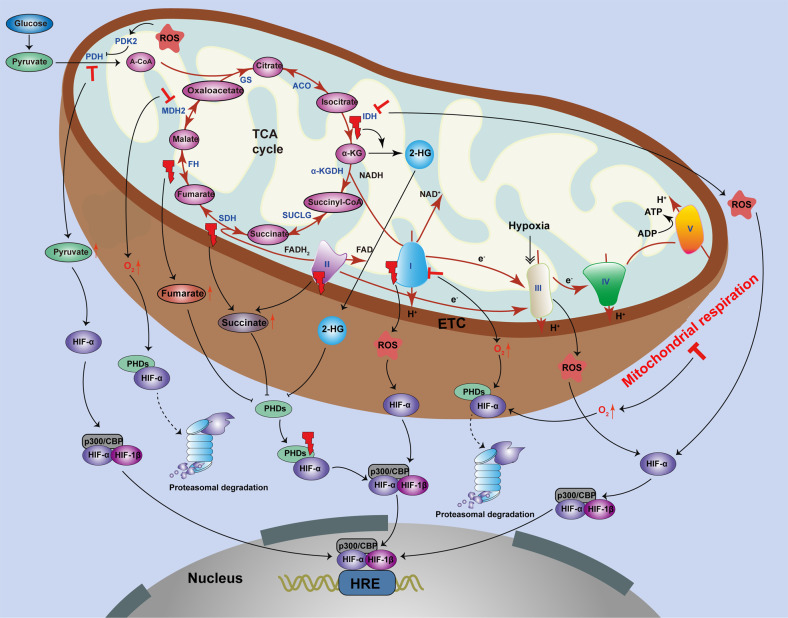
Mitochondria are essential cellular organelles that are involved in regulating cellular energy, metabolism, survival, and proliferation. To some extent, cancer is a genetic and metabolic disease that is closely associated with mitochondrial dysfunction. Hypoxia-inducible factors (HIFs), which are major molecules that respond to hypoxia, play important roles in cancer development by participating in multiple processes, such as metabolism, proliferation, and angiogenesis. The Warburg phenomenon reflects a pseudo-hypoxic state that activates HIF-1α. In addition, a product of the Warburg effect, lactate, also induces HIF-1α. However, Warburg proposed that aerobic glycolysis occurs due to a defect in mitochondria. Moreover, both HIFs and mitochondrial dysfunction can lead to complex reprogramming of energy metabolism, including reduced mitochondrial oxidative metabolism, increased glucose uptake, and enhanced anaerobic glycolysis. Thus, there may be a connection between HIFs and mitochondrial dysfunction. In this review, we systematically discuss the crosstalk between HIFs and mitochondrial dysfunctions in cancer development. Above all, the stability and activity of HIFs are closely influenced by mitochondrial dysfunction related to tricarboxylic acid cycle, electron transport chain components, mitochondrial respiration, and mitochondrial-related proteins. Furthermore, activation of HIFs can lead to mitochondrial dysfunction by affecting multiple mitochondrial functions, including mitochondrial oxidative capacity, biogenesis, apoptosis, fission, and autophagy. In general, the regulation of tumorigenesis and development by HIFs and mitochondrial dysfunction are part of an extensive and cooperative network.


Conflict of interest statement

The authors declare no competing interests.

Figures

Fig. 1
Fig. 1. Schema of regulation of HIF-α degradation and transcriptional activity.
Under conditions of normoxia, HIF-1α and HIF-2α are continuously degraded through the key oxygen sensor PHD1–3, especially PHD2 which enables HIF-α to bind to the pVHL. FIH also inhibits HIF-α by binding to HIF-α and impeding thecombination of HIF-α to the transcriptional coactivators CBP and p300. Under hypoxic conditions, the hydroxylation of HIFα is restrained, leading to stabilization of HIF-α. Next, HIF-α dimerizes with HIF-1β to comprise a transcriptional activation complex, which binds to HRE and stimulates the transactivation of target genes.
Fig. 2
Fig. 2. Role of HIFs in cancer progression.
HIFs have a wide range of target genes which function to manage different types of signaling pathways in cancer. As an example, HIFs modulate cellular metabolic reprogramming, cell proliferation and survival, angiogenesis, apoptosis, and EMT.
Fig. 3
Fig. 3. The effect of mitochondrial dysfunction on HIFs.
The stability and activity of HIF-α are closely influenced by mitochondrial dysfunction related to the TCA cycle, ETC components, and mitochondrial respiration. First, dysregulation of the TCA cycle affects HIF stability and activity. Mutations of the TCA cycle enzymes, including IDH, SDH, and FH, cause stabilization and accumulation of HIF by inhibiting PHDs. Loss of IDH2 also leads to ROS-dependent stabilization of HIF-1α under normoxic conditions. However, inhibition of MDH2 stimulates HIF-1α degradation in cancer cells. Second, ETC components can have an effect on the stability and activity of HIFs. Mutations of complex I and complex II (also known as SDH) leads to HIF-1α stabilization by increasing ROS and succinate levels, respectively. The mitochondrial complex III can sense hypoxic conditions and produce ROS, which stabilizes the HIF-1α protein. However, deficiency and inhibition of complex I cause decreased HIF-1α stabilization using PHD-mediated degradation. In addition, suppression of mitochondrial respiration impedes stabilization of HIF-1α by reactivating PHD enzymes.
Fig. 4
Fig. 4. Function of HIFs on mitochondrial dysfunction.
HIFs have an effect on multiple mitochondrial functions, including mitochondrial oxidative capacity, OXPHOS, biogenesis, apoptosis, fission, and autophagy. HIF-1 activation downregulates mitochondrial oxidation by inhibiting ATGL-mediated lipolysis via HIG2, which leads to LD storage and subsequently declines mitochondrial FA oxidation under hypoxic conditions, and by inducing PDK, which inhibits PDH, thereby inhibiting the flow of pyruvate into the TCA cycle. HIF-1 also regulates mitochondrial redox by inducing mitochondrial serine catabolism and the production of NADPH. Furthermore, miR-210, which is upregulated by HIF-1, restrains expression of the Fe–S cluster assembly proteins ISCU-1 and ISCU-2, which further restricts mitochondrial ROS generation and induces a shift of energy metabolism from OXPHOS to glycolysis. HIF-1 stimulates the less active ETC components, including NDUFA4L2, COX4-1, COX4-2, complex I, and complex IV, to delay the electron transfer through the ETC, therefore blocking the accumulation of ROS and reducing ROS-mediated apoptosis. HIF-1α reduces mitochondrial biogenesis by targeting PGC-1α, MXI, and HEY1. Meanwhile, NPAS2 increases the expression of glycolytic genes by transcriptionally upregulating HIF-1α. HIF-1 also regulates apoptosis by promoting the formation of VDAC1-∆C from VDAC1. Mitochondrial fission, which results in the activation of mitochondria-associated apoptosis, is negatively regulated by the HIF/miR-125a/Mfn2 pathways during carcinogenesis. Mitochondrial proteins can also be inhibited by suppression of the mTORC1/p70SK/S6 signaling pathway via action of HIF-1α. HIF-1α also induces BNIP3 to promote mitochondrial autophagy.

References

    1. Momcilovic M, et al. In vivo imaging of mitochondrial membrane potential in non-small-cell lung cancer. Nature. 2019;575:380–384. doi: 10.1038/s41586-019-1715-0. - DOI - PMC - PubMed
    1. Nakhle J, Rodriguez A-M, Vignais M-L. Multifaceted roles of mitochondrial components and metabolites in metabolic diseases and cancer. Int. J. Mol. Sci. 2020;21:4405–4436. doi: 10.3390/ijms21124405. - DOI - PMC - PubMed
    1. Han Y, et al. Mitochondrial fission causes cisplatin resistance under hypoxic conditions via ROS in ovarian cancer cells. Oncogene. 2019;38:7089–7105. doi: 10.1038/s41388-019-0949-5. - DOI - PubMed
    1. Sun C, et al. Endocytosis-mediated mitochondrial transplantation: transferring normal human astrocytic mitochondria into glioma cells rescues aerobic respiration and enhances radiosensitivity. Theranostics. 2019;9:3595–3607. doi: 10.7150/thno.33100. - DOI - PMC - PubMed
    1. Yang J, et al. The enhancement of glycolysis regulates pancreatic cancer metastasis. Cell. Mol. Life Sci. 2019;77:305–321. doi: 10.1007/s00018-019-03278-z. - DOI - PMC - PubMed

Publication types

MeSH terms

Substances