. 2021 Feb 26;11(1):4789.
doi: 10.1038/s41598-021-84213-w.

Cyclase-associated protein 2 (CAP2) controls MRTF-A localization and SRF activity in mouse embryonic fibroblasts

Affiliations

Cyclase-associated protein 2 (CAP2) controls MRTF-A localization and SRF activity in mouse embryonic fibroblasts

Lara-Jane Kepser et al. Sci Rep. .

Abstract

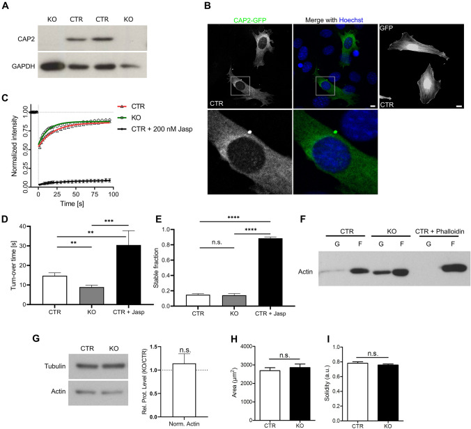
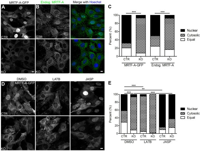
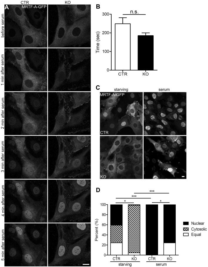
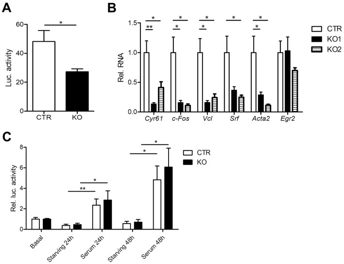
Recent studies identified cyclase-associated proteins (CAPs) as important regulators of actin dynamics that control assembly and disassembly of actin filaments (F-actin). While these studies significantly advanced our knowledge of their molecular functions, the physiological relevance of CAPs largely remained elusive. Gene targeting in mice implicated CAP2 in heart physiology and skeletal muscle development. Heart defects in CAP2 mutant mice were associated with altered activity of serum response factor (SRF), a transcription factor involved in multiple biological processes including heart function, but also skeletal muscle development. By exploiting mouse embryonic fibroblasts (MEFs) from CAP2 mutant mice, we aimed at deciphering the CAP2-dependent mechanism relevant for SRF activity. Reporter assays and mRNA quantification by qPCR revealed reduced SRF-dependent gene expression in mutant MEFs. Reduced SRF activity in CAP2 mutant MEFs was associated with altered actin turnover, a shift in the actin equilibrium towards monomeric actin (G-actin) as well as and reduced nuclear levels of myocardin-related transcription factor A (MRTF-A), a transcriptional SRF coactivator that is shuttled out of the nucleus and, hence, inhibited upon G-actin binding. Moreover, pharmacological actin manipulation with jasplakinolide restored MRTF-A distribution in mutant MEFs. Our data are in line with a model in which CAP2 controls the MRTF-SRF pathway in an actin-dependent manner. While MRTF-A localization and SRF activity was impaired under basal conditions, serum stimulation induced nuclear MRTF-A translocation and SRF activity in mutant MEFs similar to controls. In summary, our data revealed that in MEFs CAP2 controls basal MRTF-A localization and SRF activity, while it was dispensable for serum-induced nuclear MRTF-A translocation and SRF stimulation.


Conflict of interest statement

The authors declare no competing interests.

Figures

Figure 1
Figure 1
CAP2 inactivation increased G-actin levels in MEFs. (A) Immunoblots showing CAP2 expression in two CTR MEF lines and CAP2 inactivation in two KO MEF lines. GAPDH was used as loading control. (B) Representative micrographs showing localization of GFP-tagged CAP2 (green) in CTR MEFs (left panel). Merge micrograph (middle panel) includes counterstaining with DNA dye Hoechst (blue). Box indicates area shown at higher magnification. Representative micrograph of GFP-transfected CTR MEF (right panel). (C) Fluorescence recovery curve after photobleaching in GFP-actin transfected CTR (red) and KO MEFs (green) as well as in CTR MEFs treated with 200 nM jasplakinolide (JASP; black). (D) Half-recovery time of actin turnover and (E) stable actin fraction in CTR and KO MEFs as well as in JASP-treated CTR MFEs. (F) Representative immunoblots showing actin in soluble (G) and insoluble (F) protein fractions. (G) Immunoblot showing total actin levels in CTR and KO MEFs (left). Quantification of relative protein levels from 5 biological replicates (see Fig. S2). Signal intensity of actin was first normalized to tubulin, and then ratio of KO vs CTR was calculated (right). (H) Area quantification of CTR and KO MEFs. (I) Solidity index of CTR and KO MEFs. Scale bar in (B): 10 µm. *P < 0.05, **P < 0.01, ***P < 0.001, ns not significant.
Figure 2
Figure 2
CAP2 controls MRTF-A localization in an actin-dependent manner. (A) Representative micrographs of CTR and KO MEFs that stably expressed GFP-tagged MRTF-A (MRTF-A-GFP). (B) Representative micrographs of CTR and KO MEFs stained with an antibody against MRTF-A (green). MEFs were counterstained with the DNA-dye Hoechst (blue). (C) Categorization of MEFs according to the localization of MRTF-A-GFP or endogenous MRTF-A, i.e. fractions with mainly nuclear or cytosolic MRTF-A-GFP and fraction with equal levels in both compartments. (D) Representative micrographs of MRTF-A-GFP-expressing CTR and KO MEFs upon treatment with either DMSO, latrunculin B (LATB) or jasplakinolide (JASP). (E) Categorization of MEFs according to the localization of MRTF-A-GFP upon treatment with either DMSO, LATB or JASP. Scale bars (in µm): 10 (A,B,D). *P < 0.05, **P < 0.01, ***P < 0.001, ns not significant.
Figure 3
Figure 3
CAP2 was dispensable for serum induced nuclear MRTF-A translocation in MEFs. (A) Image sequence of MRTF-A-GFP-expressing MEFs before and during serum stimulation. (B) Latency of nuclear MRTF-A translocation. (C) Representative micrographs of MRTF-A-GFP-expressing CTR and KO MEFs during serum starvation and upon serum stimulation. (D) Categorization of MEFs according to the localization of MRTF-A-GFP or endogenous MRTF-A during serum starvation and upon serum stimulation. Scale bars (in µm): 10 (A,C). *P < 0.05, **P < 0.01, ***P < 0.001, ns not significant.
Figure 4
Figure 4
CAP2 inactivation reduced SRF activity in MEFs. (A) Firefly luciferase activity in CTR and KO MEFs that stably expressed a SRF reporter in which firefly luciferase expression was under control of SRF activity. (B) mRNA levels of selected SRF target genes in two KO MEF lines as determined by qPCR. (C) Luciferase expression in CTR and KO MEFs under basal conditions, during starving and upon serum stimulation for either 24 or 48 h. Values are normalized to basal levels. *P < 0.05, **P < 0.01, ***P < 0.001, ns not significant.

Similar articles

Cited by

References

    1. Balcer HI, Goodman AL, Rodal AA, Smith E, Kugler J, Heuser JE, Goode BL. Coordinated regulation of actin filament turnover by a high-molecular-weight Srv2/CAP complex, cofilin, profilin, and Aip1. Curr. Biol. 2003;13:2159–2169. doi: 10.1016/j.cub.2003.11.051. - DOI - PubMed
    1. Bertling E, Hotulainen P, Mattila PK, Matilainen T, Salminen M, Lappalainen P. Cyclase-associated protein 1 (CAP1) promotes cofilin-induced actin dynamics in mammalian nonmuscle cells. Mol. Biol. Cell. 2004;15:2324–2334. doi: 10.1091/mbc.e04-01-0048. - DOI - PMC - PubMed
    1. Freeman NL, Field J. Mammalian homolog of the yeast cyclase associated protein, CAP/Srv2p, regulates actin filament assembly. Cell Motil. Cytoskeleton. 2000;45:106–120. doi: 10.1002/(SICI)1097-0169(200002)45:2<106::AID-CM3>3.0.CO;2-3. - DOI - PubMed
    1. Hubberstey AV, Mottillo EP. Cyclase-associated proteins: CAPacity for linking signal transduction and actin polymerization. FASEB J. 2002;16:487–499. doi: 10.1096/fj.01-0659rev. - DOI - PubMed
    1. Chaudhry F, Little K, Talarico L, Quintero-Monzon O, Goode BL. A central role for the WH2 domain of Srv2/CAP in recharging actin monomers to drive actin turnover in vitro and in vivo. Cytoskeleton (Hoboken, NJ) 2010;67:120–133. - PMC - PubMed

Publication types