. 2021 Feb 26;11(1):4767.
doi: 10.1038/s41598-021-84081-4.

Autophagy-enhancing drugs limit mucosal HIV-1 acquisition and suppress viral replication ex vivo

Affiliations

Autophagy-enhancing drugs limit mucosal HIV-1 acquisition and suppress viral replication ex vivo

Alexandra P M Cloherty et al. Sci Rep. .

Abstract

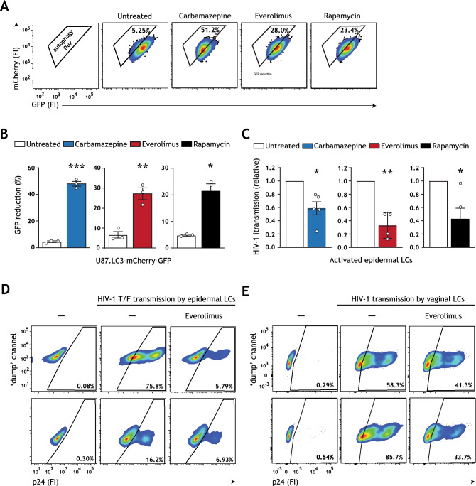
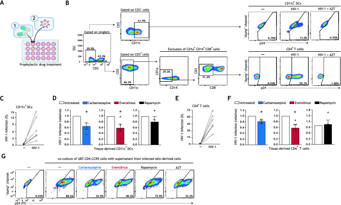
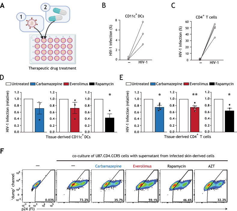
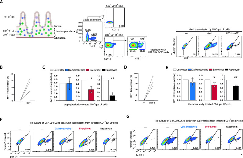
Current direct-acting antiviral therapies are highly effective in suppressing HIV-1 replication. However, mucosal inflammation undermines prophylactic treatment efficacy, and HIV-1 persists in long-lived tissue-derived dendritic cells (DCs) and CD4+ T cells of treated patients. Host-directed strategies are an emerging therapeutic approach to improve therapy outcomes in infectious diseases. Autophagy functions as an innate antiviral mechanism by degrading viruses in specialized vesicles. Here, we investigated the impact of pharmaceutically enhancing autophagy on HIV-1 acquisition and viral replication. To this end, we developed a human tissue infection model permitting concurrent analysis of HIV-1 cellular targets ex vivo. Prophylactic treatment with autophagy-enhancing drugs carbamazepine and everolimus promoted HIV-1 restriction in skin-derived CD11c+ DCs and CD4+ T cells. Everolimus also decreased HIV-1 susceptibility to lab-adapted and transmitted/founder HIV-1 strains, and in vaginal Langerhans cells. Notably, we observed cell-specific effects of therapeutic treatment. Therapeutic rapamycin treatment suppressed HIV-1 replication in tissue-derived CD11c+ DCs, while all selected drugs limited viral replication in CD4+ T cells. Strikingly, both prophylactic and therapeutic treatment with everolimus or rapamycin reduced intestinal HIV-1 productive infection. Our findings highlight host autophagy pathways as an emerging target for HIV-1 therapies, and underscore the relevancy of repurposing clinically-approved autophagy drugs to suppress mucosal HIV-1 replication.


Conflict of interest statement

The authors declare no competing interests.

Figures

Figure 1
Figure 1
Carbamazepine, everolimus, and rapamycin induce autophagy flux and decrease HIV-1 susceptibility. (A,B) U87 cell line expressing tandem fluorescent-tagged LC3 (mCherry-GFP-LC3) was incubated with carbamazepine (1000 μM), everolimus (30 nM), or rapamycin (100 nM) for 48 h. The GFP tag is acid-sensitive and is quenched upon autophagosome acidification. Autophagy flux was determined by the % GFP signal reduction. Representative flow cytometry plots (A) and quantification (B), data are mean ± SE of n = 3 experiments measured in duplicate. Open circles represent the mean of duplicates from each independent experiment. *P < 0.05, **P < 0.01, ***P < 0.001, Student's t test. (C) Human epidermal biopsies were prophylactically treated with carbamazepine (100 μM), everolimus (30 nM), or rapamycin (100 nM), or left untreated, and subsequently infected with HIV-1 NL4.3 BaL for 72 h. Emigrated LCs were washed, and co-cultured with U87.CD4.CCR5 cells. HIV-1 transmission by LCs was assessed in LC-U87.CD4.CCR5 co-culture for 72 h, determined by intracellular p24 staining by flow cytometer. LC-marker CD1a was used to exclude single LCs and LC-U87 conjugates from analysis. A detailed graphical depiction of HIV-1 transmission across epidermal tissue is available in Figure S2A, Supplementary Information. Data are mean ± SE of n = 5 experiments measured in duplicate. Open circles represent the mean of duplicates from each independent experiment. *P < 0.05, **P < 0.01, one-sample t test. (D) Human epidermal biopsies were pre-treated with everolimus (30 nM), followed by infection with transmitted/founder HIV-1 THRO for 72 h. Emigrated LCs were washed, and co-cultured with U87.CD4.CCR5 cells. HIV-1 transmission by LCs was assessed in LC-U87.CD4.CCR5 co-culture for 72 h, determined by intracellular p24 staining by flow cytometer. LC-marker CD1a was used to exclude single LCs and LC-U87 conjugates from analysis. Flow cytometry plots of HIV-1 infection of U87.CD4.CCR5 cells for each epidermal tissue donor is shown. (E) Isolated human immature vaginal LCs, obtained by CD1a magnetic cell isolation, were pre-treated with everolimus (30 nM) and subsequently infected with HIV-1 NL4-3 BaL for 72 h. Vaginal LCs were washed, and co-cultured with U87.CD4.CCR5 cells. HIV-1 transmission by LCs was assessed in LC-U87.CD4.CCR5 co-culture for 72 h, determined by intracellular p24 staining by flow cytometer. LC-marker CD1a was used to exclude single LCs and LC-U87 conjugates from analysis. Flow cytometry plots of HIV-1 infection of U87.CD4.CCR5 cells for each vaginal LC donor is shown.
Figure 2
Figure 2
Prophylactic treatment with autophagy drugs reduces HIV-1 acquisition by tissue-derived DCs and CD4+ T cells. (A) Brief schematic representation of the novel HIV-1 tissue infection model for drug screening; an extended graphical representation of the prophylactic treatment model is available in Figure S3A, Supplementary Information. Biopsies including epithelium and subepithelium were taken from skin and cultured in a 24 well plate. Skin biopsies were prophylactically treated with autophagy drugs and subsequently infected with HIV-1. HIV-1 infection of different cell types and subsets was quantified using multiparameter flow cytometry. (B) Gating strategy utilized to discriminate HIV-1 infection in the emigrated CD11c+ DCs and CD4+ T cells. Treatment of tissue biopsies with HIV-1 replication inhibitor AZT (zidovudine, 20 μM) confirmed productive HIV-1 infection of these cell types. A detailed gating strategy can be found in Figure S4, Supplementary Information. (CF) Human tissue biopsies were prophylactically treated with carbamazepine (100 μM), everolimus (5 nM), or rapamycin (100 nM) for 15 h, or left untreated, and subsequently infected with HIV-1 NL4.3BaL for 48 h. Emigrated cells were then harvested, washed, and replated for an additional 72 h. HIV-1 infection of emigrated CD11c+DCs was determined as the percentage of CD3CD11c+p24+ cells (C,D), and HIV-1 infection of emigrated CD4+ T cells as the percentage of CD3+CD11cCD14CD1aCD8p24+ cells (E,F), determined by flow cytometer. (CF) Data are mean ± SE of n = 4–5 donors measured in duplicate. Open circles represent the mean of duplicates from each independent experiment. (D,F) Untreated HIV-1 infected cells was set at 1 *P < 0.05; one-sample t test. (G) Skin biopsies including epithelium and subepithelium were prophylactically treated for 15 h with autophagy drugs carbamazepine (100 μM), everolimus (5 nM), rapamycin (100 nM), HIV-1 replication inhibitor AZT (zidovudine, 20 μM), or left untreated, followed by infection with HIV-1 NL4.3BaL. 36 h post-infection, emigrated tissue-derived cells were extensively washed to remove input virus, and replated in new medium in a 96-well plate. Supernatant from infected tissue-derived cells was collected 120 h after replating and co-cultured with U87.CD4.CCR5 cells for 72 h, to further confirm productive HIV-1 infection. A detailed graphical representation of this extracellular virus release assay is available in Figure S3B, Supplementary Information. HIV-1 infection of U87.CD4.CCR5 cells was determined by intracellular p24 staining by flow cytometer. Representative (n = 2 tissue donors) flow cytometry plots of HIV-1 infection of U87.CD4.CCR5 cells incubated with supernatants from HIV-1 infected skin-derived cells is shown.
Figure 3
Figure 3
Therapeutic treatment with autophagy drugs reduces HIV-1 replication in a cell-specific manner. (A) Brief schematic representation of HIV-1 tissue infection model for drug screening, an extended graphical representation of the therapeutic treatment model is available in Figure S3C, Supplementary Information. Biopsies including epithelium and subepithelium were taken from human skin tissue, infected with HIV-1 NL4.3BaL for 15 h, and subsequently treated with autophagy drugs. HIV-1 infection of different cell types and subsets was quantified using multiparameter flow cytometry. A detailed gating strategy can be found in Supplementary Information Figure S4. (BE) Human tissue biopsies were therapeutically treated with carbamazepine (100 μM), everolimus (5 nM), or rapamycin (100 nM), or left untreated. HIV-1 infection of emigrated CD11c+DCs was determined as the percentage of CD3CD11c+p24+ cells (B,D), and HIV-1 infection of emigrated CD4+ T cells as the percentage of CD3+CD11cCD14CD1aCD8p24+ cells (C,E), determined by flow cytometer. (BE) Data are mean ± SE of n = 3–5 donors measured in duplicate. Open circles represent the mean of duplicates from each independent experiment. (D,E) Untreated, HIV-1 infected cells was set at 1, *P < 0.05, **P < 0.01, one-sample t test. (F) Skin biopsies including epithelium and subepithelium were infected with HIV-1 NL4.3BaL for 15 h. Subsequently, skin biopsies were therapeutically treated with autophagy drugs carbamazepine (100 μM), everolimus (5 nM), rapamycin (100 nM), HIV-1 replication inhibitor AZT (zidovudine, 20 μM), or left untreated. 36 h after treatment, emigrated tissue-derived cells were extensively washed to remove input virus, and replated in new medium in a 96-well plate. Supernatant from infected tissue-derived cells was collected 120 h following replating and co-cultured with U87.CD4.CCR5 cells for 72 h, to further confirm productive HIV-1 infection. A detailed graphical representation of this extracellular virus release assay is available in Figure S3D, Supplementary Information. HIV-1 infection of U87.CD4.CCR5 cells was determined by intracellular p24 staining by flow cytometer. Representative (n = 2 tissue donors) flow cytometry plots of HIV-1 infection of U87.CD4.CCR5 cells incubated with supernatants from HIV-1 infected skin-derived cells is shown.
Figure 4
Figure 4
Autophagy-enhancing drugs everolimus and rapamycin limit intestinal HIV-1 transmission and reduce ongoing HIV-1 replication in intestinal CD4 + T cells. (A) Human intestinal lamina propria lymphocytes (LPLs) were isolated as previously described. Gating strategy employed to identify gut CD4 + T cells is shown; an extended gating strategy is available in Figure S7D, Supplementary Information. Following HIV-1 infection, LPLs were washed extensively to remove input virus, and subsequently co-cultured with permissive U87.CD4.CCR5 cells for 72 h. HIV-1 transmission by gut LPLs was determined by intracellular p24 staining; treatment of LPLs with HIV-1 replication inhibitor AZT (zidovudine, 20 μM) confirmed productive intestinal HIV-1 infection. (B,C) LPLs were prophylactically treated with carbamazepine (100 μM), everolimus (5 nM), or rapamycin (100 nM) for 15 h, or left untreated, followed by infection with HIV-1 NL4.3BaL for 24 h. Gut LPLs were harvested and washed to remove input virus, and subsequently co-cultured with U87.CD4.CCR5 cells. HIV-1 transmission by LPLs was assessed in LPL-U87.CD4.CCR5 co-culture for 72 h, determined by intracellular p24 staining by flow cytometer. T cell-marker CD3 was used to exclude single LPLs and LPL-U87.CD4.CCR5 conjugates from analysis. A detailed graphical depiction of HIV-1 transmission by gut LPLs is available in Supplementary Information Figure S7E. Data are mean ± SE of n = 3 donors measured in duplicate. Open circles represent the mean of duplicates from each independent experiment. Untreated, HIV-1 infected cells was set at 1, *P < 0.05, **P < 0.01, one-sample t test. (D,E) LPLs were infected with HIV-1 NL4.3BaL for 15 h, and subsequently therapeutically treated with carbamazepine (100 μM), everolimus (5 nM), or rapamycin (100 nM) for 24 h, or left untreated. Gut LPLs were harvested and washed to remove input virus, and subsequently co-cultured with U87.CD4.CCR5 cells. HIV-1 transmission by LPLs was assessed in LPL-U87.CD4.CCR5 co-culture for 72 h, determined by intracellular p24 staining by flow cytometer. T cell-marker CD3 was used to exclude single LPLs and LPL-U87.CD4.CCR5 conjugates from analysis. A detailed graphical depiction of HIV-1 transmission by gut LPLs is available in Figure S7F, Supplementary Information. (F) Following prophylactic treatment and infection with HIV-1 NL4.3BaL (as in B,C), LPLs were extensively washed to remove input virus and replated in new medium in a 96-well plate. (G) Following infection with HIV-1 NL4.3BaL and therapeutic treatment (as in D,E), LPLs were extensively washed to remove input virus and replated in new medium in a 96-well plate. (F,G) Supernatants from infected intestinal prophylactically—(F) or therapeutically—(G) treated LPLs were collected 24 h after replating and co-cultured with U87.CD4.CCR5 cells for 72 h, to further confirm productive intestinal HIV-1 infection. HIV-1 infection of U87.CD4.CCR5 cells was determined by intracellular p24 staining by flow cytometer. Representative (n = 2 tissue donors) flow cytometry plots of HIV-1 infection of U87.CD4.CCR5 cells incubated with supernatants from HIV-1 infected gut LPLs.

References

    1. HIV/AIDS Factsheet. World health organization.https://www.who.int/news-room/fact-sheets/detail/hiv-aids (2019).
    1. Tebit DM, Ndembi N, Weinberg A, Quinones-Mateu ME. Mucosal transmission of human immunodeficiency virus. Curr. HIV Res. 2012;10:3–8. doi: 10.2174/157016212799304689. - DOI - PMC - PubMed
    1. Cavarelli M, Scarlatti G. HIV-1 infection: The role of the gastrointestinal tract. Am. J. Reprod. Immunol. 2014;71:537–542. doi: 10.1111/aji.12245. - DOI - PubMed
    1. Trifonova RT, Bollman B, Barteneva NS, Lieberman J. Myeloid cells in intact human cervical explants capture HIV and can transmit it to CD4 T cells. Front. Immunol. 2018;9:1–12. doi: 10.3389/fimmu.2018.02719. - DOI - PMC - PubMed
    1. Patterson BK, et al. Susceptibility to human immunodeficiency virus-1 infection of human foreskin and cervical tissue grown in explant culture. Am. J. Pathol. 2002;161:867–873. doi: 10.1016/S0002-9440(10)64247-2. - DOI - PMC - PubMed

Publication types

Substances