. 2021 Feb 26;4(1):250.
doi: 10.1038/s42003-021-01779-x.

Identification of a conserved virion-stabilizing network inside the interprotomer pocket of enteroviruses

Affiliations

Identification of a conserved virion-stabilizing network inside the interprotomer pocket of enteroviruses

Justin W Flatt et al. Commun Biol. .

Abstract

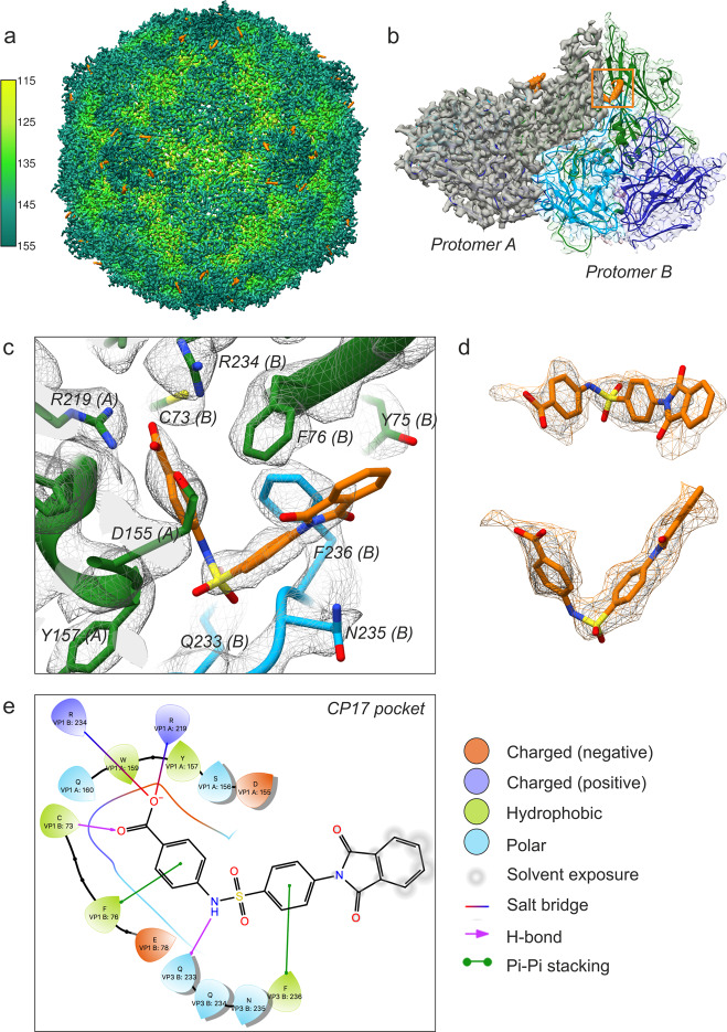
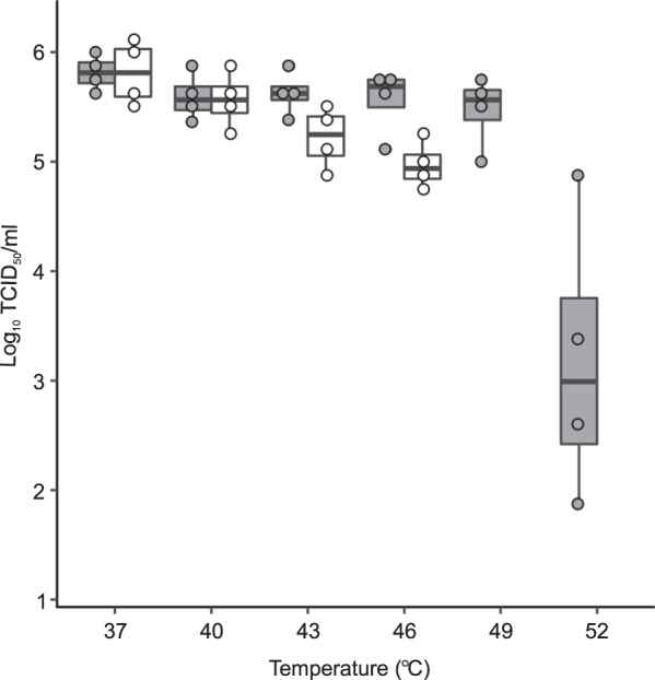
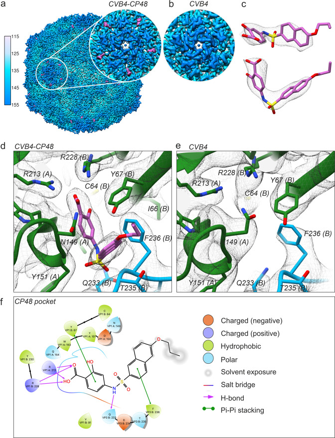
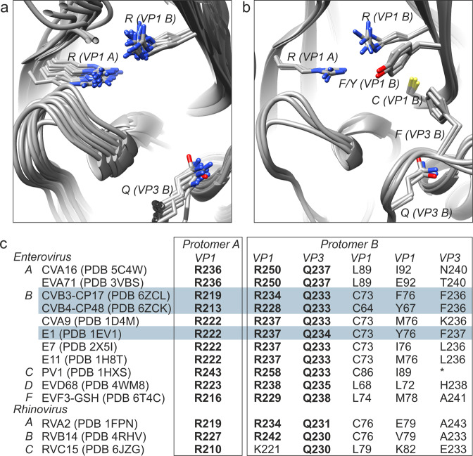
Enteroviruses pose a persistent and widespread threat to human physical health, with no specific treatments available. Small molecule capsid binders have the potential to be developed as antivirals that prevent virus attachment and entry into host cells. To aid with broad-range drug development, we report here structures of coxsackieviruses B3 and B4 bound to different interprotomer-targeting capsid binders using single-particle cryo-EM. The EM density maps are beyond 3 Å resolution, providing detailed information about interactions in the ligand-binding pocket. Comparative analysis revealed the residues that form a conserved virion-stabilizing network at the interprotomer site, and showed the small molecule properties that allow anchoring in the pocket to inhibit virus disassembly.


Conflict of interest statement

The authors declare no competing interests.

Figures

Fig. 1
Fig. 1. Cryo-EM structure of CP17-bound CVB3.
a Three-dimensional reconstruction of CVB3 after incubation with a saturating amount of capsid binder. The virion is viewed along the icosahedral twofold axis and colored according to radial distance in Å from the particle center. Density for CP17 is shown in orange. The map was resolved to 2.8 Å upon reprocessing raw data from a previous publication (see ref. ; EMPIAR-10199). b CP17 binds in a pocket between neighboring protomers. VP1, green; VP2, dark blue; VP3, light blue. c Inhibitor and pocket residues at a display contour level of 2.5σ (σ is the standard deviation of the density map). d CP17 shown in density contoured to 1.6σ. e Ligand interactions diagram for CP17 generated by Schrödinger Maestro v12.02.
Fig. 2
Fig. 2. A box and whisker plot showing the effect of CP48 on CVB4 thermal stability.
Thermostability assay in the presence (gray) or absence (white) of interprotomer-targeting CP48. No virus was detected in control samples at 49 and 52 °C at the TCID50/mL detection limit of 2 log10. N = 4 independent experiments, the middle bar is the median, the boxes represent quartile data distribution, and individual data points are shown as circles. Figure generated with the command ggplot in R.
Fig. 3
Fig. 3. Cryo-EM of the CP48–CVB4 complex in comparison to CVB4 alone.
a Visualization of CVB4 in the presence of CP48. The view is along the twofold axis with radial coloring, and the inset shows clear inhibitor density at the interprotomer site near the fivefold axis of symmetry. Density for the 385.44 Da CP48 is displayed in magenta. b The corresponding region of the cryo-EM density of the CVB4 control. c CP48 fits well into the additional density detected in the cryo-EM map of the CP48–CVB4 complex. d A close-up view of the capsid binder (magenta) within the interprotomer pocket. e A close-up view of the interprotomer pocket in the cryo-EM map of CVB4 alone shows no density for the compound. d, e Color coding for the viral proteins VP1, VP2, and VP3 is the same as in Fig. 1. f Interaction diagram of CP48 with CVB4 viral proteins generated in Schrödinger Maestro software v12.02.
Fig. 4
Fig. 4. Stabilization inside the interprotomer pocket of enteroviruses.
a Structural alignment of the three key residues that form the core of the virion-stabilizing network inside the interprotomer pocket. Atomic models used for the alignment are listed in C. b Alignment of PDB IDs 6ZCK, 6ZCL, and 1EV1 with the same view as a but with CP17/48-stabilizing hydrophobic residues of the interprotomer pocket added to the visual. c Conservation of anchor residues in the pocket based on the structural data presented in this study. The three residues that are highly conserved are in bold whereas the other major elements (hydrophobic and cysteine) vary. Enterovirus A, B, C, D, and F and Rhinovirus A, B, and C species are indicated in column 1 along with the wwPDB IDs. The numbering of the residues in columns 2–6 were taken from the wwPDB files listed in column 1. (*) not modeled in the coordinates for 1HXS.

References

    1. Kearney MT, Cotton JM, Richardson PJ, Shah AM. Viral myocarditis and dilated cardiomyopathy: mechanisms, manifestations, and management. Postgrad. Med. J. 2001;77:4–10. doi: 10.1136/pmj.77.903.4. - DOI - PMC - PubMed
    1. Massilamany C, Gangaplara A, Reddy J. Intricacies of cardiac damage in coxsackievirus B3 infection: implications for therapy. Int. J. Cardiol. 2014;177:330–339. doi: 10.1016/j.ijcard.2014.09.136. - DOI - PMC - PubMed
    1. Euscher E, Davis J, Holzman I, Nuovo GJ. Coxsackie virus infection of the placenta associated with neurodevelopmental delays in the newborn. Obstet. Gynecol. 2001;98:1019–1026. - PubMed
    1. Ornoy A, Tenenbaum A. Pregnancy outcome following infections by coxsackie, echo, measles, mumps, hepatitis, polio and encephalitis viruses. Reprod. Toxicol. 2006;21:446–457. doi: 10.1016/j.reprotox.2005.12.007. - DOI - PubMed
    1. Notkins AL, Yoon J, Onodera T, Jenson AB. Virus-induced diabetes mellitus: infection of mice with variants of encephalomyocarditis virus, coxsackievirus B4, and reovirus type 3. Adv. Exp. Med. Biol. 1979;119:137–146. doi: 10.1007/978-1-4615-9110-8_21. - DOI - PubMed

Publication types

MeSH terms