

Novel Application of Photogrammetry to Quantify Fascicle Orientations of Female Cadaveric Pelvic Floor Muscles
- PMID: 33638030
- PMCID: PMC8376743
- DOI: 10.1007/s10439-021-02747-6
Novel Application of Photogrammetry to Quantify Fascicle Orientations of Female Cadaveric Pelvic Floor Muscles
Abstract
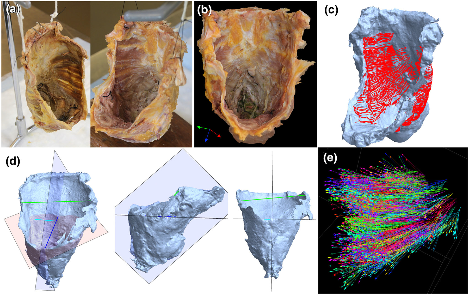
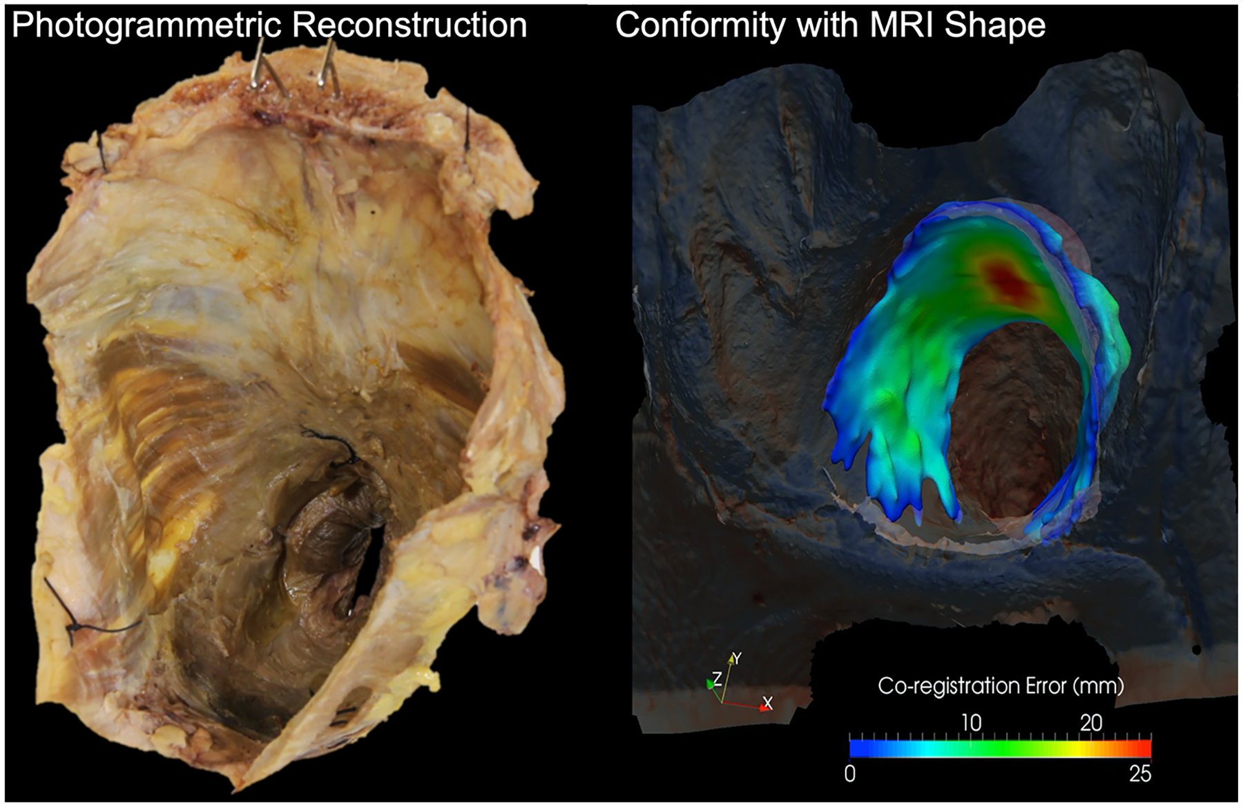
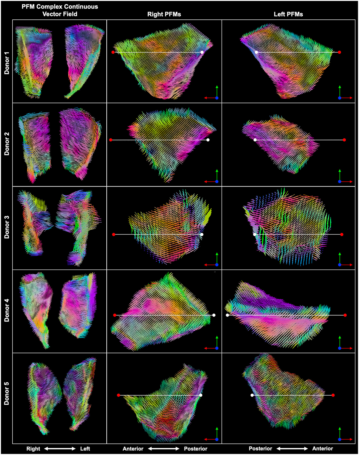
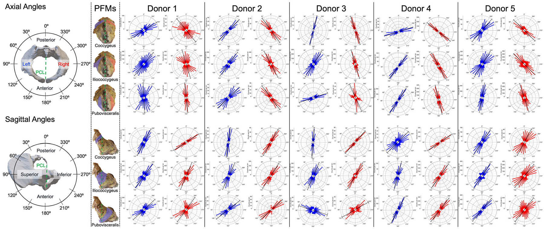
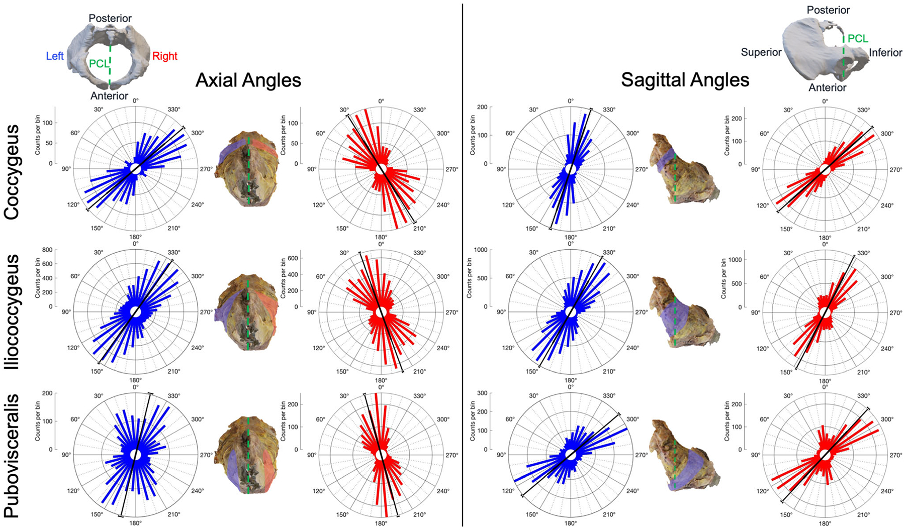
Although critical for understanding and simulating pelvic floor muscle function and pathophysiology, the fascicle arrangements of the coccygeus and levator ani remain mostly undetermined. We performed close-range photogrammetry on cadaveric pelvic floor muscles to robustly quantify surface fascicle orientations. The pelvic floor muscles of 5 female cadavers were exposed through anatomic dissections, removed en bloc, and photographed from every required angle. Overlapping images were mapped onto in silico geometries and muscle fascicles were traced manually. Tangent vectors were calculated along each trace; interpolated to define continuous, 3D vector fields; and projected onto axial and sagittal planes to calculate angles with respect to the pubococcygeal line. Contralateral and ipsilateral pelvic floor muscles were compared within each donor (Kuiper's tests) and using mean values from all donors (William-Watsons tests). Contralateral muscles and all but one ipsilateral muscle pair differed significantly within each donor (p < 0.001). When mean values were considered collectively, no contralateral or ipsilateral statistical differences were found but all muscles compared differed by more than 10° on average. Close-range photogrammetry and subsequent analyses robustly quantified surface fascicle orientations of the pelvic floor muscles. The continuous, 3D vector fields provide data necessary for improving simulations of the female pelvic floor muscles.
Keywords: Close-range photogrammetry; Coccygeus; Iliococcygeus; Muscle fascicles; Pubovisceralis; Vector fields.
© 2021. Biomedical Engineering Society.
Conflict of interest statement
CONFLICT OF INTEREST
WB is employed by Bayer U.S. LLC, Radiology R&D at the time of submission/publication, SA receives investigator-initiated research funding from Renovia Inc. for work unrelated to this project, and MA is on the Medical Advisory Board, Renovia, Inc. and receives an Editorial stipend from the American Journal of Obstetrics and Gynecology (AJOG). All other authors report no conflicts of interest.
Figures
Similar articles
-
Impact of vaginal parity and aging on the architectural design of pelvic floor muscles.Am J Obstet Gynecol. 2016 Sep;215(3):312.e1-9. doi: 10.1016/j.ajog.2016.02.033. Epub 2016 Mar 5. Am J Obstet Gynecol. 2016. PMID: 26953079 Free PMC article.
-
Localization of the nerves innervating the pelvic floor muscles: An application to pelvic pain treatment.Clin Anat. 2022 Oct;35(7):979-986. doi: 10.1002/ca.23935. Epub 2022 Aug 3. Clin Anat. 2022. PMID: 35842771
-
Age-associated changes in the mechanical properties of human cadaveric pelvic floor muscles.J Biomech. 2020 Jan 2;98:109436. doi: 10.1016/j.jbiomech.2019.109436. Epub 2019 Oct 31. J Biomech. 2020. PMID: 31708240 Free PMC article.
-
Anatomy and Physiology of the Pelvic Floor.Phys Med Rehabil Clin N Am. 2017 Aug;28(3):455-460. doi: 10.1016/j.pmr.2017.03.003. Epub 2017 May 27. Phys Med Rehabil Clin N Am. 2017. PMID: 28676358 Review.
-
Clinical anatomy of the pelvic floor.Adv Anat Embryol Cell Biol. 2004;175:III-IX, 1-64. doi: 10.1007/978-3-642-18548-9. Adv Anat Embryol Cell Biol. 2004. PMID: 15152384 Review.
Cited by
-
Special Issue on the Advances in Engineering for Women's Health.Ann Biomed Eng. 2021 Aug;49(8):1785-1787. doi: 10.1007/s10439-021-02837-5. Ann Biomed Eng. 2021. PMID: 34379234 No abstract available.
-
Twisted orientation of the muscle bundles in the levator ani functional parts in women: Implications for pelvic floor support mechanism.J Anat. 2024 Mar;244(3):486-496. doi: 10.1111/joa.13968. Epub 2023 Oct 26. J Anat. 2024. PMID: 37885272 Free PMC article.
References
-
- Aber JS, Marzolff I, Ries JB, and Aber SEW. Principles of Photogrammetry. In: Small-Format Aerial Photography and UAS Imagery. Elsevier, 2019, pp. 19–38.
-
- Agur AM, Ng-Thow-Hing V, Ball KA, Fiume E, and McKee NH. Documentation and three-dimensional modelling of human soleus muscle architecture. Clin. Anat 16:285–293, 2003. - PubMed
-
- Berens P. CircStat: Circular Statistics Toolbox (Directional Statistics)., 2020. https://www.mathworks.com/matlabcentral/fileexchange/10676-circular-stat....
MeSH terms
Grants and funding
LinkOut - more resources
Full Text Sources
Other Literature Sources
Research Materials
