. 2021 Jan-Jun:296:100467.
doi: 10.1016/j.jbc.2021.100467. Epub 2021 Feb 25.

An electrochemical method for detecting the biomarker 4-HPA by allosteric activation of Acinetobacterbaumannii reductase C1 subunit

Affiliations

An electrochemical method for detecting the biomarker 4-HPA by allosteric activation of Acinetobacterbaumannii reductase C1 subunit

Somjai Teanphonkrang et al. J Biol Chem. 2021 Jan-Jun.

Abstract

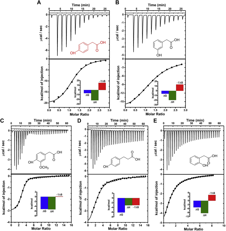
The C1 (reductase) subunit of 4-hydroxy-phenylacetate (4-HPA) 3-hydroxylase (HPAH) from the soil-based bacterium Acinetobacterbaumannii catalyzes NADH oxidation by molecular oxygen, with hydrogen peroxide as a by-product. 4-HPA is a potent allosteric modulator of C1, but also a known urinary biomarker for intestinal bacterial imbalance and for some cancers and brain defects. We thus envisioned that C1 could be used to facilitate 4-HPA detection. The proposed test protocol is simple and in situ and involves addition of NADH to C1 in solution, with or without 4-HPA, and direct acquisition of the H2O2 current with an immersed Prussian Blue-coated screen-printed electrode (PB-SPE) assembly. We confirmed that cathodic H2O2 amperometry at PB-SPEs is a reliable electrochemical assay for intrinsic and allosterically modulated redox enzyme activity. We further validated this approach for quantitative NADH electroanalysis and used it to evaluate the activation of NADH oxidation of C1 by 4-HPA and four other phenols. Using 4-HPA, the most potent effector, allosteric activation of C1 was related to effector concentration by a simple saturation function. The use of C1 for cathodic biosensor analysis of 4-HPA is the basis of the development of a simple and affordable clinical routine for assaying 4-HPA in the urine of patients with a related disease risk. Extension of this principle to work with other allosteric redox enzymes and their effectors is feasible.

Keywords: allosteric regulation; amperometry; biosensor; disease biomarker; electroanalysis; hydroxylase; oxidase; redox signaling; reduced nicotinamide adenine dinucleotide (NADH); reductase.


Conflict of interest statement

Conflict of interest The authors declare no conflicts of interest in regard to this article.

Figures

Figure 1
Figure 1
Anodic and cathodic electrochemical detection of H2O2with test of interference.A and B are the amperometric current responses of a Pt disk and a PB-SPE WE to serial additions of 50 μM of 4-HPA, NADH, and H2O2 at detection potentials of +600 and –100 mV versus RE, respectively. C, is a typical amperogram from a H2O2 calibration trial with a PB-SPE at –100 mV vs. RE. D, is the H2O2 calibration plot derived from the measurement shown in (C). The electrolyte in (A), (B), and (C) was 50 mM sodium phosphate buffer, pH 7. Data points and error bars represent the means and standard deviations of triplicate analyses.
Figure 2
Figure 2
A, Cathodic H2O2amperometry in the standard addition mode for quantification of the analyte in a model sample of 100 μM concentration. The target, H2O2, was measured with a PB-SPE at a potential of –100 mV versus RE. The electrolyte was 50 mM sodium phosphate buffer, pH 7. B, Standard addition plot constructed with the data presented in (A).
Figure 3
Figure 3
Measurement of NADH oxidation by C1, with and without allosteric activation, by cathodic H2O2amperometry. The blue trace is the amperometric current response of a PB-SPE at –100 mV versus RE in phosphate buffer, pH 7 containing 1 μM C1, with addition of 50 μM NADH about 360 s after the start of the trial. The red trace is the amperometric current response of a PB-SPE at –100 mV versus Ag/AgCl in sodium phosphate buffer containing 1 μM C1 and 100 μM 4-HPA with addition of 50 μM NADH about 360 s after the start of the trial. The steady signal increase after addition of NADH shows the development of the cathodic current for the reduction of enzymically produced H2O2; the curve slopes (ΔIt) thus are indicators of normal (blue trace) and accelerated (red trace) NADH oxidation by C1. The plateau signifies full consumption of the substrate in each case.
Figure 4
Figure 4
Quantitative detection of NADH by C1 with cathodic H2O2amperometry.A, is the amperometric H2O2 current response of a PB-SPE at –100 mV versus RE in trial buffer with 1 μM C1 and 100 μM 4-HPA with sequential additions of small aliquots of NADH. B, is the NADH calibration plot derived from the measurement shown in (A). Data points and error bars represent the means and standard deviations of triplicate analyses. C, is an amperometric recording of NADH quantification in the standard addition mode for a sample with 50 μM NADH and three additions of 50 μM NADH. D, standard addition curve constructed with the data of the trace in C. Electrolyte for the measurements in (A) and (C) was 50 mM sodium phosphate buffer, pH 7.
Figure 5
Figure 5
Evaluation of concentration-dependent allosteric C1 activation by 4-HPA by cathodic H2O2amperometry.A, the amperometric H2O2 current responses of a PB-SPE at –100 mV versus RE in sodium phosphate buffer, pH 7 containing 1 μM C1 and 5–500 μM 4-HPA, with 50 μM NADH added about 360 s after the start of recording. B, changes of the slopes (ΔIt) of the H2O2 current traces (ΔS = S4-HPA – Sno 4-HPA) in (A) plotted against the concentration of the allosteric effector, 4-HPA. Data points and error bars represent the means and standard deviations from triplicate analyses. The line through the data points is a fit based on the Michaelis–Menten function. The maximum rate change of allosterically enhanced enzymatic H2O2 production and the effector concentration for half-maximal activation (apparent dissociation constant, Kd) are 37 nA s-1 and 103 μM, respectively. The inset shows the first six points of the plot on an expanded scale.
Figure 6
Figure 6
Allosteric activation of C1 by hydroxyphenyl analogues, measured by cathodic H2O2amperometry. The plots show amperometric H2O2 current responses of a PB-SPE at –100 mV versus RE in sodium phosphate buffer, pH 7 containing 1 μM C1 plus 100 μM allosteric effector, with 50 μM NADH added about 360 s after the start of recording. The chemical structures of the phenolic allosteric effectors are shown on the right.
Figure 7
Figure 7
ITC assessment of the interaction between reductase C1 and four allosteric activators: the top panels show the raw data for (A) 4-HPA, (B) 3-HPA, (C) 4-hydroxy-3-methoxy phenylacetate, (D) 3-(4-hydroxyphenyl) propionate and (E) 2-HPA and, as insets, the chemical structures of the five organic compounds. The bottom panels show the binding isotherms created by plotting the integrated heat peaks against the molar ratio between enzyme and activator. Data points were fitted for a one-site model with variable stoichiometry, N. The insets of the bottom panels of A–E are the thermodynamic profiles for binding of the various phenols to C1.

Similar articles

References

    1. Fenton A.W. Allostery: An illustrated definition for the ‘second secret of life’. Trends Biochem. Sci. 2008;33:420–425. - PMC - PubMed
    1. Goodey N.M., Benkovic S.J. Allosteric regulation and catalysis emerge via a common route. Nat. Chem. Biol. 2008;4:474–482. - PubMed
    1. Motlagh H.N., Wrabl J.O., Li J., Hilser V.J. The ensemble nature of allostery. Nature. 2014;508:331–339. - PMC - PubMed
    1. Campitelli P., Modi T., Kumar S., Ozkan S.B. The Role of conformational Dynamics and allostery in modulating protein evolution. Annu. Rev. Biophys. 2020;49:267–288. - PubMed
    1. Changeux J.-P., Edelstein S.J. Allosteric mechanism of signal Transduction. Science. 2005;308:1424–1428. - PubMed

Publication types

LinkOut - more resources