

Transcriptome dynamics in early in vivo developing and in vitro produced porcine embryos
- PMID: 33639836
- PMCID: PMC7913449
- DOI: 10.1186/s12864-021-07430-7
Transcriptome dynamics in early in vivo developing and in vitro produced porcine embryos
Abstract
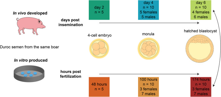
Background: The transcriptional changes around the time of embryonic genome activation in pre-implantation embryos indicate that this process is highly dynamic. In vitro produced porcine blastocysts are known to be less competent than in vivo developed blastocysts. To understand the conditions that compromise developmental competence of in vitro embryos, it is crucial to evaluate the transcriptional profile of porcine embryos during pre-implantation stages. In this study, we investigated the transcriptome dynamics in in vivo developed and in vitro produced 4-cell embryos, morulae and hatched blastocysts.
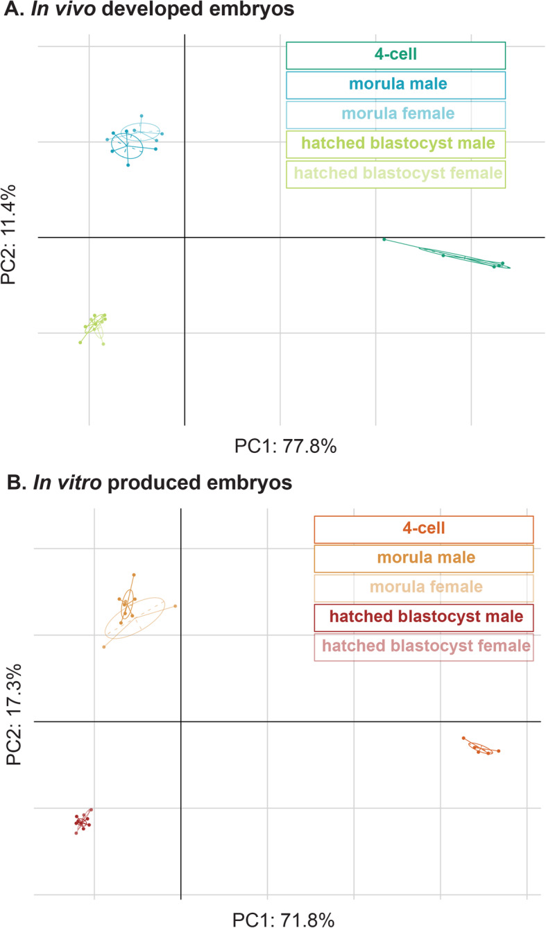
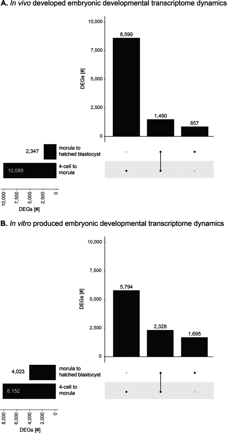
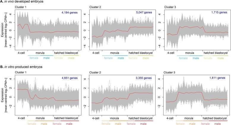
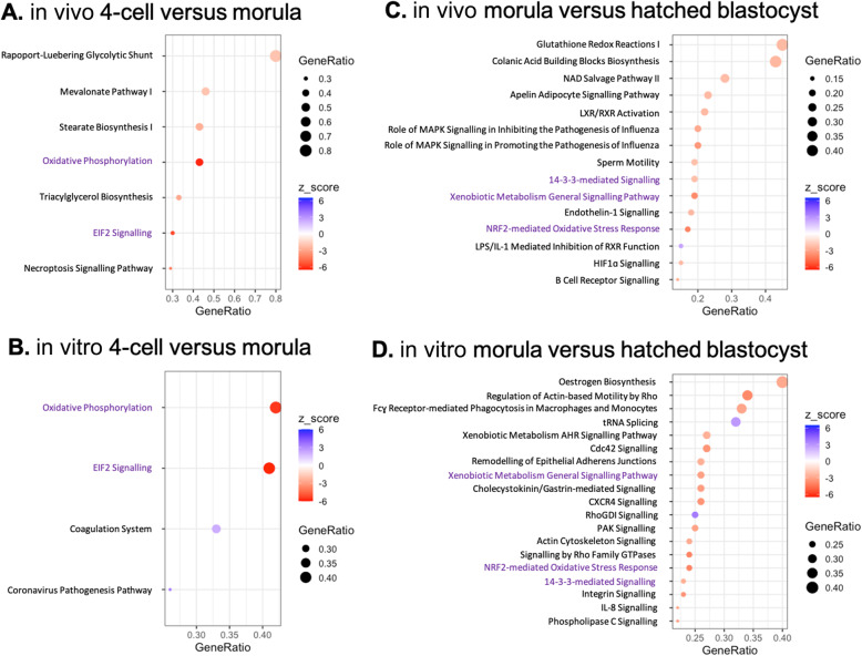
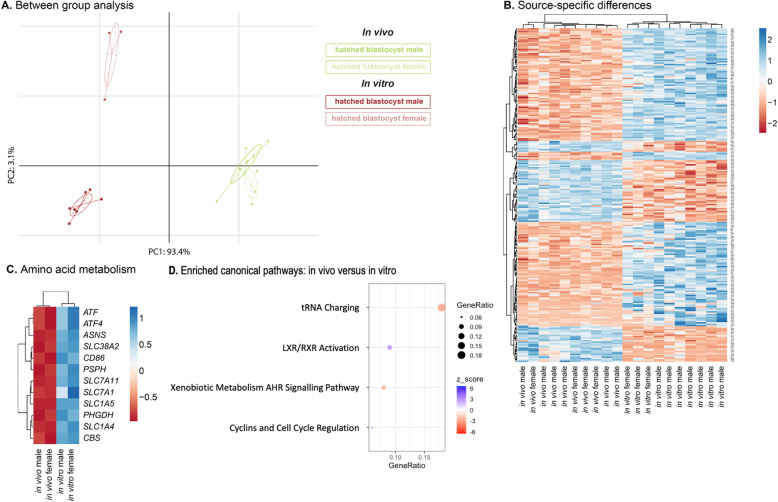
Results: In vivo developed and in vitro produced embryos displayed largely similar transcriptome profiles during development. Enriched canonical pathways from the 4-cell to the morula transition that were shared between in vivo developed and in vitro produced embryos included oxidative phosphorylation and EIF2 signaling. The shared canonical pathways from the morula to the hatched blastocyst transition were 14-3-3-mediated signaling, xenobiotic metabolism general signaling pathway, and NRF2-mediated oxidative stress response. The in vivo developed and in vitro produced hatched blastocysts further were compared to identify molecular signaling pathways indicative of lower developmental competence of in vitro produced hatched blastocysts. A higher metabolic rate and expression of the arginine transporter SLC7A1 were found in in vitro produced hatched blastocysts.
Conclusions: Our findings suggest that embryos with compromised developmental potential are arrested at an early stage of development, while embryos developing to the hatched blastocyst stage display largely similar transcriptome profiles, irrespective of the embryo source. The hatched blastocysts derived from the in vitro fertilization-pipeline showed an enrichment in molecular signaling pathways associated with lower developmental competence, compared to the in vivo developed embryos.
Keywords: Embryo development; In vivo embryo development; Porcine; Transcriptomics; in vitro fertilization.
Conflict of interest statement
The authors declare that there are no conflicts of interest.
Figures
Similar articles
-
Relationship between timing of development, morula morphology, and cell allocation to inner cell mass and trophectoderm in in vitro-produced bovine embryos.Mol Reprod Dev. 1997 May;47(1):47-56. doi: 10.1002/(SICI)1098-2795(199705)47:1<47::AID-MRD7>3.0.CO;2-Q. Mol Reprod Dev. 1997. PMID: 9110314
-
Substrate utilization in porcine embryos cultured in NCSU23 and G1.2/G2.2 sequential culture media.Mol Reprod Dev. 2001 Mar;58(3):269-75. doi: 10.1002/1098-2795(200103)58:3<269::AID-MRD4>3.0.CO;2-L. Mol Reprod Dev. 2001. PMID: 11170267
-
Effect of protein supplementation on development to the hatching and hatched blastocyst stages of cat IVF embryos.Reprod Fertil Dev. 2002;14(5-6):291-6. doi: 10.1071/rd01135. Reprod Fertil Dev. 2002. PMID: 12467353
-
Review: Overview of the transcriptomic landscape in bovine blastocysts and elongated conceptuses driving developmental competence.Animal. 2023 May;17 Suppl 1:100733. doi: 10.1016/j.animal.2023.100733. Animal. 2023. PMID: 37567651 Review.
-
Human blastoid as an in vitro model of human blastocysts.Curr Opin Genet Dev. 2024 Feb;84:102135. doi: 10.1016/j.gde.2023.102135. Epub 2023 Dec 4. Curr Opin Genet Dev. 2024. PMID: 38052115 Review.
Cited by
-
ROCK Inhibitor (Y-27632) Abolishes the Negative Impacts of miR-155 in the Endometrium-Derived Extracellular Vesicles and Supports Embryo Attachment.Cells. 2022 Oct 10;11(19):3178. doi: 10.3390/cells11193178. Cells. 2022. PMID: 36231141 Free PMC article.
-
Comparison of the developmental competence of in vitro-produced mouse embryos cultured under 5 versus 2% O2 with in vivo-derived blastocysts.J Assist Reprod Genet. 2024 Nov;41(11):3089-3103. doi: 10.1007/s10815-024-03267-7. Epub 2024 Sep 23. J Assist Reprod Genet. 2024. PMID: 39313714
-
Profiling the transcriptomic signatures and identifying the patterns of zygotic genome activation - a comparative analysis between early porcine embryos and their counterparts in other three mammalian species.BMC Genomics. 2022 Nov 24;23(1):772. doi: 10.1186/s12864-022-09015-4. BMC Genomics. 2022. PMID: 36434523 Free PMC article.
-
Maslinic Acid Supplementation during the In Vitro Culture Period Ameliorates Early Embryonic Development of Porcine Embryos by Regulating Oxidative Stress.Animals (Basel). 2023 Mar 13;13(6):1041. doi: 10.3390/ani13061041. Animals (Basel). 2023. PMID: 36978582 Free PMC article.
-
Fisetin alleviates oxidative stress and promotes porcine early embryonic development via activation of the NRF2-ARE signalling pathway.Anim Biosci. 2025 Jun;38(6):1160-1174. doi: 10.5713/ab.24.0691. Epub 2025 Feb 27. Anim Biosci. 2025. PMID: 40045619 Free PMC article.
References
MeSH terms
Grants and funding
LinkOut - more resources
Full Text Sources
Other Literature Sources
Molecular Biology Databases
