. 2021 Feb 27;19(1):61.
doi: 10.1186/s12951-021-00808-5.

Macrophage migration inhibitory factor facilitates the therapeutic efficacy of mesenchymal stem cells derived exosomes in acute myocardial infarction through upregulating miR-133a-3p

Affiliations

Macrophage migration inhibitory factor facilitates the therapeutic efficacy of mesenchymal stem cells derived exosomes in acute myocardial infarction through upregulating miR-133a-3p

Wenwu Zhu et al. J Nanobiotechnology. .

Abstract

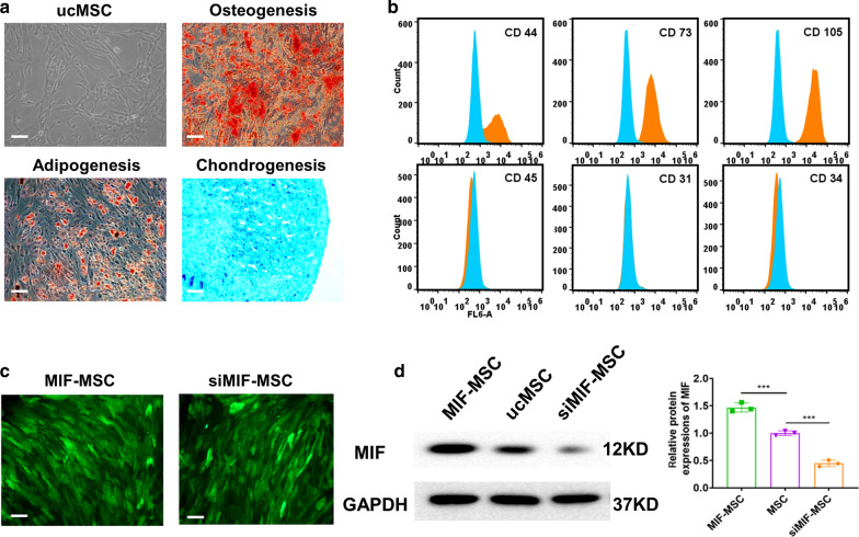
Background: Exosome transplantation is a promising cell-free therapeutic approach for the treatment of ischemic heart disease. The purpose of this study was to explore whether exosomes derived from Macrophage migration inhibitory factor (MIF) engineered umbilical cord MSCs (ucMSCs) exhibit superior cardioprotective effects in a rat model of AMI and reveal the mechanisms underlying it.

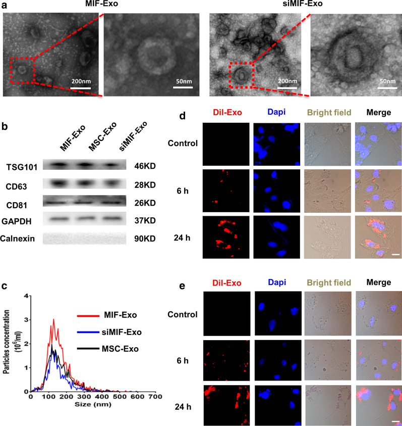
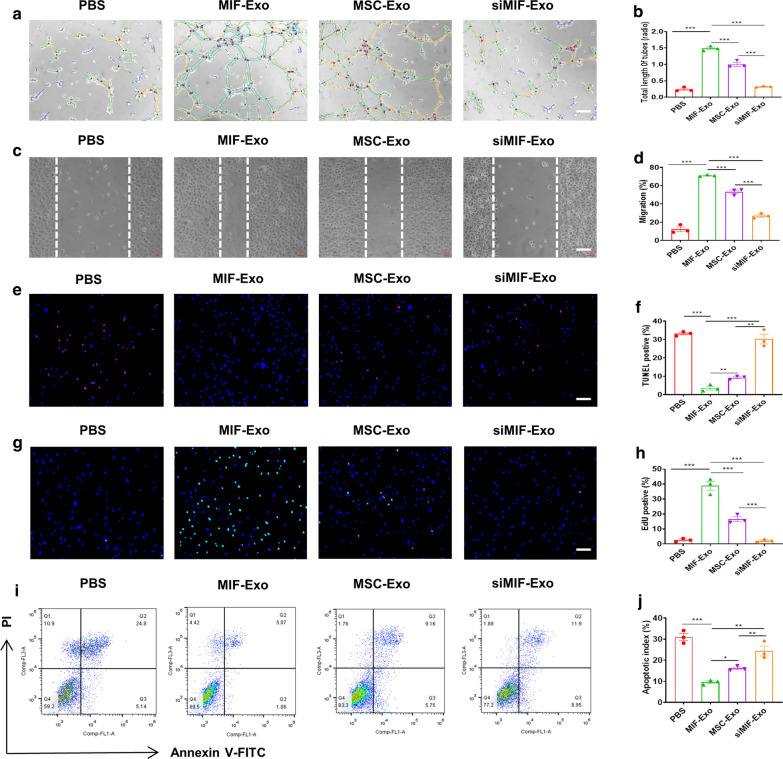
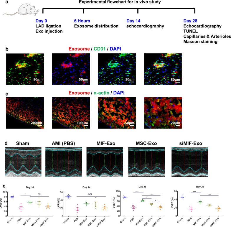
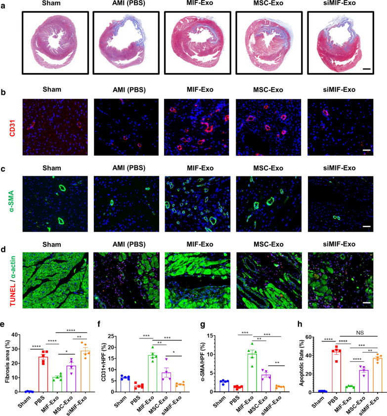
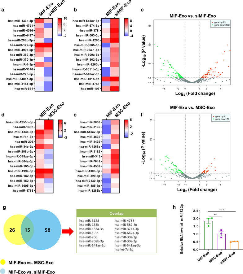
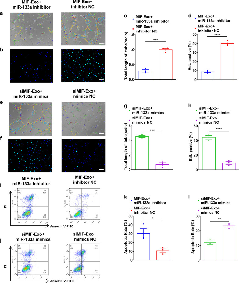
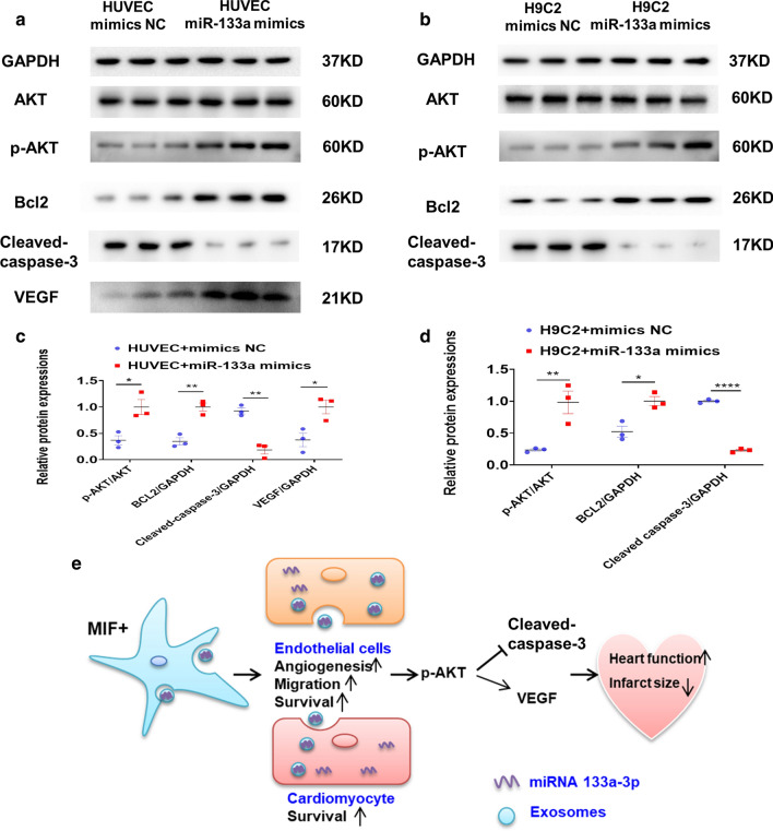
Results: Exosomes isolated from ucMSCs (MSC-Exo), MIF engineered ucMSCs (MIF-Exo) and MIF downregulated ucMSCs (siMIF-Exo) were used to investigate cellular protective function in human umbilical vein endothelial cells (HUVECs) and H9C2 cardiomyocytes under hypoxia and serum deprivation (H/SD) and infarcted hearts in rats. Compared with MSC-Exo and siMIF-Exo, MIF-Exo significantly enhanced proliferation, migration, and angiogenesis of HUVECs and inhibited H9C2 cardiomyocyte apoptosis under H/SD in vitro. MIF-Exo also significantly inhibited cardiomyocyte apoptosis, reduced fibrotic area, and improved cardiac function as measured by echocardiography in infarcted rats in vivo. Exosomal miRNAs sequencing and qRT-PCR confirmed miRNA-133a-3p significantly increased in MIF-Exo. The biological effects of HUVECs and H9C2 cardiomyocytes were attenuated with incubation of MIF-Exo and miR-133a-3p inhibitors. These effects were accentuated with incubation of siMIF-Exo and miR-133a-3p mimics that increased the phosphorylation of AKT protein in these cells.

Conclusion: MIF-Exo can provide cardioprotective effects by promoting angiogenesis, inhibiting apoptosis, reducing fibrosis, and preserving heart function in vitro and in vivo. The mechanism in the biological activities of MIF-Exo involves miR-133a-3p and the downstream AKT signaling pathway.

Keywords: Exosomes; Macrophage migration inhibitory factor; MiR-133a-3p; Myocardial infarction; UcMSCs.


Conflict of interest statement

The authors have declared that no competing interest exists.

Figures

Fig. 1
Fig. 1
Identification of human umbilical cord mesenchymal stem cell. a Morphology and multiple differentiation potential of ucMSCs. Scale bar = 100 μm. b Surface markers profiling of ucMSCs. c Successful lentiviral transfection was confirmed by positive green fluorescence under microscope in both MIF-MSC and siMIF-MSC groups. d Western blot images showed MIF protein levels in MIF-MSC, MSC and siMIF-MSC groups. ***P < 0.001, MIF-MSC vs. ucMSCs; ***P < 0.001, ucMSCs vs. siMIF-MSC
Fig. 2
Fig. 2
Characterization of exosomes derived from ucMSCs. a Cup-shaped morphology of purified MIF-Exo and siMIF-Exo (arrowhead) assessed by TEM. b Representative images of western blot showing the exosomal protein markers in MIF-MSC, ucMSCs and siMIF-MSC groups. c The particle size distribution and particle concentration were analyzed by nanoparticle tracking analysis (n = 3 biological replicates for each group). Confocal images showed that red fluorescence of dye Dil labeled exosomes were endocytosed by H9C2 (d) and HUVECs (e) 6 and 24 h after incubation. Scale bar = 20 μm
Fig. 3
Fig. 3
MIF-Exo exhibited more significant protective effects on HUVECs and H9C2 cardiomycytes than MSC-Exo in vitro. Tube formation of HUVECs incubated with PBS, MIF-Exo, MSC-Exo and siMIF-Exo (a), and quantification analysis (b). Scale bar = 100 μm. (n = 3 biological replicates for each group). Migration was monitored for 12 h after scratching in HUVECs cultured with PBS, MIF-Exo, MSC-Exo and siMIF-Exo (c), and quantification analysis (d). Scale bar = 100 μm (n = 3 biological replicates for each group). In H/SD, DAPI nucleic acid stained for apoptosis of HUVECs after culturing with PBS, MIF-Exo, MSC-Exo and siMIF-Exo. Red point indicated apoptotic cells (e), and quantification analysis (f). Scale bar = 100 μm. (n = 3 biological replicates for each group; 5 random fields for each biological replicate). EdU positive cells were in PBS, MIF-Exo, MSC-Exo and siMIF-Exo (g), and quantification analysis (h). Scale bar = 100 μm. (n = 3 biological replicates for each group; 5 random fields for each biological replicate). In H/SD, apoptosis of H9C2 after incubating with PBS, MIF-Exo, MSC-Exo and siMIF-Exo (i), and quantification analysis (j). Continuous variables and categorical variables were described by means ± SEM and percentages. (n = 3 biological replicates for each group). *P < 0.05; **P < 0.01; ***P < 0.001
Fig. 4
Fig. 4
MIF-Exo effectively preserved cardiac function in rats with MI in vivo. a The flowchart of experimental design in vivo. The protein expression level of CD31 (b) and a-actin (c) was detected by immunofluorescence after Dil labeled exosomes taken by endothelial cells and cardiomyocytes 6 h after intramyocardial injection. (n = 3 biological replicates; 4 random fields for each animal). LVEF and LVFS were measured 2 and 4 weeks post MI (d), and quantification analysis (f). (2 weeks: n = 5 animals for each group; 4 weeks: n = 4 animals for MSC-Exo group, n = 5 animals for the other groups). Continuous variables and categorical variables were described by means ± SEM and percentages. *P < 0.05; ***P < 0.001; NS, not significant
Fig. 5
Fig. 5
MIF-Exo promoted angiogenesis and cardiomyocyte survival in infarcted hearts. Fibrosis area was in Sham, PBS, MIF-Exo, MSC-Exo and siMIF-Exo groups (a), and quantification analysis (e). Scale bar = 2 mm. CD31 positively stained capillaries at the border zone 4 weeks post MI. Left ventricle was selected in Sham group (b), and quantification analysis (f). Scale bar = 50 μm. α-SMA positively stained arterioles in the infarct area week 4 after MI. Left ventricle was selected (c), and quantification analysis (g). Scale bar = 50 μm. TUNEL staining at the border zone 4 weeks after MI (d), and quantification analysis (h). Scale bar = 50 μm. (n = 4 animals for MSC-Exo group, n = 5 animals for the other groups, 4 random fields per animal). Continuous variables and categorical variables were described by means ± SEM and percentages. *P < 0.05; **P < 0.01; ***P < 0.001; ****P < 0.0001; NS, not significant
Fig. 6
Fig. 6
miR-133a-3p level increased in MIF-Exo. Heat map based on exosomal miRNAs sequence values (red represents high expression and blue represents low expression) between MIF-Exo and siMIF-Exo. Top 15 upregulated miRNAs (a) and top 15 downregulated miRNAs (bB) were shown in MIF-Exo group. c Volcano plot showed log2 (Fold change) on x-axis and -log10 (P value) on y-axis. Heat map based on exosomal miRNAs sequence values between MIF-Exo and MSC-Exo group. Top 15 upregulated miRNAs (d) and top 15 downregulated miRNAs (e) in MIF-Exo group are shown. f Volcano plot showed log2 (Fold change) on the x-axis and -log10 (P value) on the y-axis. g In these upregulated miRNAs, 15 miRNAs were overlapped. h miR-133a-3p levels in MIF-Exo, MSC-Exo and siMIF-Exo groups were measured by qRT-PCR. (n = 3 biological replicates, 3 technical replicates for each biological replicate). All data were mean ± SEM. **P < 0.01; ***P < 0.001; NS, not significant
Fig. 7
Fig. 7
Gain and loss function of exosomal miR-133a-3p on pro-angiogenisis, proliferation, and apoptosis in HUVECs and H9c2 cells in vitro. Proangiogenic effects of HUVECs were diminished with incubation of MIF-Exo and miR-133a-3p inhibitor (a), and quantification analysis (c). Scale bar: 100 μm. (n = 3 biological replicates for each group). Proliferation effects of HUVECs were attenuated with incubation of MIF-Exo and miR-133a-3p inhibitor (b), and quantification analysis (d). Scale bar: 100 μm. (n = 3 biological replicates for each group; 5 random fields for each biological replicate). Proangiogenic activity of HUVECs restored with incubation of siMIF-Exo and miR-133a-3p mimics (e), and quantification analysis (g). Scale bar: 100 μm. (n = 3 biological replicates for each group). Proliferation of HUVECs rescued with incubation of siMIF-Exo and miR-133a-3p mimics (f), and quantification analysis (h). Scale bar: 100 μm. (n = 3 biological replicates; 5 random fields for each biological replicate). Anti-apoptotic ability of H9c2 cells reduced with incubation of MIF-Exo and miR-133a-3p inhibitor (i), and quantification analysis (k). Scale bar: 100 μm C. (n = 3 biological replicates for each group). Anti-apoptotic ability of H9c2 cells rescued with incubation of siMIF-Exo and miR-133a-3p mimics (j), and quantification analysis (l). (n = 3 biological replicates for each group). Continuous variables and categorical variables were described by means ± SEM and percentages. *P < 0.05; **P < 0.01; ***P < 0.001; ****P < 0.0001
Fig. 8
Fig. 8
Exosomal miR-133a-3p inhibited cardiomyocyte apoptosis, promoted angiogenesis by AKT signal pathway, and improved the protein expression of VEGF in HUVECs. The protein levels of p-AKT, AKT, Bcl-2, cleaved caspase-3 and VEGF in HUVECs (a), and quantification analysis (c). (n = 3 biological replicates). The protein levels of p-AKT, AKT, Bcl-2, and cleaved caspase-3 in H9C2 cells (b), and quantification analysis (d). (n = 3 biological replicates). Continuous variables and categorical variables were described by means ± SEM and percentages. *P < 0.05; **P < 0.01; ****P < 0.0001. e Schematic showed the working model of this study

Similar articles

Cited by

References

    1. Colombo M, Raposo G, Théry C. Biogenesis, secretion, and intercellular interactions of exosomes and other extracellular vesicles. Annu Rev Cell Dev Biol. 2014;30:255–289. doi: 10.1146/annurev-cellbio-101512-122326. - DOI - PubMed
    1. Singla DK. Stem cells and exosomes in cardiac repair. Curr Opin Pharmacol. 2016;27:19–23. doi: 10.1016/j.coph.2016.01.003. - DOI - PubMed
    1. Min PK, Chan SY. The biology of circulating microRNAs in cardiovascular disease. Eur J Clin Invest. 2015;45:860–874. doi: 10.1111/eci.12475. - DOI - PMC - PubMed
    1. Taylor DD, Gercel-Taylor C. MicroRNA signatures of tumor-derived exosomes as diagnostic biomarkers of ovarian cancer. Gynecol Oncol. 2008;110:13–21. doi: 10.1016/j.ygyno.2008.04.033. - DOI - PubMed
    1. Lamichhane TN, Sokic S, Schardt JS, Raiker RS, Lin JW, Jay SM. Emerging roles for extracellular vesicles in tissue engineering and regenerative medicine. Tissue Eng B Rev. 2015;21:45–54. doi: 10.1089/ten.teb.2014.0300. - DOI - PMC - PubMed

MeSH terms