. 2021 Feb 27;18(1):46.
doi: 10.1186/s12985-021-01515-1.

Inhibitors of endosomal acidification suppress SARS-CoV-2 replication and relieve viral pneumonia in hACE2 transgenic mice

Affiliations

Inhibitors of endosomal acidification suppress SARS-CoV-2 replication and relieve viral pneumonia in hACE2 transgenic mice

Chao Shang et al. Virol J. .

Abstract

Background: Coronavirus disease 2019 (COVID-19) is caused by SARS-CoV-2 and broke out as a global pandemic in late 2019. The acidic pH environment of endosomes is believed to be essential for SARS-CoV-2 to be able to enter cells and begin replication. However, the clinical use of endosomal acidification inhibitors, typically chloroquine, has been controversial with this respect.

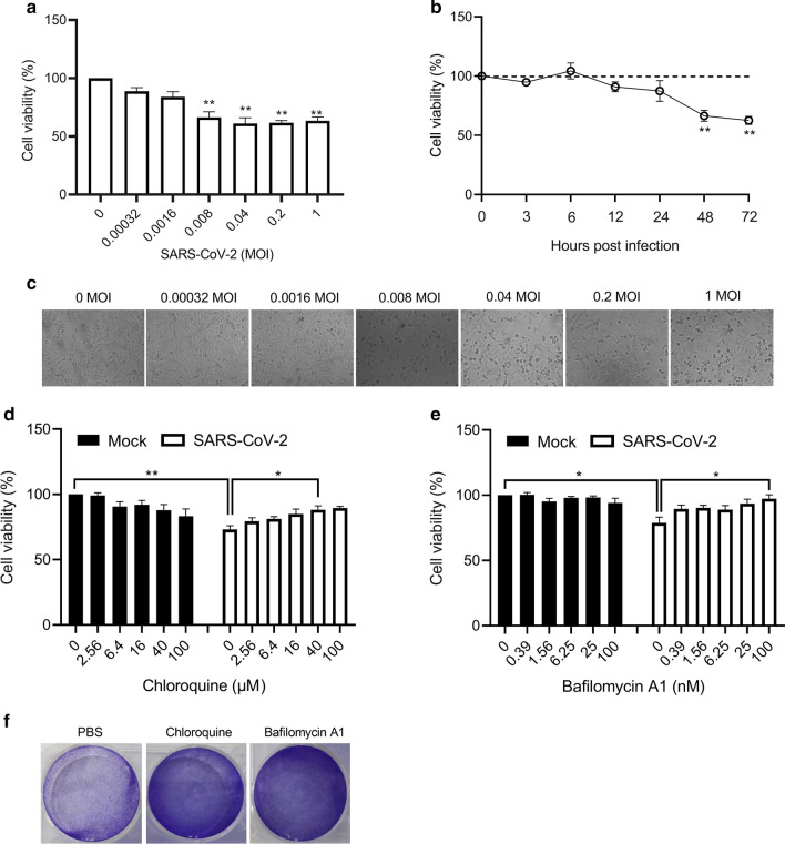
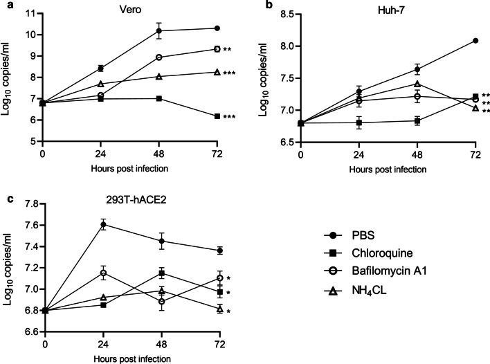
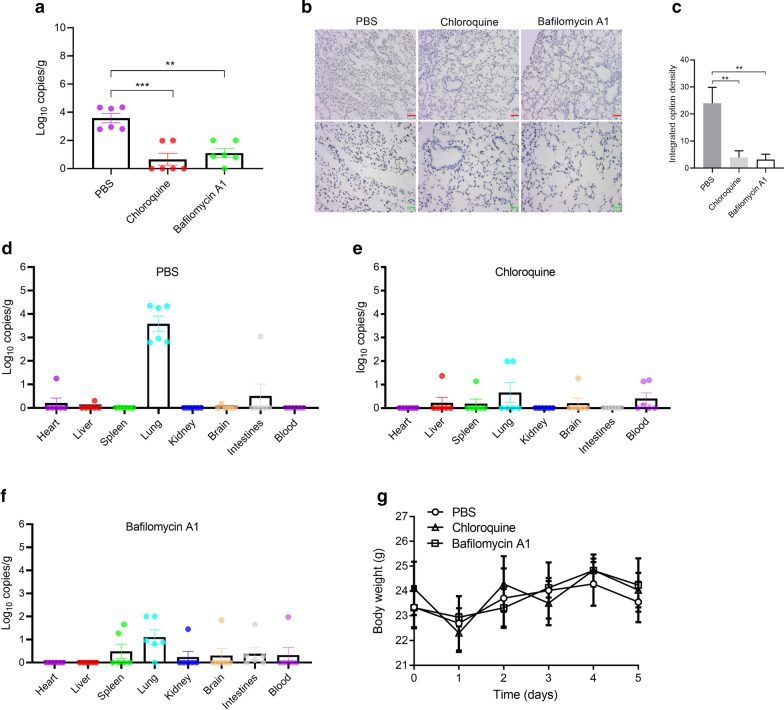
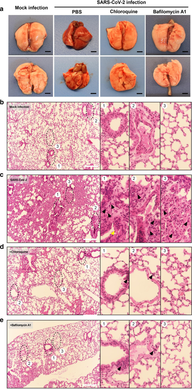
Methods: In this study, RT-qPCR method was used to detect the SARS-CoV-2N gene to evaluate viral replication. The CCK-8 assay was also used to evaluate the cytotoxic effect of SARS-CoV-2. In situ hybridization was used to examine the distribution of the SARS-CoV-2 gene in lung tissues. Hematoxylin and eosin staining was also used to evaluate virus-associated pathological changes in lung tissues.

Results: In this study, analysis showed that endosomal acidification inhibitors, including chloroquine, bafilomycin A1 and NH4CL, significantly reduced the viral yields of SARS-CoV-2 in Vero E6, Huh-7 and 293T-ACE2 cells. Chloroquine and bafilomycin A1 also improved the viability and proliferation of Vero E6 cells after SARS-CoV-2 infection. Moreover, in the hACE2 transgenic mice model of SARS-CoV-2 infection, chloroquine and bafilomycin A1 reduced viral replication in lung tissues and alleviated viral pneumonia with reduced inflammatory exudation and infiltration in peribronchiolar and perivascular tissues, as well as improved structures of alveolar septum and pulmonary alveoli.

Conclusions: Our research investigated the antiviral effects of endosomal acidification inhibitors against SARS-CoV-2 in several infection models and provides an experimental basis for further mechanistic studies and drug development.

Keywords: Bafilomycin A1; Chloroquine; Endosomal acidification; SARS-CoV-2.


Conflict of interest statement

The authors declare no competing interests.

Figures

Fig. 1
Fig. 1
Inhibitors of endosomal acidification improved the viability of Vero E6 cells after SARS-CoV-2 infection. a Viability of Vero E6 cells infected with different doses of SARS-CoV-2 for 48 h. b Cell viability after SARS-CoV-2 infection (0.008 MOI) at different time points. c Images of Vero E6 cells challenged with 0.008 MOI of SARS-CoV-2 for 48 h. d, e Cell viability of Vero E6 cells treated with chloroquine or bafilomycin after SARS-CoV-2 infection (0.008 MOI) for 48 h. f Colony formation assays of Vero E6 cells treated with chloroquine (40 μM) or bafilomycin A1 (100 nM) after SARS-CoV-2 infection (0.008 MOI) for 48 h. Three experiments were performed (n = 6 each group). *p < 0.05, **p < 0.01
Fig. 2
Fig. 2
Inhibitors of endosomal acidification suppressed SARS-CoV-2 replication in vitro. Cells were infected with SARS-CoV-2 (0.008 MOI) and cell supernatant was harvested on indicated time points post infection. SARS-CoV-2 (0.008 MOI) was diluted in cell supernatant and harvested immediately as samples of 0 h post infection. Viral genome copies in Vero E6 cells (a), Huh-7 (b), and 293T-ACE2 cells (c) treated with PBS, chloroquine (40 μM), bafilomycin A1 (100 nM) or NH4CL (12.5 mM). Three experiments were performed (n = 3 each group). *p < 0.05, **p < 0.01, ***p < 0.001
Fig. 3
Fig. 3
Inhibitors of endosomal acidification suppressed SARS-CoV-2 replication in hACE2 transgenic mice. hACE2 transgenic mice were inoculated with 6.7 × 103 PFU of SARS-CoV-2 intratracheally in 30 μl of PBS. PBS, chloroquine (60 mg kg−1 once daily), or bafilomycin A1 (0.1 mg kg−1 once daily), were then given intraperitoneally for 5 days. Organ tissues were collected on day 5. a Viral genome copies in lung tissues. b, c In situ hybridization for SARS-CoV-2 RNA in lung tissues. df Viral genome copies in primary organs. g Body weight changes. Three experiments were performed (n = 6 each group). **p < 0.01, ***p < 0.001. Green bar = 25 μm. Red bar = 50 μm
Fig. 4
Fig. 4
Inhibitors of endosomal acidification alleviated viral pneumonia in hACE2 transgenic mice. a Images of gross pathology of lungs from hACE2 transgenic mice subjected to indicated treatments. b, c Representative images for H&E staining of histopathological changes in lung tissues from Mock-infected (b) and SARS-CoV-2-challenged hACE2 transgenic mice (c). The numbered panels display magnified images of indicated black circles in the left. SARS-CoV-2 induced histopathological changes as follows: (1) bronchiolar epithelium cell death (yellow arrow) and peribronchiolar infiltration of segmented granulocytes or lymphocytes (black arrow). (2) Inflammatory exudation and infiltration in perivascular tissues (black arrow). (3) Collapse of alveolar structure and widened alveolar septum with inflammatory cell infiltration (black arrow). d, e Representative H&E staining images of lung tissues from SARS-CoV-2-challenged hACE2 transgenic mice that are subjected to chloroquine (d) and bafilomycin A1 (e) treatments. Reduced inflammatory exudation and infiltration in peribronchiolar (1) and perivascular (2) tissues, as well as improved structures of alveolar septum and pulmonary alveoli (3), were observed in chloroquine and bafilomycin A1 treatment groups. Representative images from 6 mice are shown. Black bar = 2 mm. White bar = 200 μm

Similar articles

Cited by

References

    1. Zhu N, Zhang D, Wang W, Li X, Tan W. A Novel Coronavirus from Patients with Pneumonia in China, 2019. N Engl J Med. 2020;382(8). - PMC - PubMed
    1. Gorbalenya AE, Baker SC, Baric RS, Groot RJD, Ziebuhr J.The species Severe acute respiratory syndrome-related coronavirus: classifying 2019-nCoV and naming it SARS-CoV-2. Nat Microbiol. 2020;5(5). - PMC - PubMed
    1. Kai H, Kai M. Interactions of coronaviruses with ACE2, angiotensin II, and RAS inhibitors—lessons from available evidence and insights into COVID-19. Hypertens Res. 2020;43(7):648–654. doi: 10.1038/s41440-020-0455-8. - DOI - PMC - PubMed
    1. Belouzard S, Millet JK, Licitra BN, Whittaker GR. Mechanisms of coronavirus cell entry mediated by the viral spike protein. Viruses. 2012;4(6):1011–1033. doi: 10.3390/v4061011. - DOI - PMC - PubMed
    1. Heald-Sargent T, Gallagher T. Ready, set, fuse! The coronavirus spike protein and acquisition of fusion competence. Viruses. 2012;4(4). - PMC - PubMed

Publication types

MeSH terms