Review
. 2021 Sep:84:100954.
doi: 10.1016/j.preteyeres.2021.100954. Epub 2021 Feb 25.

VEGFR1 signaling in retinal angiogenesis and microinflammation

Affiliations
Review

VEGFR1 signaling in retinal angiogenesis and microinflammation

Akiyoshi Uemura et al. Prog Retin Eye Res. 2021 Sep.

Abstract

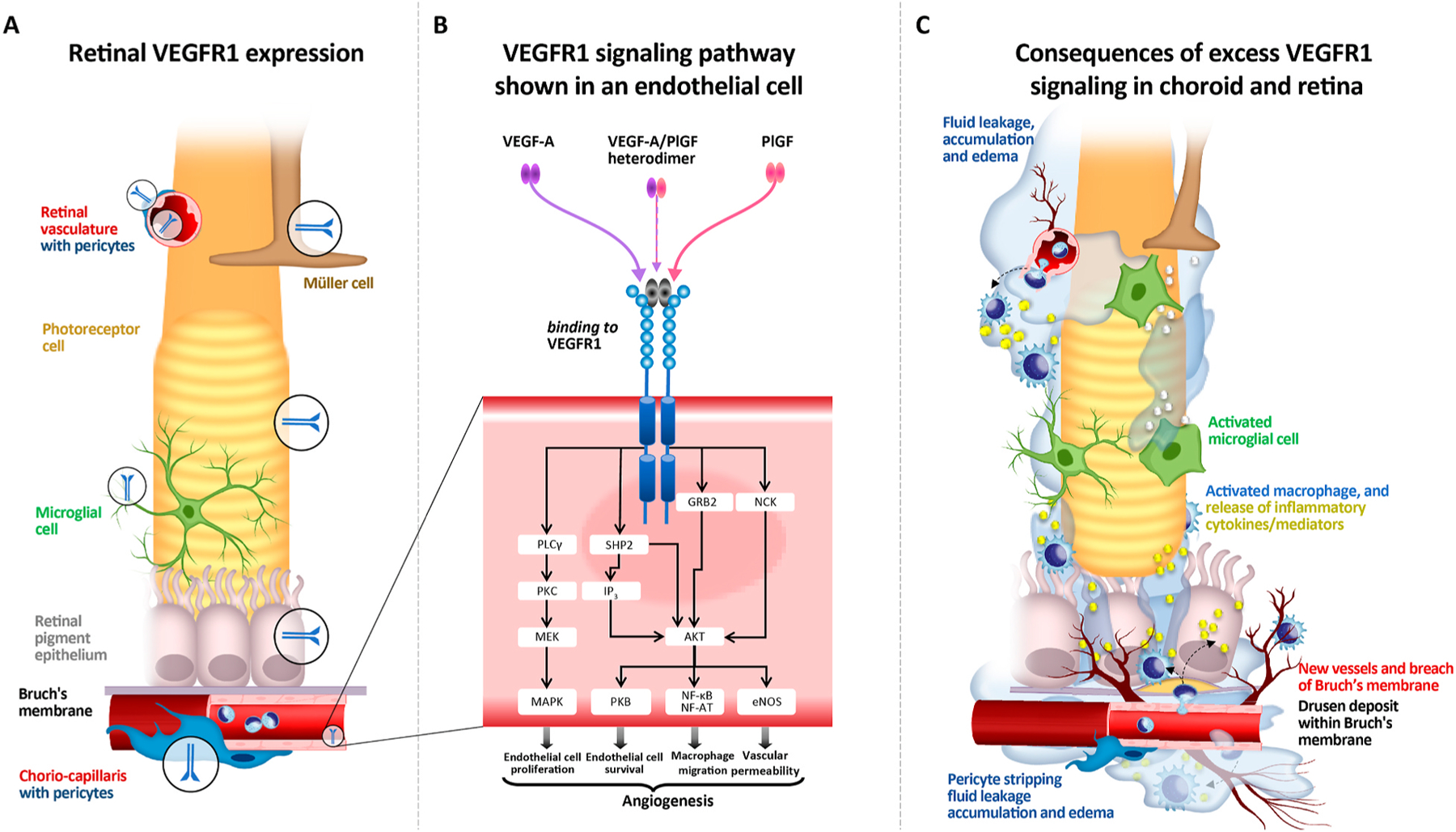
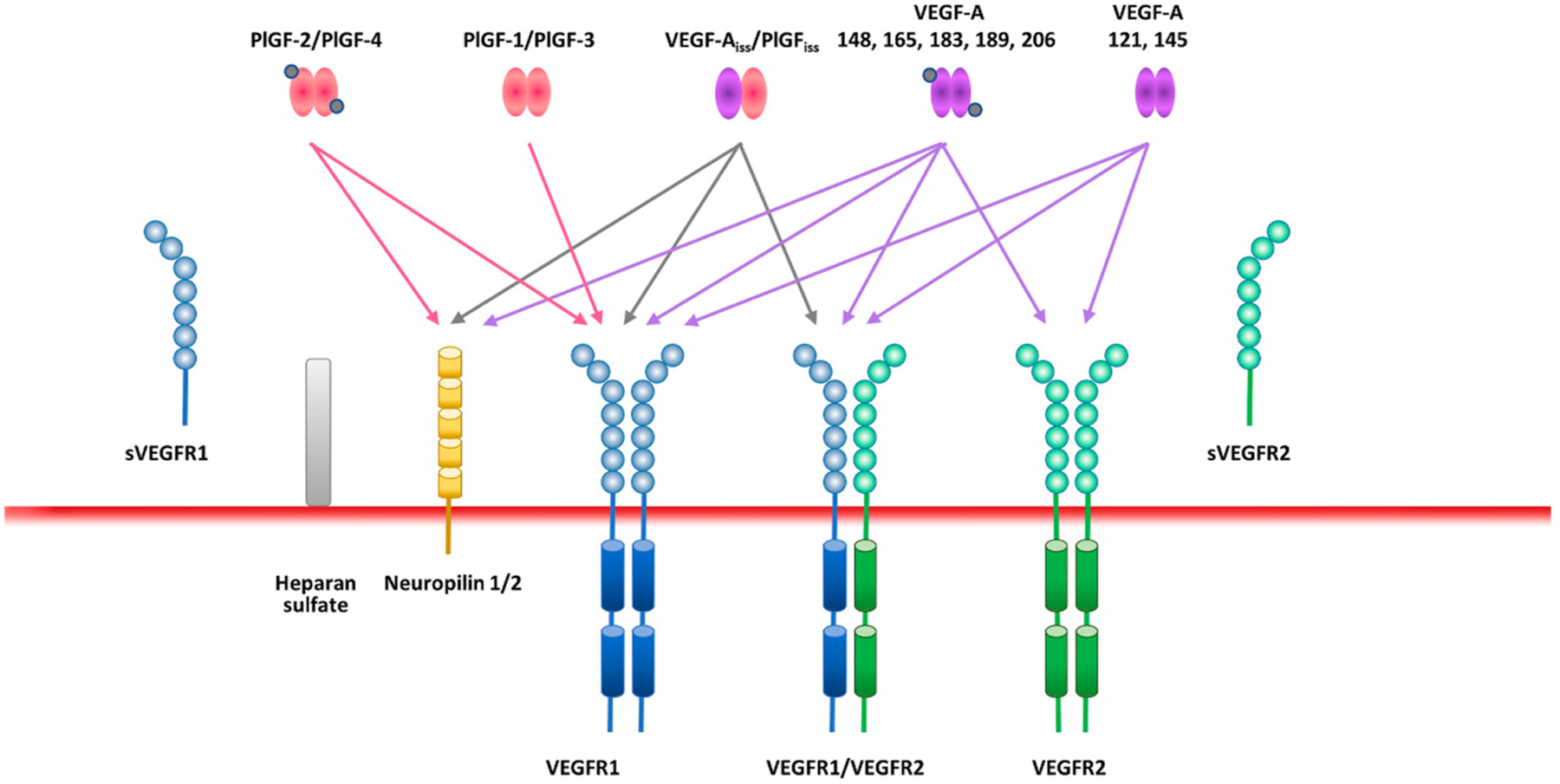
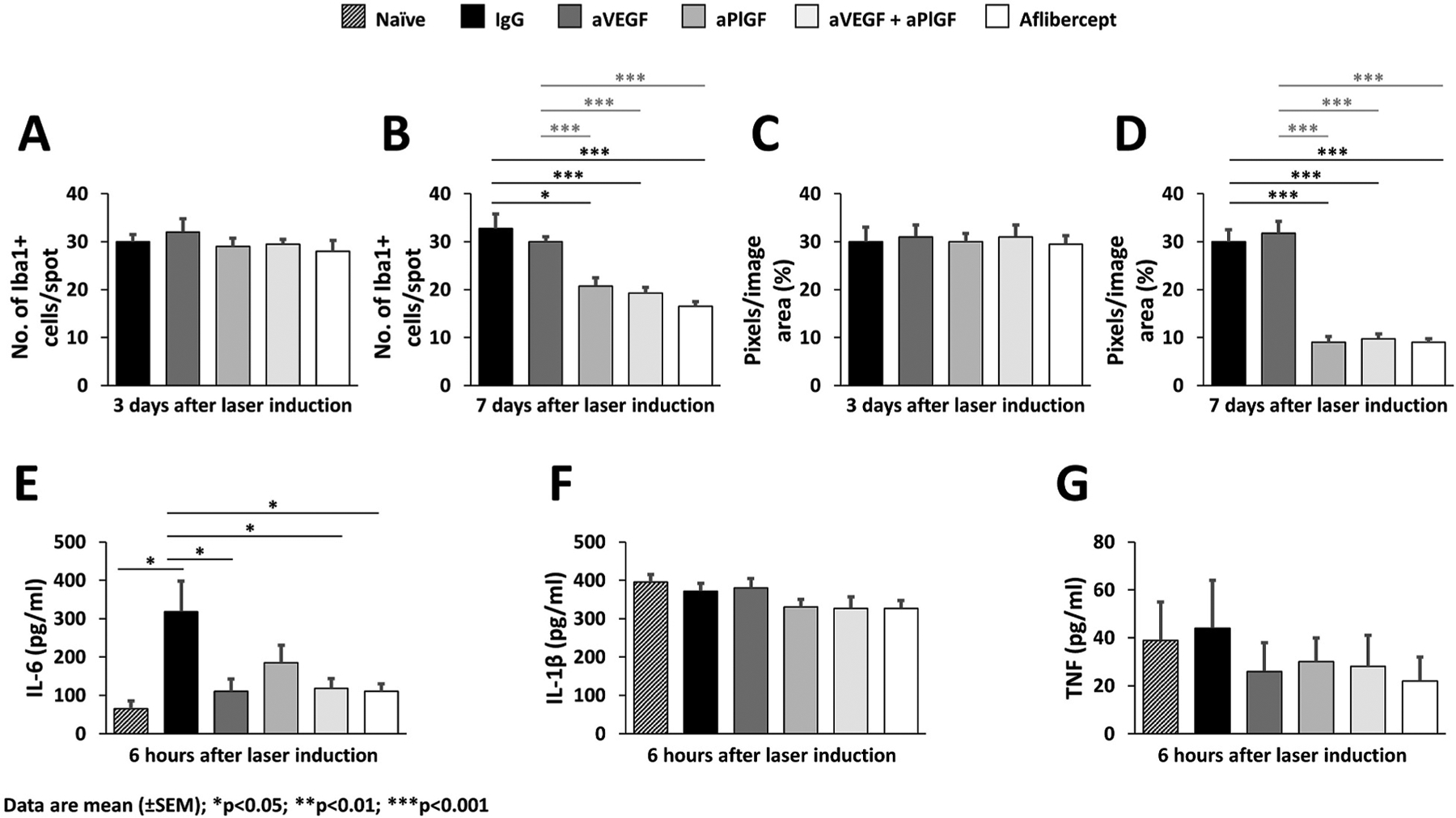
Five vascular endothelial growth factor receptor (VEGFR) ligands (VEGF-A, -B, -C, -D, and placental growth factor [PlGF]) constitute the VEGF family. VEGF-A binds VEGF receptors 1 and 2 (VEGFR1/2), whereas VEGF-B and PlGF only bind VEGFR1. Although much research has been conducted on VEGFR2 to elucidate its key role in retinal diseases, recent efforts have shown the importance and involvement of VEGFR1 and its family of ligands in angiogenesis, vascular permeability, and microinflammatory cascades within the retina. Expression of VEGFR1 depends on the microenvironment, is differentially regulated under hypoxic and inflammatory conditions, and it has been detected in retinal and choroidal endothelial cells, pericytes, retinal and choroidal mononuclear phagocytes (including microglia), Müller cells, photoreceptor cells, and the retinal pigment epithelium. Whilst the VEGF-A decoy function of VEGFR1 is well established, consequences of its direct signaling are less clear. VEGFR1 activation can affect vascular permeability and induce macrophage and microglia production of proinflammatory and proangiogenic mediators. However the ability of the VEGFR1 ligands (VEGF-A, PlGF, and VEGF-B) to compete against each other for receptor binding and to heterodimerize complicates our understanding of the relative contribution of VEGFR1 signaling alone toward the pathologic processes seen in diabetic retinopathy, retinal vascular occlusions, retinopathy of prematurity, and age-related macular degeneration. Clinically, anti-VEGF drugs have proven transformational in these pathologies and their impact on modulation of VEGFR1 signaling is still an opportunity-rich field for further research.

Keywords: Angiogenesis; Microinflammation; Placental growth factor (PlGF); Vascular endothelial growth factor receptor 1 (VEGFR1); Vascular endothelial growth factor-A (VEGF-A).


Conflict of interest statement

Conflict of interest disclosures

Akiyoshi Uemura, Marcus Fruttiger and Patricia A. D’Amore declare no conflicts of interest. Sandro De Falco is co-founder of the startup AnBition s.r.l., Naples, Italy and co-inventor of the patents PCT/IB2018/057636, Peptides and medical uses thereof, Priority date September 11, 2019 and Italian patents n. 102018000008493 and n. 102018000008507, Peptidi ed usi medici correlati, priority date September 11, 2018. Antonia M. Joussen is a consultant for Allergan, Bayer, Novartis and Roche and has received research funding from Bayer and Novartis. Florian Sennlaub declares no conflict of interest. Lynne R. Brunck, Kristian T. Johnson, George N. Lambrou and Kay D. Rittenhouse are employees of Bayer Consumer Care AG. Thomas Langmann has participated in advisory boards from Bayer HealthCare AG.

Figures

Fig. 1.
Fig. 1.
Schematic representation of VEGFR1 in the choroid and retina and VEGFR1 signaling (for illustrative purposes and not to scale). A. VEGFR1 expression in various types of cells, including vascular endothelial cells, pericytes, mononuclear phagocytes, Müller cells, photoreceptor cells, and the retinal pigment epithelium. B. VEGFR1 signaling through VEGF-A and/or PlGF, via a variety of different pathways, contributing to numerous pathologic processes in endothelial cells and pericytes in the choroid and retina: pericyte ablation, loss of tight junctions between endothelial cells, vasodilation, breakdown of the blood-retinal-barrier, increased permeability and leakage, edema and hemorrhage in surrounding tissue, neutrophil migration and monocyte migration and differentiation into macrophages, influx of pro-inflammatory cytokines e.g. tumor necrosis factor-α and interleukin-6 into surrounding tissue, increased angiogenic sprouts and neoangiogenesis. C. Consequences of excess VEGFR1 signaling in the choroid and retina: in retinal pigment cells: neoangiogenesis of vessels through Bruch’s membrane into the retinal pigment epithelium, loss of retinal pigment cells; in photoreceptor cells: loss of photoreceptor integrity, rod death and cone segment loss; in Müller cells: Müller cell activation; in microglial cells: recruitment, accumulation, and activation of microglial cells and other retinal macrophages, release of pro-inflammatory cytokines e.g. platelet-derived growth factor-A, soluble intracellular adhesion molecule-1, CC chemokine ligand 2, and interleukin-8, leading to the development of hyperreflective foci.
Fig. 2.
Fig. 2.
VEGFR1 and VEGFR2 and the family of ligands and co-receptors. There are five VEGFR ligands, of which VEGF-A binds to both VEGFR1 and VEGFR2, and PlGF only binds VEGFR1. Splicing creates isoforms of both VEGF-A and PlGF. In addition, soluble/secreted versions of VEGFR1 and VEGFR2 can be produced via alternative splicing or proteolytic cleavage retaining the extracellular ligand–binding domains. Furthermore, VEGF-A and PlGF are able to bind neuropilin (NRP) 1 and 2, bridging VEGFRs and NRP1 or NRP2 to create holoreceptor complexes. VEGF-A and PlGF ligands and the VEGFR1 and VEGFR2 receptors can form heterodimers as well as homodimers. Functional synergistic effects of PlGF and VEGF-A are due to sharing of the common receptor, VEGFR1, and the ability to heterodimerize.
Fig. 3.
Fig. 3.
Pgf-DE-Ki mice, a fully functional Pgf-KO model achieved by knocking in the Pgf-DE variant unable to bind and activate VEGFR1, show robust reduction of CNV and protection from vascular leakage. A. CNV volumes measured 7 days after laser-induced damage by Isolectin B4 staining of RPE-choroid flat mounts. B. Q ualitative fundus fluorescein angiography in C57BL6/J, Pgf-KO, and Pgf-DE-Knock-in mice acquired at three different times (early – 1 min, intermediate – 5 min, late – 15 min) after intraperitoneal delivery of fluorescein at Days 3, 7, and 14 after laser-induced damage. Reproduced under Creative Common CC-BY license (Apicella et al., 2018).
Fig. 4.
Fig. 4.
VEGFR1 signal in a mouse model of pericyte-deficient retinopathy (pups intraperitoneally injected with an anti-platelet-derived growth factor receptor β monoclonal antibody [clone APB5 in A–H] or control phosphate-buffered saline [A–E]) at postnatal day [P]1). A. Labeling of retinal endothelial cells (ECs) and pericytes (PCs) at P5 by whole-mount immunohistochemistry (WIHC) for CD31 and NG2, respectively. Note the absence of PCs and disorganized vascular networks in the APB5-treated retina. B. Hematoxylin and eosin (HE) staining of paraffin sections from P10 retinas showing edema and hemorrhage in the APB5-treated retina. C. Flow cytometry in P8 retinas. Tissue-resident microglia and inflammatory mononuclear phagocytes (MPs) are represented by CD45loloCD11b+ and CD45hiCD11b+ cells, respectively. Note the high VEGFR1 expression level in CD45hiCD11b+Ly6C+ MPs from the APB5-treated retinas. D. Retinal whole-mount in situ hybridization for Vegfa (left) and Pgf (right) at P8 in conjunction with labeling of vascular basement membranes and MPs by WIHC for type IV collagen and Iba1, respectively. Note the upregulation of Vegfa and Pgf in perivascular MPs of the APB5-treated retinas. E. VEGFR1 reporter expression in P8 retinas from Vegfr1-BAC-DsRed mice in conjunction with WIHC for Iba1. Note the VEGFR1-expressing MP (arrowhead) in the APB5-treated retina. F. Retinal cups (upper) and WIHC for CD31 and Iba1 (lower) at P11 in APB5-treated VEGFR1-TK mice. Note the suppression of retinal edema and MP infiltration even without PC coverage in VEGFR1-TK/mice. The graphs show the number of Iba1+ cells per area and the vessel density (n = 20). G. The trajectory of MPs in APB5-treated retinas from P8 Cx3cr1-GFP mice. After 3 h ex vivo imaging, retinas were treated with control IgG or aflibercept, and further monitored for 3 h. The graphs show quantification of cell body movement velocity (Pre IgG, n = 68; Post IgG, n = 56; Pre VEGF Trap, n = 52; Post VEGF Trap, n = 47) and total dendrite length per cell (Pre IgG, n = 40; Post IgG, n = 34; Pre VEGF Trap, n = 43; Post VEGF Trap, n = 33). H. Labeling for isolectin B4 (IB4), ICAM-1, and Iba1 at P13 in APB5-treated retinas after intravitreal injections of control IgG or aflibercept at P7. Note the normalization of vascular networks with reduced MP infiltration after aflibercept injection. I. Schematic diagram of EC-MP interactions in PC-deficient retina. In ECs, activation of nuclear factor of activated T cells (NFAT) leads to upregulation of CCL2, which subsequently facilitates the influx of circulating CCR2+ monocytes. The infiltrating monocytes and activated microglia contribute to generation of inflammatory MPs, which secrete VEGF-A and PlGF, and activate VEGFR1 in MPs and VEGFR2 in ECs. The VEGF-A–VEGFR2 signal further activates NFAT. This positive feedback loop sustains breakdown of the blood-retina barrier. In box-and-whisker plots, median (line within the box), upper and lower quartile (bounds of the box), with minimum and maximum values (bars) are shown. ***p < 0.001; NS, not significant, by Student’s t-test. Scale bars, 50 μm (A and B); 20 μm (D and E); 100 μm (F and H). Adapted from Ogura et al. (2017) with permission from American Society for Clinical Investigation.
Fig. 5.
Fig. 5.
Effects of PlGF and VEGF inhibition on mononuclear phagocytes in retinal flat mounts in the laser-induced mouse model of CNV. A and B. Quantification of microglia/macrophages per laser spot in retinal flat mounts 3 and 7 days, respectively, after laser-induced damage. C and D. Quantification of ionized calcium-binding adaptor molecule 1 signals 3 and 7 days, respectively, after laser coagulation in retinal flat mounts by counting the mean of colored pixels per image. E, F, and G. Interleukin-6, interleukin-1β, and tumor necrosis factor levels, respectively, in retinal flat mounts 6 h after laser damage quantified by enzyme-linked immunosorbent assay with naive (not lasered) animals used as controls. Reproduced under Creative Common CC-BY license (Balser et al., 2019).

References

    1. Achen MG, Jeltsch M, Kukk E, Makinen T, Vitali A, Wilks AF, Alitalo K, Stacker SA, 1998. Vascular endothelial growth factor D (VEGF-D) is a ligand for the tyrosine kinases VEGF receptor 2 (Flk1) and VEGF receptor 3 (Flt4). Proc. Natl. Acad. Sci. U. S. A 95, 548–553. - PMC - PubMed
    1. Adamis AP, Miller JW, Bernal MT, D’Amico DJ, Folkman J, Yeo TK, Yeo KT, 1994. Increased vascular endothelial growth factor levels in the vitreous of eyes with proliferative diabetic retinopathy. Am. J. Ophthalmol 118, 445–450. - PubMed
    1. Adini A, Kornaga T, Firoozbakht F, Benjamin LE, 2002. Placental growth factor is a survival factor for tumor endothelial cells and macrophages. Cancer Res 62, 2749–2752. - PubMed
    1. Agarwal K, Jalali S, 2018. Classification of retinopathy of prematurity: from then till now. Community Eye Health 31, S4–S7. - PMC - PubMed
    1. Aiello LP, Avery RL, Arrigg PG, Keyt BA, Jampel HD, Shah ST, Pasquale LR, Thieme H, Iwamoto MA, Park JE, et al., 1994. Vascular endothelial growth factor in ocular fluid of patients with diabetic retinopathy and other retinal disorders. N. Engl. J. Med 331, 1480–1487. - PubMed

Substances