. 2021 Feb 26;13(5):7300-7313.
doi: 10.18632/aging.202584. Epub 2021 Feb 26.

Maf1 suppression of ATF5-dependent mitochondrial unfolded protein response contributes to rapamycin-induced radio-sensitivity in lung cancer cell line A549

Affiliations

Maf1 suppression of ATF5-dependent mitochondrial unfolded protein response contributes to rapamycin-induced radio-sensitivity in lung cancer cell line A549

Chen Lai et al. Aging (Albany NY). .

Abstract

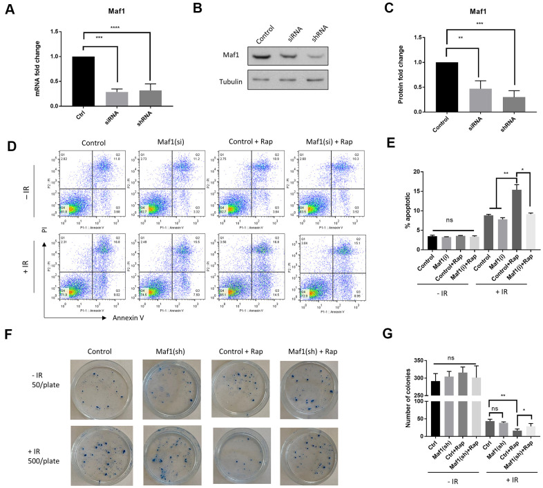
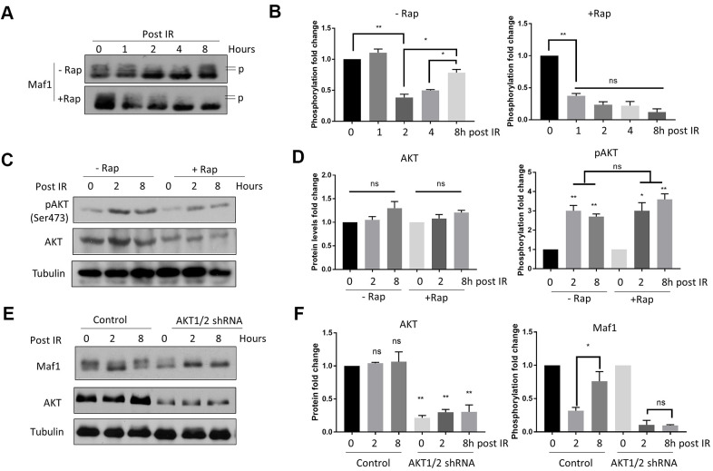
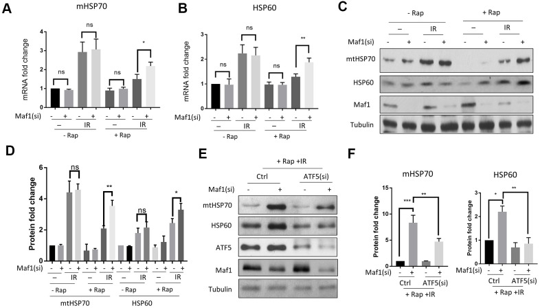
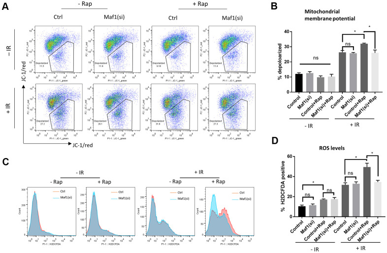
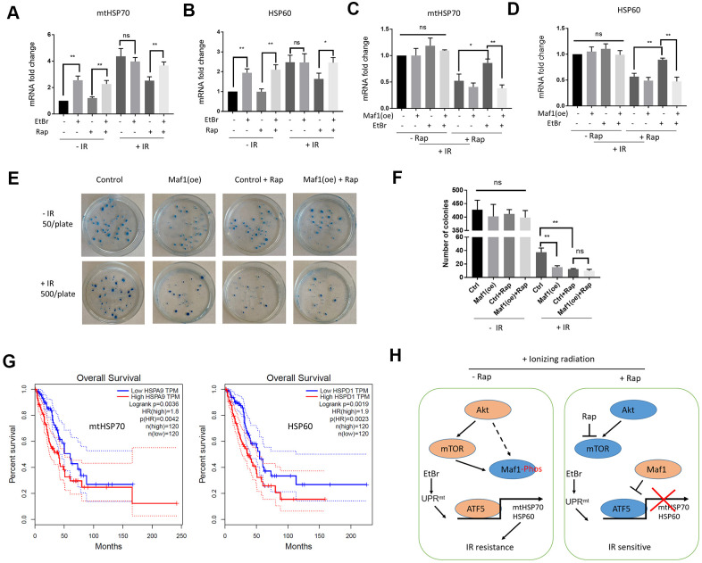
mTOR is well known to promote tumor growth but its roles in enhancing chemotherapy and radiotherapy have not been well studied. mTOR inhibition by rapamycin can sensitize cancer cells to radiotherapy. Here we show that Maf1 is required for rapamycin to increase radio-sensitivity in A549 lung cancer cells. In response to ionizing radiation (IR), Maf1 is inhibited by Akt-dependent re-phosphorylation, which activates mitochondrial unfolded protein response (UPRmt) through ATF5. Rapamycin suppresses IR-induced Maf1 re-phosphorylation and UPRmt activation in A549 cells, resulting in increased sensitivity to IR-mediated cytotoxicity. Consistently, Maf1 knockdown activates ATF5-transcription of mtHSP70 and HSP60, enhances mitochondrial membrane potential, reduces intracellular ROS levels and dampens rapamycin's effect on increasing IR-mediated cytotoxicity. In addition, Maf1 overexpression suppresses ethidium bromide-induced UPRmt and enhances IR-mediated cytotoxicity. Supporting our cell-based studies, elevated expression of UPRmt makers (mtHSP70 and HSP60) are associated with poor prognosis in patients with lung adenocarcinoma (LAUD). Together, our study reveals a novel role of Maf1-UPRmt axis in mediating rapamycin's enhancing effect on IR sensitivity in A549 lung cancer cells.

Keywords: Maf1; mTOR; mitochondrial unfolded protein response; non-small cell lung cancer cell; radio-resistance.


Conflict of interest statement

CONFLICTS OF INTEREST: The authors declare that there are no conflicts of interest.

Figures

Figure 1
Figure 1
Maf1 is required for rapamycin to increase radio-sensitivity in A549 cells. (AC) Maf1 mRNA (A) and protein levels (C) were knocked down by siRNA and shRNA. Representative western blot results are shown in (B). Experiments were performed for ≥3 times with replicates. Data were normalized to non-transfected control (Ctrl) and expressed as fold change. (D, E) Maf1 is required for rapamycin to increase apoptosis in A549 cells in response to radiation. Maf1 was knocked down by siRNA (si) in A549 cells. Cells were then irradiated (IR) with 6 Gy x-ray and treated with 100 nM rapamycin (Rap) as indicated. Apoptosis (Annexin V) and cell death (PI) were analyzed by flow cytometry after 48 hours. Experiments were performed for 2 times and representative results (D) and the quantifications of apoptotic cells (E) are shown. (F, G) Maf1 is required for rapamycin to enhance radiosensitivity in A549 cells. Cells treated with radiation and rapamycin were plated at 500 cells/plate and irradiated (+IR). Non-irradiated cells were plated at 50 cells/plate as controls (-IR). Colonies were counted after 2 weeks of incubation. Experiments were performed for 3 times and representative results are shown in (F) and the quantifications in (G). In all panels, the error bars stand for Standard Deviation (SD) of the mean. Statistical significance was evaluated by 2-tailed, paired student’s t-test (ns, not significant, *, P<0.05, **, P<0.01, ***, P<0.001, ****, P<0.0001).
Figure 2
Figure 2
Rapamycin suppresses Akt-mediated Maf1 re-phosphorylation in response to IR in A549 cells. (A, B) Maf1 phosphorylation (P) was decreased by IR but was re-phosphorylated after 8 hours (Post IR). Rapamycin inhibited Maf1 re-phosphorylation. A549 cells were irradiated (IR) at 6 Gy then treated with 100 nM rapamycin (Rap) for 48 hours. Maf1 phosphorylation was detected by slow migration by Western blot. (C, D) Akt phosphorylation at Ser473 was increased by IR. Akt phosphorylation was detected with phospho-specific antibody. (E, F) Akt was required for Maf1 re-phosphorylation in response to IR. A549 cells with stable Akt knockdown were analyzed by Western blot at indicated time points. In all panels, representative data and the quantifications of at least 2 biological repeats are shown. Data were normalized to time 0. The error bars stand for Standard Deviation (SD) of the mean. Statistical significance was evaluated by 2-tailed, paired student’s t-test (ns, not significant, *, P<0.05, **, P<0.01).
Figure 3
Figure 3
Rapamycin suppresses IR-induced UPRmt through Maf1 in A549 cells. (A, B) A549 cells treated with IR and rapamycin were examined for UPRmt marker genes (mHSP70 and HSP60) expression by RT-qPCR. Experiments were performed for 3 biological repeats and data were normalized to non-treated control. (C, D) HSP70 and HSP60 protein levels were analyzed by Western blot. Representative results are shown in (C) and quantification of 3 biological repeats in (D). Data were normalized to non-treated control. (E, F) Maf1 knockdown activated UPRmt in an ATF5-dependent manner. A549 cells were knocked down for Maf1, ATF5 or both, then treated with rapamycin and IR. mHSP70 and HSP60 protein levels were analyzed by Western blot. Representative results are shown in (E). Experiment were performed for 3 times. Data were normalized to non-transfected control. For all bar graph, the error bars stand for Standard Deviation (SD) of the mean. Statistical significance was evaluated by 2-tailed, paired student’s t-test (ns, not significant, *, P<0.05, **, P<0.01, ***, P<0.001).
Figure 4
Figure 4
Maf1 knockdown mitigates rapamycin inhibition of mitochondrial function in response to IR in A549 cells. (A, B) Mitochondrial membrane potential was evaluated by JC-1 staining followed by flow cytometry. Representative results are shown in (A) and the quantifications of 2 biological replicates are shown in (B). (C, D) Intracellular ROS levels were detected by H2DCFCA probes, followed by flow cytometry. Representative results are shown in (C) and the quantifications of 2 biological replicates are shown in (D). For all bar graph, the error bars stand for Standard Deviation (SD) of the mean. Statistical significance was evaluated by 2-tailed, paired student’s t-test (ns, not significant, *, P<0.05).
Figure 5
Figure 5
Maf1 overexpression suppresses UPRmt-induced IR resistance in A549 cells. (A, B) EtBr activates UPRmt in the presence of rapamycin. A549 cells were treated with indicated drugs or/and IR. UPRmt marker genes (HSP60 and mtHSP70) were examined by RT-qPCR. (C, D) Maf1 overexpression (oe) prevents EtBr from increasing HSP60 and mtHSP70 expression in the present of rapamycin and IR. (E, F) Maf1(oe) sensitizes A549 cells to IR and is not additive to rapamycin. A549 cells (500 cells/plate) stably expressed Maf1 were treated with rapamycin and IR, then allowed to form colonies for 2 weeks. A549 cells were plated at 50 cells/plate as non-irradiated controls. Representative data are shown in (E) and the quantifications of 3 biological replicates in (F). (G) High expression of UPRmt marker genes (HSP60 and mtHSP70) are significantly associated with poor prognosis in lung adenocarcinoma (LAUD) patients. (H) A working model showing the role of Maf1-UPRmt in mediating rapamycin’s enhancing effect on IR sensitivity. For all bar graph, the error bars stand for Standard Deviation (SD) of the mean. Statistical significance was evaluated by 2-tailed, paired student’s t-test (ns, not significant, *, P<0.05, **, P<0.01).

Similar articles

Cited by

References

    1. Xie J, Wang X, Proud CG. mTOR inhibitors in cancer therapy. F1000Res. 2016; 5:F1000. 10.12688/f1000research.9207.1 - DOI - PMC - PubMed
    1. Cargnello M, Tcherkezian J, Roux PP. The expanding role of mTOR in cancer cell growth and proliferation. Mutagenesis. 2015; 30:169–76. 10.1093/mutage/geu045 - DOI - PMC - PubMed
    1. Li Y, Liu F, Wang Y, Li D, Guo F, Xu L, Zeng Z, Zhong X, Qian K. Rapamycin-induced autophagy sensitizes A549 cells to radiation associated with DNA damage repair inhibition. Thorac Cancer. 2016; 7:379–86. 10.1111/1759-7714.12332 - DOI - PMC - PubMed
    1. Nagata Y, Takahashi A, Ohnishi K, Ota I, Ohnishi T, Tojo T, Taniguchi S. Effect of rapamycin, an mTOR inhibitor, on radiation sensitivity of lung cancer cells having different p53 gene status. Int J Oncol. 2010; 37:1001–10. 10.3892/ijo_00000751 - DOI - PubMed
    1. Liang SQ, Bührer ED, Berezowska S, Marti TM, Xu D, Froment L, Yang H, Hall SR, Vassella E, Yang Z, Kocher GJ, Amrein MA, Riether C, et al.. mTOR mediates a mechanism of resistance to chemotherapy and defines a rational combination strategy to treat KRAS-mutant lung cancer. Oncogene. 2019; 38:622–36. 10.1038/s41388-018-0479-6 - DOI - PubMed

Publication types

MeSH terms