Review
. 2021 Jun 4;48(3-4):kuab018.
doi: 10.1093/jimb/kuab018.

Docking domain-mediated subunit interactions in natural product megasynth(et)ases

Affiliations
Review

Docking domain-mediated subunit interactions in natural product megasynth(et)ases

Helen G Smith et al. J Ind Microbiol Biotechnol. .

Abstract

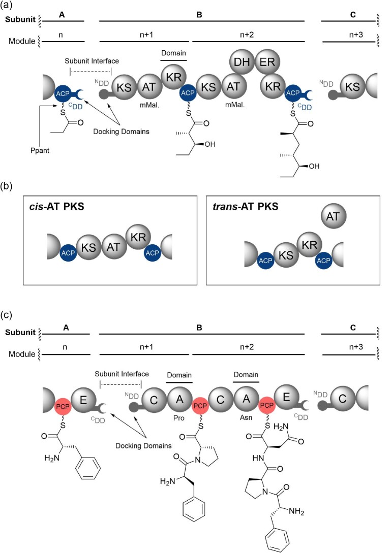
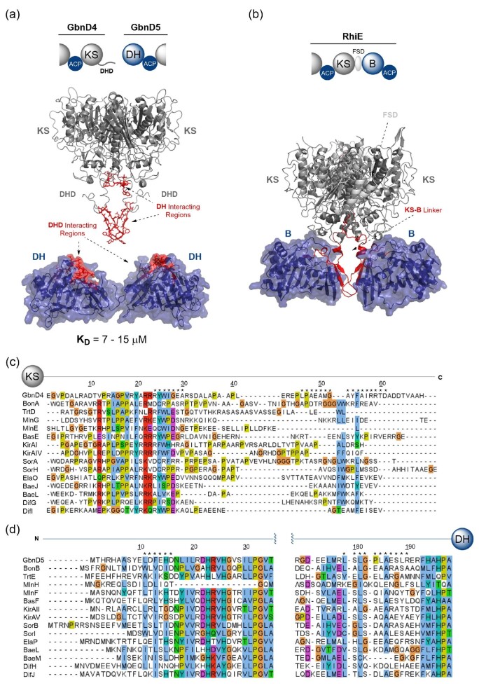
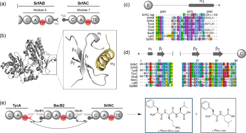
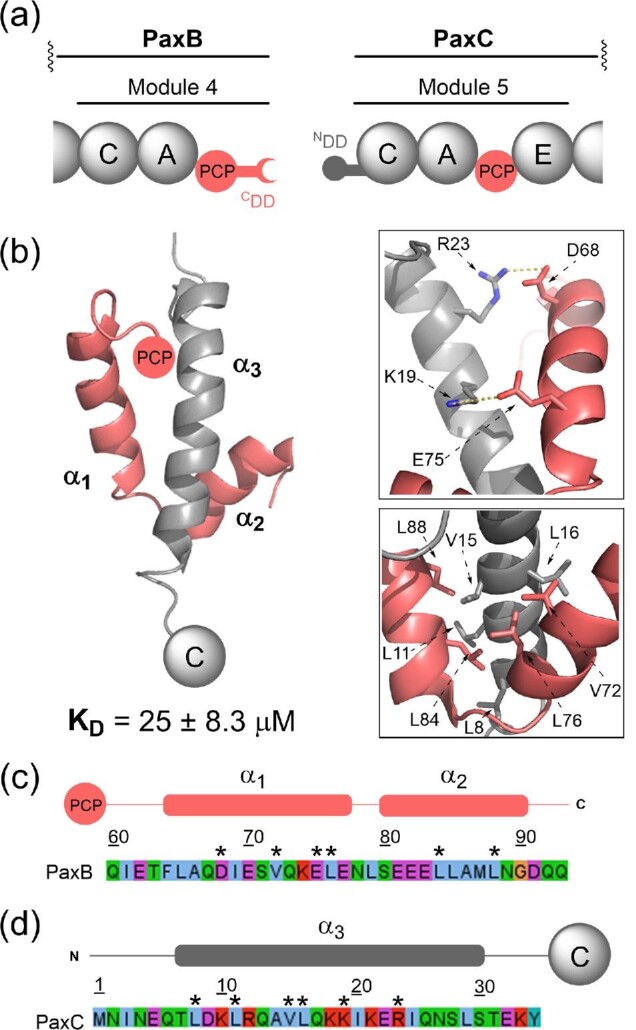
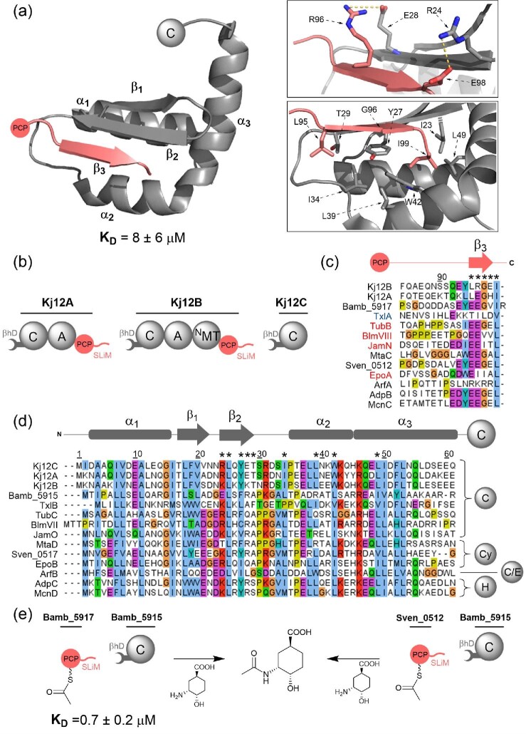
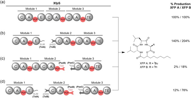
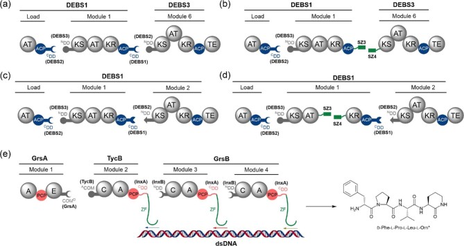
Polyketide synthase (PKS) and non-ribosomal peptide synthetase (NRPS) multienzymes produce numerous high value metabolites. The protein subunits which constitute these megasynth(et)ases must undergo ordered self-assembly to ensure correct organisation of catalytic domains for the biosynthesis of a given natural product. Short amino acid regions at the N- and C-termini of each subunit, termed docking domains (DDs), often occur in complementary pairs, which interact to facilitate substrate transfer and maintain pathway fidelity. This review details all structurally characterised examples of NRPS and PKS DDs to date and summarises efforts to utilise DDs for the engineering of biosynthetic pathways.

Keywords: Biosynthesis; Non-ribosomal peptide synthetase; Polyketide synthase.


Figures

Fig. 1.
Fig. 1.
(a) Partial domain organisation of a hypothetical PKS assembly line. Enzymatic domains are represented by spheres and biosynthetic intermediates are shown appended to each ACP domain. Subunit and module labelling conventions are highlighted above the PKS, and features relating to the main text are highlighted. (b) Domain organisation of cis-AT (left) and trans-AT (right) PKS modules. The stand-alone AT domain in trans-AT PKSs loads extender units onto multiple ACP domains, whereas the AT domain in cis-AT PKSs loads the ACP domain within its module. (c) Partial domain organisation of a hypothetical NRPS assembly line. Enzymatic domains are represented by spheres and biosynthetic intermediates are shown appended to each PCP domain. Subunit and module labelling conventions are highlighted above the NRPS, and features relating to the main text are highlighted. Domain abbreviations are as follows: KS, ketosynthase; AT, acyltransferase; DH, dehydratase; ER, enoylreductase; KR, ketoreductase; ACP, acyl carrier protein; C, condensation; A, adenylation; PCP, peptidyl carrier protein; CDD, C-terminal docking domain; NDD, N-terminal docking domain.
Fig. 2.
Fig. 2.
Structural features, sequence alignments and use in biosynthetic engineering of Class 1 PKSs DDs. (a) Domain architecture of the DEBS2–DEBS3 intersubunit junction from the PKS involved in the biosynthesis of erythromycin A. (b) Solution state NMR structure of the 4-α-helix bundle docked complex formed by the covalently tethered DEBS2 C-terminal and DEBS3 N-terminal DDs (PDB accession code: 1PZR). A dimerisation motif is found upstream of the docking interface and comprises four additional helices (PDB accession code: 1PZQ). Inset (top): Key electrostatic interactions between helix α3 and helices α4 and α4′ that confer specificity to the docking interface. Inset (bottom): Hydrophobic interface formed between the α3, α4 and α4′ helices. (c) Sequence alignment of select 4-α-helix bundle C-terminal docking domains (DDs), including the dimerisation motif. (d) Sequence alignment of select 4-α-helix bundle N-terminal DDs. Asterisks (*) denote the positions of the interfacial residues highlighted in (b). Red chevrons (v) denote interacting electrostatic residues highlighted in (b) where charge is not conserved across DDs. Above the alignments, a schematic displaying the positions of the secondary structural elements from the solution state NMR structure of the DEBS2–DEBS3 DD complex is provided. Residue numbering provided in (b), (c) and (d) is relative to that from PDB accession code 1PZQ. (e) Artificial splitting of the PikAI subunit from the PKS responsible for pikromycin biosynthesis using four α-helix bundle DDs from the Plm1–Plm2 subunit junction in phoslactomycin biosynthesis. Quantification of pikromycin production was determined by HPLC analysis of culture extracts from a Streptomyces venezuelae ΔpikAI mutant complemented with the engineered PikAI proteins in trans.
Fig. 3.
Fig. 3.
Structural features, sequence alignments and use in biosynthetic engineering of PKS Class 2 DDs. (a) Domain architecture of the CurG-CurH, CurK-CurL and Bamb_5925-Bamb_5924 intersubunit junctions from the hybrid PKS-NRPSs responsible for the biosynthesis of curacin A and enacyloxin IIa. Cognate DD pairs are depicted using complimentary fitting shapes. (b) Sequence alignment of select eight α-helix bundle C-terminal DDs. (c) Sequence alignment of select eight α-helix bundle N-terminal DDs. Asterisks (*) denote the positions of the conserved hydrophobic interfacial residues highlighted in (d). Above the alignments, schematics displaying the positions of the secondary structural elements observed in the structures of each of the DD complexes is provided. (d) X-ray crystal structure of the eight α-helix bundle docked complex formed by the covalently tethered CurG C-terminal and CurH N-terminal DDs (PDB accession code: 4MYY). Inset: Hydrophobic residues across α1, α2, α4, α3′ and α4′ implicated in the formation of the CurG–CurH DD interface. (e) X-ray crystal structure of the docked complex formed by the covalently tethered CurK C-terminal and CurL N-terminal DDs (PDB accession code: 4MYZ) Inset: Electrostatic interactions between the α2′ and α4 helices. The same interactions are not conserved in the CurG–CurH interface. (f) Solution state NMR structure of the docked complex formed by the covalently tethered Bamb_5925 C-terminal and Bamb_5924 N-terminal DDs (PDB accession code: 6TDN) Inset: Electrostatic interactions between α1 and α4 (top) and α1 and α3 (bottom). The former is also observed in the CurG–CurH interface. Residue numbering in (b)–(e) is relative to that of PDB entry 4MYY. (g). Engineering of the PikAIII–IV intersubunit junction from the pikromycin PKS. This PKS produces two products, pikromycin, of which narbolide (nbl) is the precursor, and methymycin, for which 10-deoxymethynolide (10-dml) is the precursor, resulting from a module-skipping mechanism. Exchanging the WT four α-helix bundle DD pair at this junction with the eight α-helix bundle CurG-H or CurK-L DD pair was demonstrated to maintain productive interaction between proteins by in vitro assays. Additionally, introducing eight α-helix bundle DDs was found to change the product profile from 50% nbl, to predominantly 10-dml, suggesting more effective delivery of the PikAIII ACP-tethered substrate to the PikAIV TE domain. Percentage production values are given with respect to the WT DD pair.
Fig. 4.
Fig. 4.
Structural features and sequence alignments of PKS four α-helix bundle DDs. (a) Sequence alignment of select CDDs. (b) Sequence alignment of select NDDs. Residue numbering throughout is relative to that of PDB entry 2N5D. Asterisks (*) denote the positions of the interfacial residues highlighted in (d). Above the alignment, a schematic displaying the positions of the secondary structural elements observed in the VirA–VirFG docked complex is provided. (c) Domain architecture of the VirA–VirFG intersubunit interface in the virginiamycin trans-AT PKS. (d) Solution state NMR structure of the docked complex formed by covalently-tethered VirA CDD and VirFG NDD (PDB accession code: 2N5D). Inset (left): Hydrophobic interface formed between all four helices. Inset (right): Electrostatic and hydrogen bonding interactions at the docking domain interface.
Fig. 5.
Fig. 5.
Structural features and sequence alignments of PKS DHD domains and corresponding DH domains. (a) Domain architecture (top) and structural model (bottom) of GbnD4–GbnD5 KS-DH intersubunit junction from the gladiolin trans-AT PKS. Regions highlighted in red on the DHD domain and DH domain have been shown to interact. (b) Domain architecture (top) and X-ray crystal structure (bottom) of RhiE KS-B di-domain from the rhizoxin trans-AT PKS (PDB accession code: 4KC5). The B domain is structurally homologous to a DH domain, and the region connecting the KS to the B domain is highlighted in red. The flanking subdomain (FSD) is highlighted, which is absent from KS domains at KS–DH junctions. (c) Sequence alignment of selected DHD domains from the C-termini of KS domains. (d) Sequence alignment of selected N-terminal DH domains corresponding to the DHD domains in (c). Asterisks (*) denote the positions of the interfacial residues highlighted in (a), as observed experimentally by NMR spectroscopy and carbene footprinting mass spectrometry. Domain abbreviation: B, branching domain.
Fig. 6.
Fig. 6.
Structural features, sequence alignments and use in biosynthetic engineering of NRPS COM domains. (a) Domain organisation of the SrfAB–SrfAC intersubunit junction. (b) X-ray crystal structure of the SrfAC condensation domain (PDB accession code: 2VSQ). Inset: The helix-hand motif is formed of a helix and a beta sheet comprising three non-contiguous strands. A portion of the C-terminal protein tag of SrfAC, shown in yellow, was found to interact with the helix-hand motif and is proposed to mimic the NDD helix. (c) Sequence alignment of selected C-terminal COMD domains. The sequence of the interacting region of the SrfAC C-terminal tag is provided and aligned as described by Tanovic et al. (d) Sequence alignment of selected N-terminal COMA domains, encompassing the two regions of protein comprising the helix-hand motif. Residue numbering throughout is relative to that of PDB entry 2VSQ. (e) Engineering of intermodular interfaces using the TycA–TycB COM domain pairs to mediate productive crosstalk between non-cognate NRPS modules from the tyrocidine, bacitracin and surfactin A assembly lines. Successive interactions of TycAΔE–BacB2 and BacB2–SrfAC indicated by the blue arrows lead to formation of the tripeptide shown in the blue box, while the direct interaction between TycAΔE–SrfAC indicated by the grey arrow leads to the dipeptide product shown in the grey box.
Fig. 7.
Fig. 7.
Structural features and sequence alignments of NRPS three α-helix bundle DDs. (a) Domain organisation of the PaxB–PaxC intersubunit junction. (b) Solution state NMR structure of the docked complex formed by the covalently tethered PaxB CDD and PaxC NDD (PDB accession code: 6TRP). Inset (top): Electrostatic interactions between the α1 and α3 helices. Inset (bottom): Hydrophobic interface formed between all three helices. (c) Sequence of the PCP domain-tethered PAX CDD. (d) Sequence of the C domain-tethered PAX NDD. Asterisks (*) denote the positions of the interfacial residues highlighted in (b). Above each sequence, a schematic displaying the positions of the secondary structural elements observed in the docked complex is provided.
Fig. 8.
Fig. 8.
Structural features, sequence alignments and use in biosynthetic engineering of βhD domains. (a) Solution state NMR structure of the docked complex formed by the covalently tethered Kj12B C-terminal SLiM and Kj12C N-terminal βhD domain (PDB accession code: 6EWV). Inset (top): Residues involved in salt bridge-forming interactions between the SLiM (β3) and β2 strand of the βhD domain. Inset (bottom): Hydrophobic interface formed between β2, β3, α2 and α3. (b) Domain architecture of the rhabdopeptide-producing NRPS. Note that this iterative system contains three βhD domains and two SLiMs, which can all interact with varying affinities. (c) Sequence alignment of select cDD SLiMs. Highlighted in red are SLiMs appended to the carrier protein domain of a PKS module, those in black are appended to the carrier protein domain of NRPS modules. TxlA, blue, contains a SLiM downstream of an E domain. (d) Sequence alignment of select N-terminal βhD domains. To the right of the alignment, the domain directly downstream of the βhD domain in each protein is shown. In (c) and (d), a schematic indicating the positions of the secondary structural elements from the solution state NMR structure is provided. Residue numbering in (a)–(d) is relative to that of PDB entry 6EWV. In (c) and (d), asterisks (*) denote the positions of the key interfacial residues highlighted in (a). (e) SLiM–βhD domain junction in the enacyloxin NRPS–PKS pathway. Crosstalk of the Sven_0512 PCP domain, a SLiM-bearing carrier protein domain from the watasemycin NRPS, with the enacyloxin Bamb_5915 βhD-C domain from the enacyloxin PKS-NRPS was able to produce N-acetyl (1S,3R,4S)-3-amino-4-hydroxycyclohexane-1-carboxylic acid. Domain abbreviations are as follows: Cy, heterocyclisation domain; C/E, dual condensation–epimerisation domain; H, flavin-dependent halogenase.
Fig. 9.
Fig. 9.
Engineering of XfpS, the single-subunit NRPS responsible for production of xefoampeptides A and B. (a) Wild-type XfpS. Bottom: XfpS engineered with DD pairs to artificially split the three modules into separate subunits. (b) As in (a) with a SLiM–βHD domain pair from the TxlA–TxlB intersubunit junction from taxlllaid biosynthesis introduced between modules 1 and 2. (c) Same as (a) with a 3-α-helix bundle DD pair from the PaxB–PaxC intersubunit junction from PAX peptide biosynthesis introduced between modules 2 and 3. (d) Same as (a) with both intermodular junctions engineered as in (b) and (c). In each case, the proteins were heterologously expressed in E. coli and XFP A/XFP B production was determined from LC–MS of the methanolic extract. Percentage production is given with respect to the wild-type system in (a). In all cases, a Strep-Tag II affinity tag is found at the N-terminus of module 1 and the C-terminus of module 3.
Fig. 10.
Fig. 10.
Engineering of PKS and NRPS biosynthetic systems using synthetic docking tools. (a) DEBS1 loading module, module 1 and DEBS3 module 6 engineered using four α-helix bundle DD pairs from DEBS2-3 and DEBS1-2 intersubunit junctions. (b) Same as (a) but with the four α-helix bundle DDs at the module 1-module 6 junction replaced by synthetic DD pair SYNZIP3-4. Higher initial rates of turnover are observed compared to (a). (c) DEBS1 loading module, module 1 and module 2 engineered using four α-helix bundle DD pairs from DEBS2-3 and DEBS1-2 intersubunit junctions. (d) Same as (c) but with module 1 artificially split at the AT–KR junction using the SYNZIP3-4 pair. Initial rates of turnover observed are comparable to (c). In (a–d), the TE domain is derived from the DEBS system and fused to the end of the terminal module. (e) Engineering of the gramicidin S NRPS using DNA templating to direct protein–protein interactions. SLiM–βHD domain pairs from the InxA–InxB intersubunit junction of the rhabdopeptide/xenortide- like peptide biosynthetic pathway from Xenorhabdus inexxi were inserted at TycB1–GrsB3 and GrsB3-4 interfaces. ZFs were inserted C-terminal to the SLiMs and allowed site-specific binding to synthetic DNA. The productivity of the engineered system was determined by monitoring production of a known tetrapeptide shunt metabolite that is terminated with a cyclic ornithine residue (denoted as l-Orn*).

Similar articles

Cited by

References

    1. Aparicio J. F., Molnár I., Schwecke T., König A., Haydock S. F., Ee Khaw L., Staunton J., Leadlay P. F. (1996). Organization of the biosynthetic gene cluster for rapamycin in Streptomyces hygroscopicus: Analysis of the enzymatic domains in the modular polyketide synthase. Gene, 169(1), 9–16. https://doi.org/10.1016/0378-1119(95)00800-4. - PubMed
    1. Bretschneider T., Heim J. B., Heine D., Winkler R., Busch B., Kusebauch B., Stehle T., Zocher G., Hertweck C. (2013). Vinylogous chain branching catalysed by a dedicated polyketide synthase module. Nature, 502(7469), 124–128. https://doi.org/10.1038/nature12588. - PubMed
    1. Broadhurst R. W., Nietlispach D., Wheatcroft M. P., Leadlay P. F., Weissman K. J. (2003). The structure of docking domains in modular polyketide synthases. Chemistry & Biology, 10(8), 723–731. https://doi.org/10.1016/s1074-5521(03)00156-x. - PubMed
    1. Buchholz T. J., Geders T. W., Bartley F. E., Reynolds K. A., Smith J. L., Sherman D. H. (2009). Structural basis for binding specificity between subclasses of modular polyketide synthase docking domains. ACS Chemical Biology, 4(1), 41–52. https://doi.org/10.1021/cb8002607. - PMC - PubMed
    1. Cai X., Zhao L., Bode H. B. (2019). Reprogramming promiscuous nonribosomal peptide synthetases for production of specific peptides. Organic Letters, 21(7), 2116–2120. https://doi.org/10.1021/acs.orglett.9b00395. - PubMed

MeSH terms