. 2021 Jul 16;42(7):940-950.
doi: 10.1093/carcin/bgab017.

DPYD, down-regulated by the potentially chemopreventive agent luteolin, interacts with STAT3 in pancreatic cancer

Affiliations

DPYD, down-regulated by the potentially chemopreventive agent luteolin, interacts with STAT3 in pancreatic cancer

Hiroyuki Kato et al. Carcinogenesis. .

Abstract

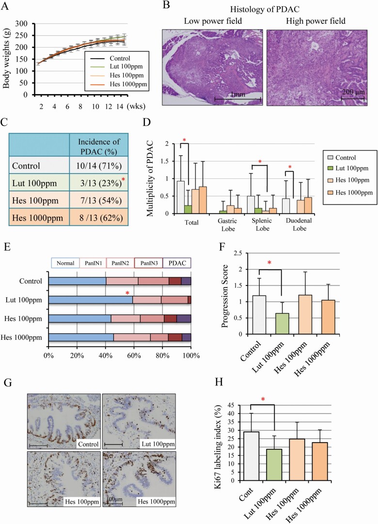
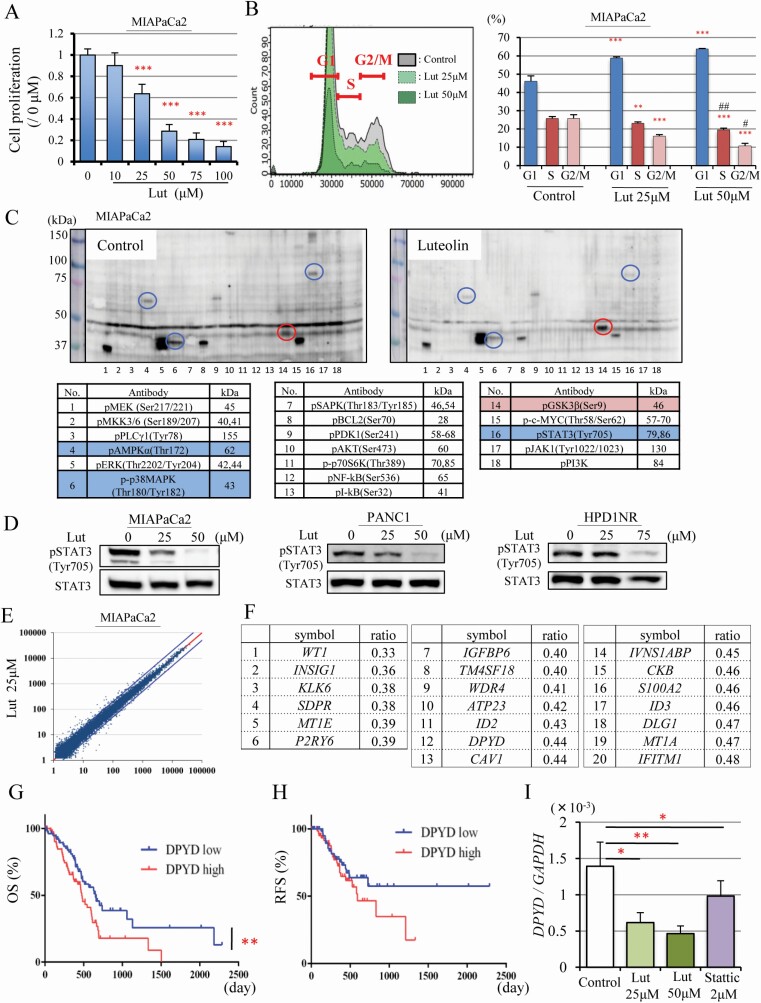
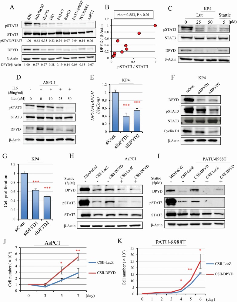
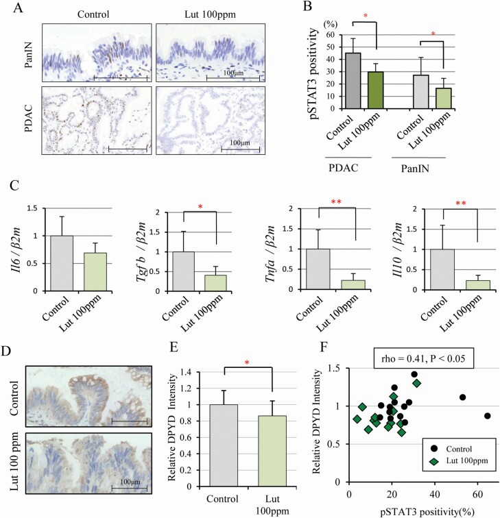
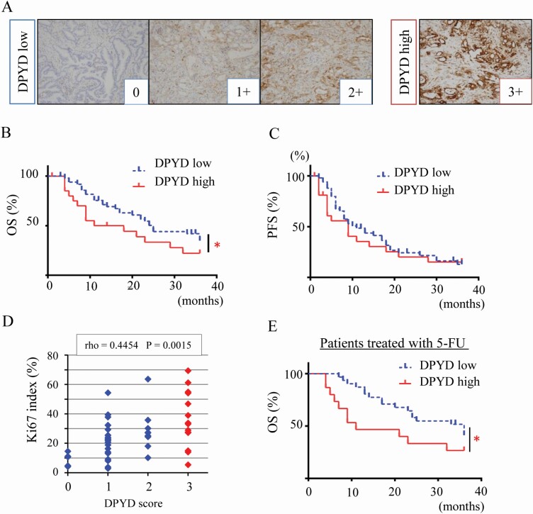
The 5-year survival rate of pancreatic ductal carcinoma (PDAC) patients is <10% despite progress in clinical medicine. Strategies to prevent the development of PDAC are urgently required. The flavonoids Luteolin (Lut) and hesperetin (Hes) may be cancer-chemopreventive, but effects on pancreatic carcinogenesis in vivo have not been studied. Here, the chemopreventive effects of Lut and Hes on pancreatic carcinogenesis are assessed in the BOP-induced hamster PDAC model. Lut but not Hes suppressed proliferation of pancreatic intraepithelial neoplasia (PanIN) and reduced the incidence and multiplicity of PDAC in this model. Lut also inhibited the proliferation of hamster and human pancreatic cancer cells in vitro. Multi-blot and microarray assays revealed decreased phosphorylated STAT3 (pSTAT3) and dihydropyrimidine dehydrogenase (DPYD) on Lut exposure. To explore the relationship between DPYD and STAT3 activity, the former was silenced by RNAi or overexpressed using expression vectors, and the latter was inactivated by small molecule inhibitors or stimulated by IL6 in human PDAC cells. DPYD knock-down decreased, and overexpression increased, pSTAT3 and cell proliferation. DPYD expression was decreased by inactivation of STAT3 and increased by its activation. The frequency of pSTAT3-positive cells and DPYD expression was significantly correlated and was decreased in parallel by Lut in the hamster PDAC model. Finally, immunohistochemical analysis in 73 cases of human PDAC demonstrated that DPYD expression was positively correlated with the Ki-67 labeling index, and high expression was associated with poor prognosis. These results indicate that Lut is a promising chemopreventive agent for PDAC, targeting a novel STAT3-DPYD pathway.


Figures

Figure 1.
Figure 1.
Luteolin inhibits pancreatic carcinogenesis and cell proliferation in a hamster model. Body-weights (A) and representative histology of hamster PDAC in controls; on the left, a low power field (×20), on the right, a high power field (×100) (B). The incidence in all lobes (C) and multiplicity in all, duodenal, splenic, and gastric lobes (D). The proportion of normal, PanIN1, PanIN2, PanIN3 and PDAC in all pancreatic ducts (diameter > 200 mm) of duodenal lobes (E) and the progression score calculated by weighting respective lesions (normal = 0, PanIN1 = 1, PanIN2 = 2, PanIN3 = 3, PDAC = 4) (F), Immunohistochemical findings of Ki-67 (G) and Ki-67 labeling index in PanINs (H). Data represented as mean ± SD, n = 14 (Control) and 13 (Lut 100 ppm, Hes 100 ppm, 1000 ppm). *P < 0.05 statistically significant compared with controls.
Figure 2.
Figure 2.
Luteolin suppresses pSTAT3 and DPYD expression. Cell proliferation quantified by WST-1 assay (n = 6 per dose) (A), and cell cycle analysis (n = 3 per dose) of MIAPaCa2 cells after Luteolin (Lut) treatment for 48 h (B). Multi-Western blotting of phosphorylated proteins associated with cell proliferation. On the left, no treatment and on the right 75 μM Lut treatment for 48 h. Circles and labels in blue or red depict Lut down- or up-regulated proteins in MIAPaCa2 cells, respectively (C). Western blotting for pSTAT3 (Tyr705), STAT3 after Lut (25, 50μM) treatment for 48 h in MIAPaCa2, PANC1, HPD1NR (D). Relative mRNA expression plotted from the result of microarrays comparing no treatment with 25 μM Lut treatment for 48 h in MIAPaCa2 cells. Blue lines are ratios of mRNA expression in controls: 25 μM Lut = 1 : 2 or 2 : 1 (E). The 20 genes most down-regulated by 25 μM Lut treatment (relative expression >100 in controls) were detected by microarray analysis (F). Overall survival (OS) (Log-rank test **P < 0.01) (G) and recurrence-free survival (RFS) (H) of PDAC patients extracted from the TCGA pancreatic cancer dataset (n = 159), which includes DPYD high expression (n = 79) and DPYD low expression (n = 80). Expression of DPYD mRNA after Lut (25, 50 μM) and Stattic (2 μM) treatment in MIAPaCa2 cells (I). Data are mean ± SD. *, **, *** P < 0.05, 0.01, 0.001 compared with no treatment. #, ##P < 0.05, 0.01 between 25 μM and 50 μM Lut treatment.
Figure 3.
Figure 3.
DPYD and pSTAT3 expression affected one other. Western blotting for pSTAT3, STAT3, DPYD and β-Actin protein in nine human pancreatic cell lines (A). DPYD and pSTAT3 expression corrected by β-Actin and total STAT3 (Spearman rho = 0.883, P < 0.01) (B). Western blotting for pSTAT3, STAT3, DPYD and β-Actin after Lut (25, 50 μM) or Stattic (5 μM) treatment in the DPYD high-expressing line (KP4) (C) and after stimulation of the STAT3 pathway by IL 6 (50 ng/ml) for 24 h followed by addition of Lut (10, 25, 50 μM) in a DPYD low-expressing line (AsPC1) (D). Expression of DPYD mRNA (n = 3 per cell) (E) and DPYD protein for pSTAT3, STAT3, Cyclin D1 and β-Actin (F), and cell proliferation (n = 6 per condition) (G) after siDPYD (siDPYD1, siDPYD2) transfection into KP4 cells. Expression of DPYD, pSTAT3, STAT3 and β-Actin protein (H, I) and proliferation (J, K) when DPYD is overexpressed with or without Stattic treatment in AsPC1 and PATU-8988T cells (MIAPaCa cell protein as a positive control for DPYD). Data are mean ± SD. *, **, *** P < 0.001 compared with control or siControl or CSII-LacZ.
Figure 4.
Figure 4.
Luteolin inhibits pSTAT3 and DPYD expression in the hamster pancreatic cancer model. Immunohistochemical findings of pSTAT3 in PanIN and PDAC (A) and nuclear pSTAT3 positivity of PanIN (Controls: n = 14, Lut 100 ppm: n = 13) and PDAC (Controls: n = 9, Lut 100 ppm: n = 3) in control and 100 ppm Lut treated animals (B). Expression of cytokine mRNA (Il6, Tgfβ, Tnfα, Il10) associated with the STAT3 pathway in hamster pancreas gastric lobe (n = 6 respectively, standardized by β2-microglobulin, Bmg) (C). DPYD expression detected in cytoplasm immunohistochemically (D) and its relative intensity in PanIN of 100 ppm Lut (n = 13) and controls (n = 14) (E). Graph of DPYD relative intensity and pSTAT3 nuclear positivity (Spearman rho = 0.41, P < 0.05) (F). Data are mean ± SD. *, ** P < 0.05, 0.01 as compared with controls.
Figure 5.
Figure 5.
DPYD expression is associated with poor prognosis in human PDAC. Immunohistochemical staining of DPYD in a PDAC tissue microarray (n = 73) stratified into four categories (0; none, 1+; weak, 2+; moderate, 3+; strong) and coded as DPYD low (0, 1+, 2+, n =50) and high expression (3+, n = 23) (A). Three-year overall survival (OS) (B) and 3-year recurrence-free survival (RFS) (C) of PDAC patients with low DPYD (n = 50) or high DPYD expression (n = 23) *P = 0.045 between low and high DPYD expression groups. DPYD expression score and Ki-67 labeling index (Spearman rho = 0.445, P < 0.05) (D). Three-year OS of PDAC patients with S-1 adjuvant therapy (n = 45) including high DPYD (n = 15) and low DPYD expression groups (n = 30) *P = 0.018 between low and high DPYD expressors (E).

Similar articles

Cited by

References

    1. Roth G and GBD 2017 Causes of Death Collaborators. (2018) Global, regional, and national age-sex-specific mortality for 282 causes of death in 195 countries and territories, 1980–2017: a systematic analysis for the Global Burden of Disease Study 2017. Lancet, 392, 1736–1788. - PMC - PubMed
    1. Ministry of Health, Labour and Welfare. (2017) Vital Statistics Japan.
    1. Moffat, G.T., et al. (2019) Pancreatic cancer-a disease in need: optimizing and integrating supportive care. Cancer, 125, 3927–3935. - PMC - PubMed
    1. Benzel, J., et al. (2018) Chemoprevention and treatment of pancreatic cancer: update and review of the literature. Digestion, 97, 275–287. - PubMed
    1. Kato, A., et al. (2015) Chemopreventive effect of resveratrol and apocynin on pancreatic carcinogenesis via modulation of nuclear phosphorylated GSK3β and ERK1/2. Oncotarget, 6, 42963–42975. - PMC - PubMed

Publication types

MeSH terms