. 2021 Apr 30;30(6):454-466.
doi: 10.1093/hmg/ddab058.

Identification of LRRK2 missense variants in the accelerating medicines partnership Parkinson's disease cohort

Affiliations

Identification of LRRK2 missense variants in the accelerating medicines partnership Parkinson's disease cohort

Nicole Bryant et al. Hum Mol Genet. .

Abstract

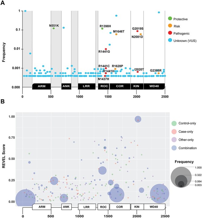
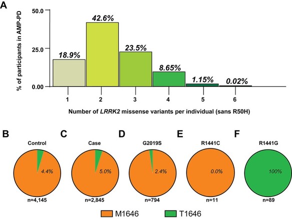
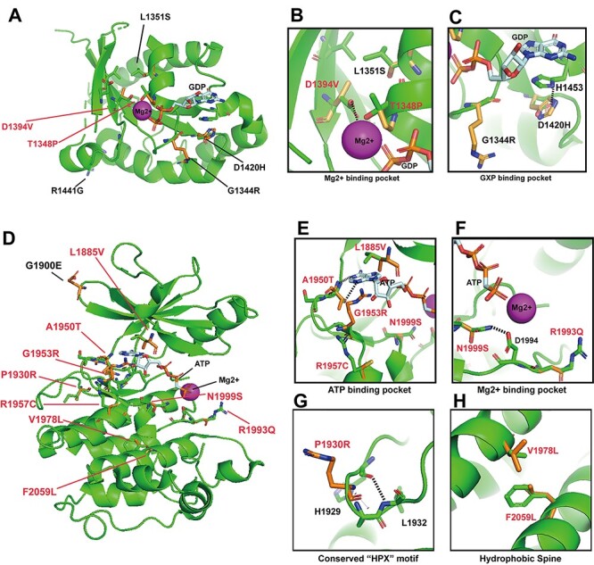
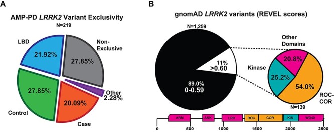
Pathogenic missense variants in the leucine-rich repeat kinase 2 (LRRK2) gene have been identified through linkage analysis in familial Parkinson disease (PD). Subsequently, other missense variants with lower effect sizes on PD risk have emerged, as well as non-coding polymorphisms (e.g. rs76904798) enriched in PD cases in genome-wide association studies. Here we leverage recent whole-genome sequences from the Accelerating Medicines Partnership-Parkinson's Disease (AMP-PD) and the Genome Aggregation (gnomAD) databases to characterize novel missense variants in LRRK2 and explore their relationships with known pathogenic and PD-linked missense variants. Using a computational prediction tool that successfully classifies known pathogenic LRRK2 missense variants, we describe an online web-based resource that catalogs characteristics of over 1200 LRRK2 missense variants of unknown significance. Novel high-pathogenicity scoring variants, some identified exclusively in PD cases, tightly cluster within the ROC-COR-Kinase domains. Structure-function predictions support that some of these variants exert gain-of-function effects with respect to LRRK2 kinase activity. In AMP-PD participants, all p.R1441G carriers (N = 89) are also carriers of the more common PD-linked variant p.M1646T. In addition, nearly all carriers of the PD-linked p.N2081D missense variant are also carriers of the LRRK2 PD-risk variant rs76904798. These results provide a compendium of LRRK2 missense variants and how they associate with one another. While the pathogenic p.G2019S variant is by far the most frequent high-pathogenicity scoring variant, our results suggest that ultra-rare missense variants may have an important cumulative impact in increasing the number of individuals with LRRK2-linked PD.


Figures

Figure 1
Figure 1
Description of disease-linked missense variants in the first iteration of the AMP-PD cohort. (A) Pie chart depicting diagnosis for 9887 participants that correspond to available WGS data. ‘Case’ indicates a diagnosis of PD. ‘Control’ indicates neurologically healthy participants that were evaluated by movement disorder specialists (see Materials and Methods). ‘Other’ indicates parkinsonism-related neurological disorders including LBD (representing 90% of this category), multiple-system atrophy (MSA), essential tremor, progressive supranuclear palsy (PSP), Alzheimer’s disease, corticobasal degeneration, psychogenic illness, possible prodromal PD (motor and non-motor) and other atypical parkinsonisms (see Supplementary Material, Table S2). (B) Distribution of PD-risk modifying non-synonymous (missense) variants (e.g. disease-linked missense mutations) identified from AMP-PD participants (see Supplementary Material, Table S1 for descriptions of the individual studies combined into the AMP-PD cohort). Subgroups include: LRRK2 variants, ‘LRRK2+’, missense variants include p.G2019S, p.R1441C, p.R1441G and p.R1441H and GBAI variants, GBA+, missense variants include, p.E365K, p.T369M and p.N370S. Smaller subgroups of participants include PD-linked (typically compound heterozygous in disease) missense variants in parkin ‘PRKN+’ that include p.T240M, p.R275W, p.G430D. α-Synuclein ‘SNCA+’ missense variants include p.A53T. ~ 0.6% of AMP-PD participants have both LRRK2 and GBA missense variants associated with PD, ‘LRRK2+/GBA+’, and four participants have PD-associated variants in both GBA and PRKN, ‘GBA+/PRKN+’. (C) Breakdown within the subgroups identified from panel (B) as positive for a disease-linked missense variant according to diagnosis. Color scheme is identical to that in panel (A). (D/E) Age at baseline and sex distribution of cases (D) and controls (E).
Figure 2
Figure 2
Identification and analysis of missense LRRK2 variants in AMP-PD. Non-synonymous missense variants in LRRK2 with respect to the LRRK2 sequence deposition in reference assembly GRCh38:CM000674.2 (ENSG00000188906). (A) In total, 219 missense LRRK2 variants are aligned with conserved domains in the LRRK2 protein (positioned below the x-axis), and the frequency in which these variants occur in the AMP-PD cohort (PD and control, y-axis, Supplementary Material, Table S3). LRRK2 domain annotation, from N- to C-terminal, are ARM = armadillo, ANK = ankyrin, LRR = leucine-rich repeat, ROC = Rab-like GTPase, COR = C-terminal of ROC, KIN = kinase and WD40 = WD40-repeat. Variants previously annotated in the literature that are associated with risk or susceptibility to PD are indicated as follows: green: suspected protective, yellow: suspected risk and red: pathogenic. Novel variants, or those with an unknown contribution to disease, are indicated as light-blue. (B) Bubble plot aligning all missense variants with protein sequence position and pathogenicity prediction score (y-axis: REVEL score, see Materials and Methods), along with frequency in the AMP-PD cohort (larger bubble is more frequent). Color indicates the diagnosis of participant(s), where green is control-only, salmon is case-only, purple is ‘other’-only and blue is found in a combination of cases, ‘other’ and controls (e.g. the incompletely penetrant pathogenic p.G2019S missense variant). Variants from all participants are included. No removal of PD-linked variant carrying participants was conducted. An interactive browsing tool is available via https://nbviewer.jupyter.org/github/west-lab/LRRK2Figures/blob/master/Interactive%20Figures/AMP-PD_LRRK2_New.html.
Figure 3
Figure 3
LRRK2 missense variant combinations in AMP-PD participants. (A) Column graph indicating the number of non-synonymous missense variants identified within AMP-PD participants with respect to LRRK2 (reference assembly GRCh38:CM000674.2). All variants included with exception of R50H (rs2256408) (see Materials and Methods). The number of individuals with no LRRK2 variants from consensus (5.2%, sans R50H) is not shown. Pie charts of (B) participants with (percentage of group) or without the p.M1646T missense variant from controls or (C) cases, (D) p.G2019S variant carriers, (E) p.R1441C variant carriers, (E) p.R1441C variant carriers and (F) p.R1441G variant carriers.
Figure 4
Figure 4
Identification and analysis of missense LRRK2 variants in gnomAD. (A) Bubble plot for missense variants in gnomAD participants (v2.1.1; https://gnomad.broadinstitute.org). The size of the bubble represents the relative frequency of the variant. The x-axis aligns the bubbles across conserved domains in LRRK2. The y-axis indicates the REVEL pathogenicity prediction score (26). LRRK2 domains are: ARM, ANK, LRR, ROC, COR, KIN and WD40. (B) Seven pathogenic LRRK2 variants (red boxes) known to cause PD and the corresponding REVEL scores. (C) Blue boxes highlight seven missense variants found only in PD cases in AMP-PD that were not identified in any participant in gnomAD, with REVEL scores indicated for each of these variants. An integrated browser is available via https://nbviewer.jupyter.org/github/west-lab/LRRK2Figures/blob/master/Interactive%20Figures/BubbleChartRaceFreq.html.
Figure 5
Figure 5
Structural models of high-REVEL LRRK2 missense variants in the ROC and kinase domains suggest both LOF and GOF variants. (A) cryo-EM structure of LRRK2 bound with GDP and Mg2+ (6VNO) (30) was used to model variants from gnomAD predicted to alter protein function (REVEL scores >0.700). ROC variants and their corresponding REVEL scores include: p.D1394V:0.977, p.L1351S:0.806, p.T1348P:0.848, p.G1344R:0.719, p.D1420H:0.786. Red-colored variants are discussed in text. (B) Blow-up model of variants predicted to interact in the Mg-binding pocket. p.T1348P and p.D1394V may interfere with Mg binding. (C) Magnification of the GXP-binding pocket in the ROC-domain. H1453 interacts with the guanosine group in GTPase activity, disrupted by p.D1420H. (D) AppCp and Mg2+ is docked into the LRRK2 cryo-EM structure(6VNO) based on the crystal structure of the G1179S Roco4 Kinase Domain bound to AppCp and Mg2+ from D. discoideum (PDB ID 4F1M) (36). REVEL scores for novel variants include p.L1885V:0.793, p.A1950T:0.866, p.G1953R:0.960, p.R1957C:0.714, p.V1978L:0.809, p.F2059L:0.952, p.R1993Q:0.924, p.N1999S:0.862, p.P1930R:0.882. (E) Blow-up model of the ATP-binding pocket with variants that may affect ATP binding. (F) The Mg-binding pocket in the LRRK2 kinase domain, demonstrating p.N1999S potentially interrupts hydrogen bond interactions (dashed lines) with the highly conserved D1994 residue required for kinase activity (H/YRD motif). p.R1993Q:0.924, another potentially disrupting variant within this loop, is also positioned in the model. (G) Magnification of the structure-altering variant p.P1930R:0.882 that lies within the conserved β-turn segment, likely disrupting bonds with residues L1932 and H1929 (dashed line). (H) Predicted function-altering variants, p.V1978L:0.809 and p.F2059L:0.952, lie on the hydrophobic spine and would likely disrupt hydrophobic interactions critical for overall conformational regulation of the kinase domain.
Figure 6
Figure 6
Summary of LRRK2 variant discoveries from both AMP-PD and gnomAD. (A) Pie chart showing the distribution of the 219 LRRK2 variant in case (PD), control, LBD or multiple participants from each group, ‘Non-exclusive’ from AMP-PD. ‘Other’ refers to either MSA-exclusive (N = 2, p.R924C and p.A75G), PSP (N = 1, p.K657I) and prodromal PD (N = 2, p.S1096C and p.T146S). Case-only (PD) variants are listed in Supplementary Material, Table S4. (B) Pie chart showing the portion of LRRK2 variants from gnomAD with less REVEL score ≤0.599 (black) or ≥0.6 (white). Smaller pie chart represents the distribution of location within LRRK2 for variants that are at or exceed the REVEL score of 0.6 in gnomAD (N = 139). List of gnomAD variants > 0.6 are described in Supplementary Material, Table S8.

References

    1. Trinh, J., Guella, I. and Farrer, M.J. (2014) Disease penetrance of late-onset parkinsonism: a meta-analysis. JAMA Neurol., 71, 1535–1539. - PubMed
    1. Heckman, M.G., Soto-Ortolaza, A.I., Aasly, J.O., Abahuni, N., Annesi, G., Bacon, J.A., Bardien, S., Bozi, M., Brice, A., Brighina, L.et al. (2013) Population-specific frequencies for LRRK2 susceptibility variants in the Genetic Epidemiology of Parkinson’s Disease (GEO-PD) Consortium. Mov. Disord., 28, 1740–1744. - PMC - PubMed
    1. Nalls, M.A., Blauwendraat, C., Vallerga, C.L., Heilbron, K., Bandres-Ciga, S., Chang, D., Tan, M., Kia, D.A., Noyce, A.J., Xue, A.et al. (2019) Identification of novel risk loci, causal insights, and heritable risk for Parkinson’s disease: a meta-analysis of genome-wide association studies. Lancet Neurol., 18, 1091–1102. - PMC - PubMed
    1. Paisán-Ruíz, C., Jain, S., Evans, E.W., Gilks, W.P., Simón, J., van derBrug, M., López de Munain, A., Aparicio, S., Gil, A.M., Khan, N.et al. (2004) Cloning of the gene containing mutations that cause PARK8-linked Parkinson’s disease. Neuron, 44, 595–600. - PubMed
    1. Zimprich, A., Biskup, S., Leitner, P., Lichtner, P., Farrer, M., Lincoln, S., Kachergus, J., Hulihan, M., Uitti, R.J., Calne, D.B.et al. (2004) Mutations in LRRK2 cause autosomal-dominant parkinsonism with pleomorphic pathology. Neuron, 44, 601–607. - PubMed

Publication types

Substances