. 2021 May;30(5):1072-1080.
doi: 10.1002/pro.4053. Epub 2021 Mar 12.

Membrane-dependent amyloid aggregation of human BAX α9 (173-192)

Affiliations

Membrane-dependent amyloid aggregation of human BAX α9 (173-192)

David A Price et al. Protein Sci. 2021 May.

Abstract

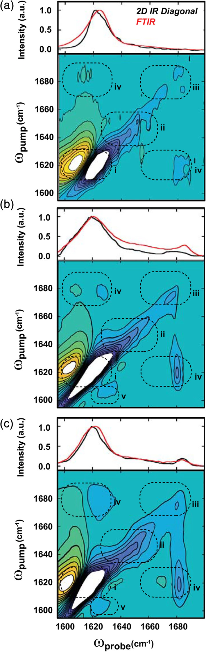
Mitochondrial outer membrane permeabilization, which is a critical step in apoptosis, is initiated upon transmembrane insertion of the C-terminal α-helix (α9) of the proapoptotic Bcl-2 family protein BAX. The isolated α9 fragment (residues 173-192) is also competent to disrupt model membranes, and the structures of its membrane-associated oligomers are of interest in understanding the potential roles of this sequence in apoptosis. Here, we used ultrafast two-dimensional infrared (2D IR) spectroscopy, thioflavin T binding, and transmission electron microscopy to show that the synthetic BAX α9 peptide (α9p) forms amyloid aggregates in aqueous environments and on the surfaces of anionic small unilamellar vesicles. Its inherent amyloidogenicity was predicted by sequence analysis, and 2D IR spectra reveal that vesicles modulate the β-sheet structures of insoluble aggregates, motivating further examination of the formation or suppression of BAX amyloids in apoptosis.

Keywords: 2D IR; BAX; amyloid; apoptosis.


Conflict of interest statement

The authors declare no conflict of interest.

Figures

FIGURE 1
FIGURE 1
Structure and aggregation propensity of wild type human BAX. (a) Solution structure of BAX (PDB ID: 1F16) 43 with helices α1–α8 in gray and α9 in blue, showing the sequence synthesized in this work (α9p). (b) Aggregation and amyloid propensities of full‐length BAX protein via sequence analysis extracted from AGGRESCAN, 35 TANGO, 36 , 37 , 38 PASTA 2.0, 39 and MetAmyl 40
FIGURE 2
FIGURE 2
Aggregation behavior of α9p in aqueous buffer. (a–c) (Bottom) two‐dimensional infrared (2D IR) spectra with (top) corresponding diagonal slices for α9p in buffer at (a) early, (b) intermediate, and (c) late time points. Spectra are normalized to the largest Amide I intensity in the stationary‐phase (>250 min) spectrum and diagonal slices, which reflect the ν(0–1) bleach signals, are reversed in sign for comparison to Fourier transform infrared (FTIR) absorbance. Horizontal lines are drawn through diagonal frequencies discussed in the main text and monitored in (d). (d) Diagonal intensities at 1,610 cm−1 (gray); 1,620 cm−1 (red); 1,645 cm−1 (black); and 1,675 cm−1 (blue) during α9p aggregation. Kinetic traces are color coded as indicated by arrows in (a–c) (right). Additional trials are shown in Figure S3
FIGURE 3
FIGURE 3
Two‐dimensional infrared (2D IR) and Fourier transform infrared (FTIR) spectra of insoluble α9p aggregates formed over 24 hr. (a–c): (Bottom) 2D IR spectra with (top) corresponding diagonal slices (black line) and FTIR spectra (red line) for mature α9p aggregates formed in the absence of small unilamellar vesicles (SUVs) (a), and in the presence of 10 mM (b) and 1 μM (c) 1‐palmitoyl‐2‐oleoyl‐sn‐glycero‐3‐phosphocholine:1‐palmitoyl‐2‐oleoyl‐sn‐glycero‐3‐phosphoglycerol (POPC:POPG) SUVs. 2D IR spectra are scaled to 50% of their respective Amide I maxima and peaks in Regions i–iv correspond to structural features discussed in the text. Vertical slices taken at ω probe = 1,680 cm−1 are shown in Figure S5 to compare the intensities of features (iii) and (iv)

References

    1. Youle RJ, Strasser A. The BCL‐2 protein family: Opposing activities that mediate cell death. Nat Rev Mol Cell Biol. 2008;9:47–59. - PubMed
    1. Chipuk JE, Moldoveanu T, Llambi F, Parsons MJ, Green DR. The BCL‐2 family reunion. Mol Cell. 2010;37:299–310. - PMC - PubMed
    1. Gillies LA, Kuwana T. Apoptosis regulation at the mitochondrial outer membrane. J Cell Biochem. 2014;115:632–640. - PubMed
    1. Xin M, Li R, Xie M, et al. Small‐molecule Bax agonists for cancer therapy. Nat Commun. 2014;5:4935–4935. - PMC - PubMed
    1. Adams JM, Cory S. The BCL‐2 arbiters of apoptosis and their growing role as cancer targets. Cell Death Differ. 2018;25:27–36. - PMC - PubMed

Publication types

MeSH terms