Review
. 2022;41(2):171-206.
doi: 10.1080/08830185.2021.1883600. Epub 2021 Feb 27.

COVID-19: Immunology, Immunopathogenesis and Potential Therapies

Affiliations
Review

COVID-19: Immunology, Immunopathogenesis and Potential Therapies

Asha Bhardwaj et al. Int Rev Immunol. 2022.

Abstract

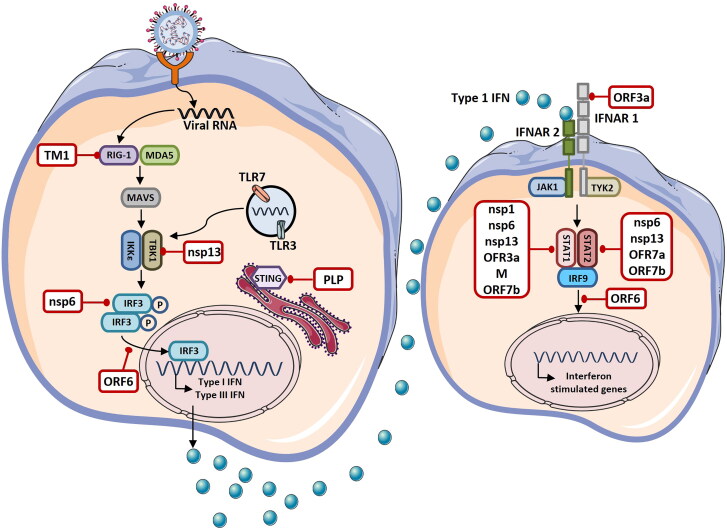
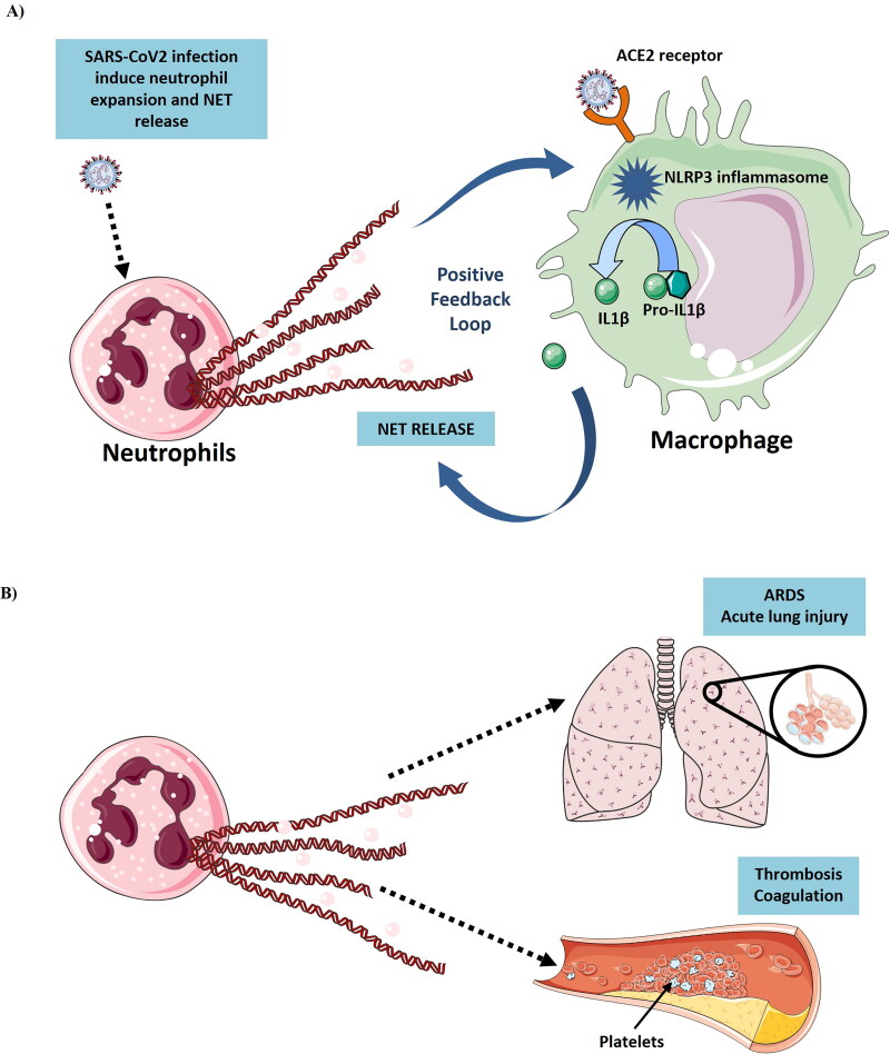
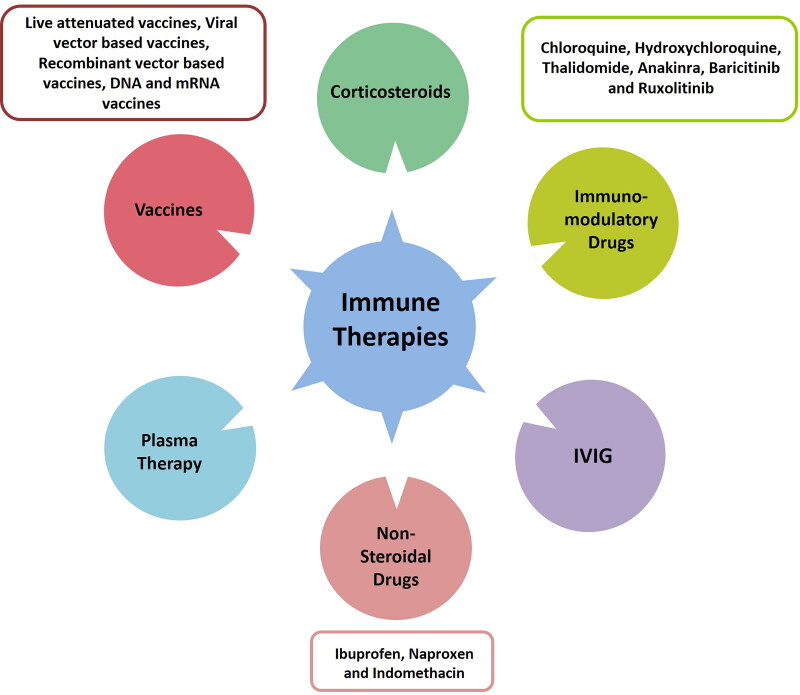
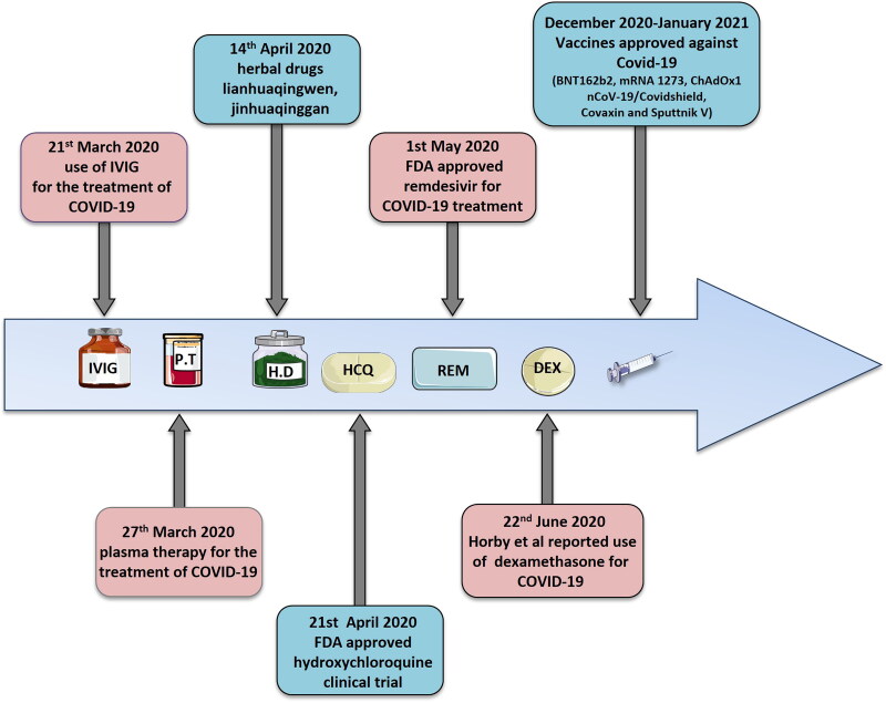
The Coronavirus Disease-2019 (COVID-19) imposed public health emergency and affected millions of people around the globe. As of January 2021, 100 million confirmed cases of COVID-19 along with more than 2 million deaths were reported worldwide. SARS-CoV-2 infection causes excessive production of pro-inflammatory cytokines thereby leading to the development of "Cytokine Storm Syndrome." This condition results in uncontrollable inflammation that further imposes multiple-organ-failure eventually leading to death. SARS-CoV-2 induces unrestrained innate immune response and impairs adaptive immune responses thereby causing tissue damage. Thus, understanding the foremost features and evolution of innate and adaptive immunity to SARS-CoV-2 is crucial in anticipating COVID-19 outcomes and in developing effective strategies to control the viral spread. In the present review, we exhaustively discuss the sequential key immunological events that occur during SARS-CoV-2 infection and are involved in the immunopathogenesis of COVID-19. In addition to this, we also highlight various therapeutic options already in use such as immunosuppressive drugs, plasma therapy and intravenous immunoglobulins along with various novel potent therapeutic options that should be considered in managing COVID-19 infection such as traditional medicines and probiotics.

Keywords: COVID-19; SARS-CoV-2; cytokine storm; immunology; immunotherapy.


Conflict of interest statement

The authors declare no conflicts of interest.

Figures

Figure 1.
Figure 1.
(A, B) Structure and genome organization of SARS-CoV-2 (Figure illustrated with the help of https://smart.servier.com/. ).
Figure 2.
Figure 2.
The schematic diagram of SARS-CoV-2 invasion and replication into host cells in step wise manner: (1) SARS-CoV-2 enter into the cell by binding to the ACE2 receptor present on the host cell membrane via S protein which mediates viral-host membrane fusion and viral entry. (2) Entry of SARS-CoV-2 results in the uncoating of viral RNA into cytoplasm which then undergoes translation to produce polyproteins pp1a and pp1b, which are further processed by virus-encoded proteinases into individual nonstructural proteins (nsps). (3) Replication transcription complex is formed by some of the nsps. Replication transcription complex uses the (+) stand genomic RNA as template. Following replication (+) stand genomic RNA is produced which becomes the genome of the new viral particle. (4) Subgenomic RNAs synthesized through transcription are translated into viral structural proteins: S (Spike) protein, M (Membrane) protein, E (Envelope) protein, N (Nucleocapsid) protein. (5) N protein combined with the (+) strand genomic RNA to form the nucleoprotein complex. S, E and M proteins enter into endoplasmic reticulum (ER) and transported to Golgi apparatus. The nucleoprotein complex and S, E and M proteins are further assembled in the ER-Golgi intermediate compartment (ERGIC) to form mature virion. (6) Virions are released from the host cells by exocytosis (Figure illustrated with the help of https://smart.servier.com/. ).
Figure 3.
Figure 3.
Schematic representation of immunopathogenesis during COVID-19 in a step wise manner: (1) SARS-CoV-2 gain entry into the lung cells through ACE2 receptor. ACE2 receptor is expressed by different organs of the body like brain, lungs, kidney, liver and intestine. SARS-CoV-2 frequently infects these organs but the pathogenesis of SARS-CoV-2 infection initiates from lungs and cause major damage to the lungs. Lung infection starts when SARS-CoV-2 enter into type 2 alveolar cells through ACE2 receptor. (2) Virus replicates into the lung cells and induce cells to undergo pyroptosis and secrete damage associated molecular patterns (DAMPs). (3) DAMPs are recognized by adjoining epithelial cells, alveolar macrophages and endothelial cells which stimulate the secretion of various pro-inflammatory cytokines and chemokines like IL-6, IL-10, MIP1-α, MIP1-1β, MCP-1. (4) Release of theses cytokines and chemokines induce the recruitment of monocytes, macrophages and neutrophils into lungs which further secrete the proinflammatory cytokines and form the inflammatory feedback loop. (5) During SARS-CoV-2 infection immune response is dysregulated which leads to the persistent recruitment of immune cells which promote overproduction of inflammatory cytokines which cause various lung disorders like ARDS and acute lung injury. SARS-CoV-2 infection induce delayed activation of dendritic cells that cause defective T cell response. Most of the T lymphocytes in SARS-CoV-2 infection are induced to become Th1 cells. SARS-CoV-2 infection also induce delayed antibody response (Figure illustrated with the help of https://smart.servier.com/. ).
Figure 4.
Figure 4.
The schematic diagram depicting immunopathogenesis of COVID-19 (Figure illustrated with the help of https://smart.servier.com/. ).
Figure 5.
Figure 5.
Immune evasion by Coronavirus. Coronavirus is sensed by various pathogen recognition receptors (PRRs) present in the host cell like toll like receptor (TLR)-3, TLR-7, retinoic acid inducible gene (RIG)-1 and melanoma differentiation associated protein 5 (MDA5). Recognition of Coronavirus by PRRs stimulate the production of type I and type 3 interferons (IFNs). IFNs are then secreted in autocrine and paracrine manner and induce the expression of interferon stimulated genes via JAK-STAT signaling pathway leading to the activation of antiviral response. Several viral proteins (shown in white boxes) encoded by the Coronavirus antagonize various steps of IFN signaling and escape the immune response. TM1: First Transmembrane Domain, MAVS: Mitochondrial Antiviral-Signaling, IKKε: IкB Kinase Epsilon, TBK1: TANK-Binding Kinase 1, IRF: Interferon Regulatory Factor, STING: Stimulator of IFN Genes, PLP: Papain Like Protease, NSPs: Nonstructural Proteins, ORFs: Open Reading Frames, IFNAR: Interferon- Alpha/Beta Receptor, JAK1: Janus Kinase 1, TYK2: Tyrosine Kinase 2, STAT: Signal Transducer and Activator of Transcription 2 (Figure illustrated with the help of https://smart.servier.com/. ).
Figure 6.
Figure 6.
(A) Induction of Neutrophil Extracellular Traps (NETs) from neutrophils by SARS-CoV-2 infection. SARS-CoV-2 induce the neutrophil expansion and NET release. NET release promote the IL-1β secretion from macrophages which further leads to inflammation. SARS-CoV-2 also induce the IL-1β secretion from macrophages by activating the NLRP3 inflammasome activation. IL-1β further stimulate NET release and forms a positive feedback loop. (B) NET release causes ARDS, thrombosis and coagulation in COVID-19 patients (Figure illustrated with the help of https://smart.servier.com/. ).
Figure 7.
Figure 7.
Different types of immunotherapies for prevention of COVID 19.
Figure 8.
Figure 8.
Timeline of COVID-19-therapies: Advancements in the COVID-19 therapies. IVIG (Intravenous immunoglobulin), P.T (Plasma therapy), H.D (Herbal drugs), HCQ (Hydroxychloroquine), REM (Remdesivir), DEX (Dexamethasone).
Figure 9.
Figure 9.
The schematic representation of SARS-CoV-2 life cycle in step wise manner depicting the potential therapeutic approaches against SARS-CoV-2: (1) SARS-CoV-2 enter into the cell by binding to the ACE2 receptor present on the host cell membrane via S protein which mediates viral-host membrane fusion and viral entry. (2) Entry of SARS-CoV-2 results in the uncoating of viral RNA into cytoplasm which then undergoes translation to produce polyproteins pp1a and pp1b, which are further processed by virus-encoded proteinases into individual nonstructural proteins (nsps). (3) Replication transcription complex is formed by some of the nsps. Replication transcription complex uses the (+) stand genomic RNA as template. Following replication (+) stand genomic RNA is produced which becomes the genome of the new viral particle. (4) Subgenomic RNAs synthesized through transcription are translated into viral structural proteins: S (Spike) protein, M (Membrane) protein, E (Envelope) protein, N (Nucleocapsid) protein. (5) N protein combined with the (+) strand genomic RNA to form the nucleoprotein complex. S, E and M proteins enter into endoplasmic reticulum (ER) and transported to Golgi apparatus. The nucleoprotein complex and S, E and M proteins are further assembled in the ER-Golgi intermediate compartment (ERGIC) to form mature virion. (6) Virions are released from the host cells by exocytosis. Many potential therapeutic approaches are considered for the management of COVID-19. These therapeutics act on various steps of the SARS-CoV-2 life cycle. Monoclonal antibodies and Convalescent plasma targets the S proteins of SARS-CoV-2 from interacting with the ACE2 receptor. Camostat mesylate and Nafomostat targets the serine protease (TMPRSS2) and prevents the S protein cleavage which is required for the viral fusion to the host cells. Lopinavir and Ritonavir targets the proteolysis of polypeptide chains. Remsdesivir prevents the replication of SARS-CoV-2 and drugs like Tocilizumab, Lenzilumab, Gimsilumab, Namilumab and Lianhuaqingwen inhibit the pro-inflammatory response.

References

    1. Lan J, Ge J, Yu J, et al. Structure of the SARS-CoV-2 spike receptor-binding domain bound to the ACE2 receptor. Nature. 2020;581(7807):215–220. doi: 10.1038/s41586-020-2180-5. - DOI - PubMed
    1. Wu JT, Leung K, Bushman M, et al. Estimating clinical severity of COVID-19 from the transmission dynamics in Wuhan, China. Nat Med. 2020;26(4):506–510. doi: 10.1038/s41591-020-0822-7. - DOI - PMC - PubMed
    1. Hassan SA, Sheikh FN, Jamal S, et al. Coronavirus (COVID-19): a review of clinical features, diagnosis, and treatment. Cureus. 2020;12(3):e7355. doi: 10.7759/cureus.7355. - DOI - PMC - PubMed
    1. Li G, Fan Y, Lai Y, et al. Coronavirus infections and immune responses. J Med Virol. 2020;92(4):424–432. doi: 10.1002/jmv.25685. - DOI - PMC - PubMed
    1. Wang C, Li W, Drabek D, et al. A human monoclonal antibody blocking SARS-CoV-2 infection. Nat Commun. 2020;11(1):2251. doi: 10.1038/s41467-020-16256-y. - DOI - PMC - PubMed

Publication types