

Contour-aware semantic segmentation network with spatial attention mechanism for medical image
- PMID: 33642659
- PMCID: PMC7898027
- DOI: 10.1007/s00371-021-02075-9
Contour-aware semantic segmentation network with spatial attention mechanism for medical image
Abstract
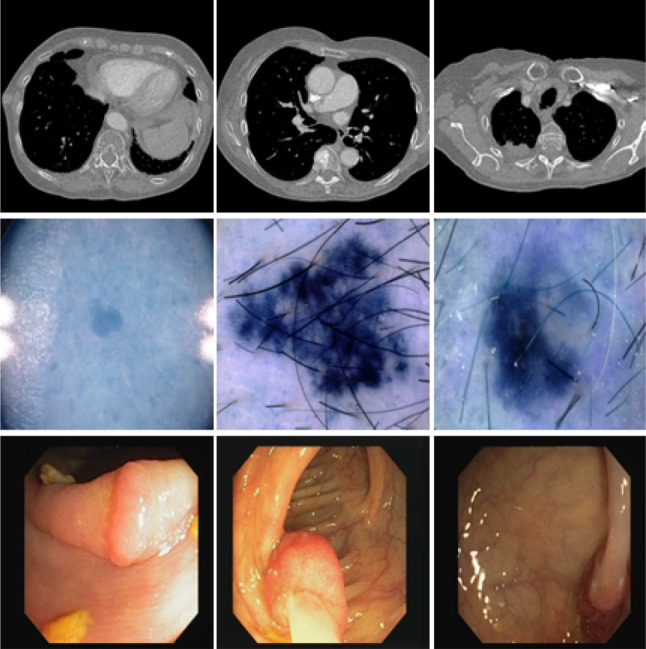
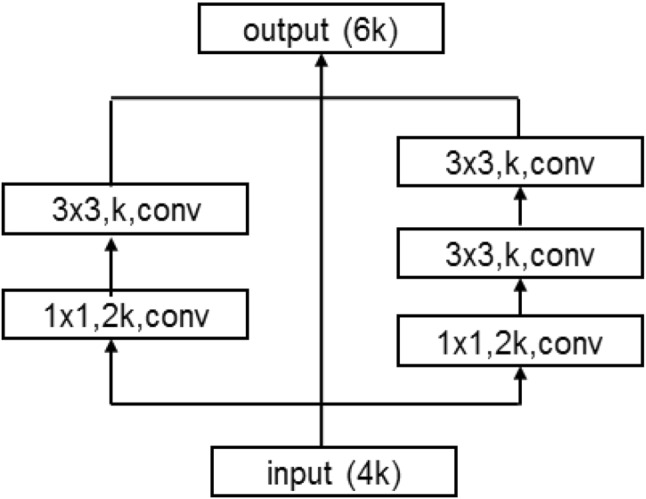
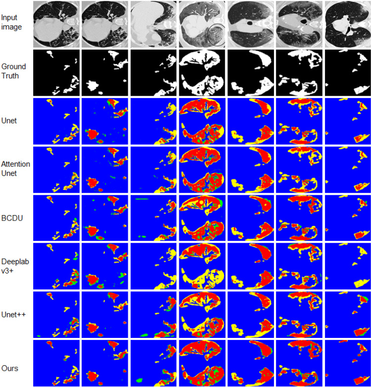
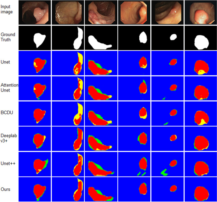
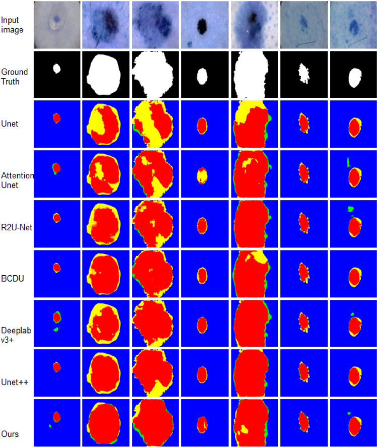
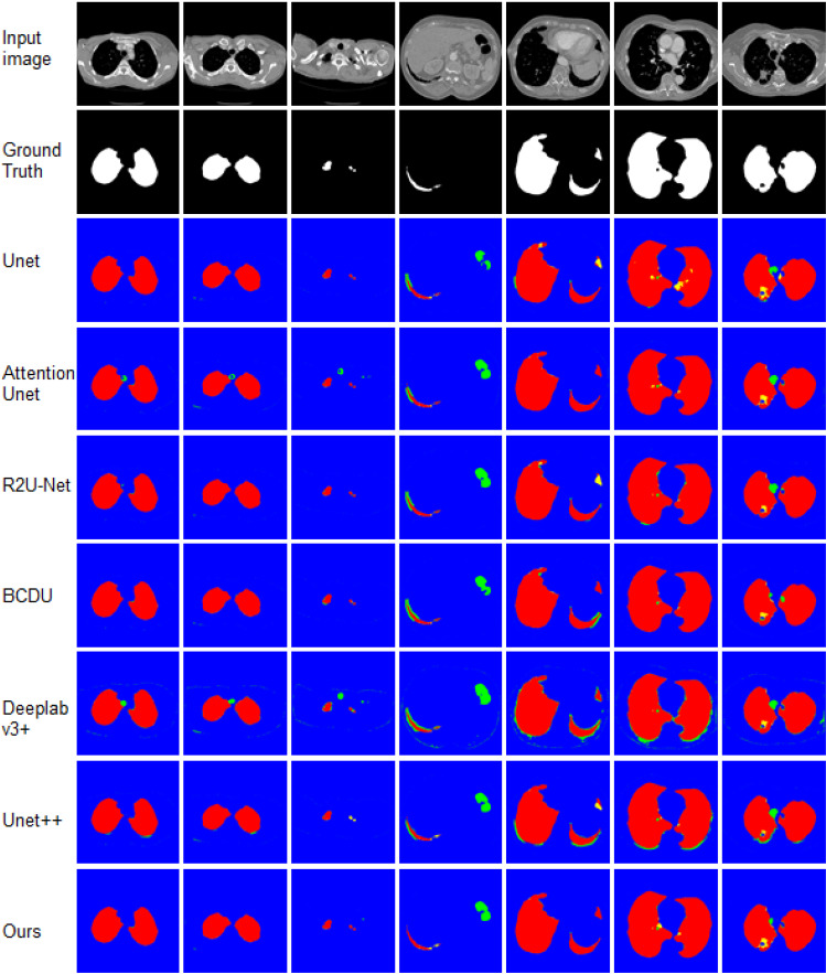
Medical image segmentation is a critical and important step for developing computer-aided system in clinical situations. It remains a complicated and challenging task due to the large variety of imaging modalities and different cases. Recently, Unet has become one of the most popular deep learning frameworks because of its accurate performance in biomedical image segmentation. In this paper, we propose a contour-aware semantic segmentation network, which is an extension of Unet, for medical image segmentation. The proposed method includes a semantic branch and a detail branch. The semantic branch focuses on extracting the semantic features from shallow and deep layers; the detail branch is used to enhance the contour information implied in the shallow layers. In order to improve the representation capability of the network, a MulBlock module is designed to extract semantic information with different receptive fields. Spatial attention module (CAM) is used to adaptively suppress the redundant features. In comparison with the state-of-the-art methods, our method achieves a remarkable performance on several public medical image segmentation challenges.
Keywords: Medical image segmentation; Neural network; Semantic segmentation.
© The Author(s), under exclusive licence to Springer-Verlag GmbH, DE part of Springer Nature 2021.
Conflict of interest statement
Conflict of interestThe authors declare that they have no conflict of interest.
Figures


References
-
- Alom, M.Z., Hasan, M., Yakopcic, C., Taha, T.M., Asari, V.K.: Recurrent residual convolutional neural network based on u-net (r2u-net) for medical image segmentation. arXiv preprint arXiv:1802.06955 (2018)
-
- Asadi-Aghbolaghi, M., Azad, R., Fathy, M., Escalera, S.: Multi-level context gating of embedded collective knowledge for medical image segmentation. arXiv preprint arXiv:2003.05056 (2020)
-
- Azad, R., Asadi-Aghbolaghi, M., Fathy, M., Escalera, S.: Bi-directional convlstm u-net with densley connected convolutions. In: 2019 IEEE/CVF International Conference on Computer Vision Workshop (ICCVW), pp. 406–415 (2019)
LinkOut - more resources
Full Text Sources
Other Literature Sources