. 2021 Mar;37(2):205-208.
doi: 10.1007/s12055-020-01070-4. Epub 2020 Oct 28.

Pulmonary embolism from cardiac hydatids

Affiliations

Pulmonary embolism from cardiac hydatids

Aamir Mohammad et al. Indian J Thorac Cardiovasc Surg. 2021 Mar.

Abstract

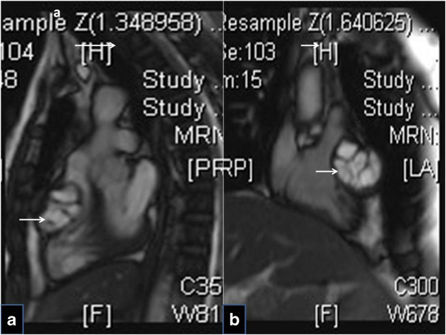
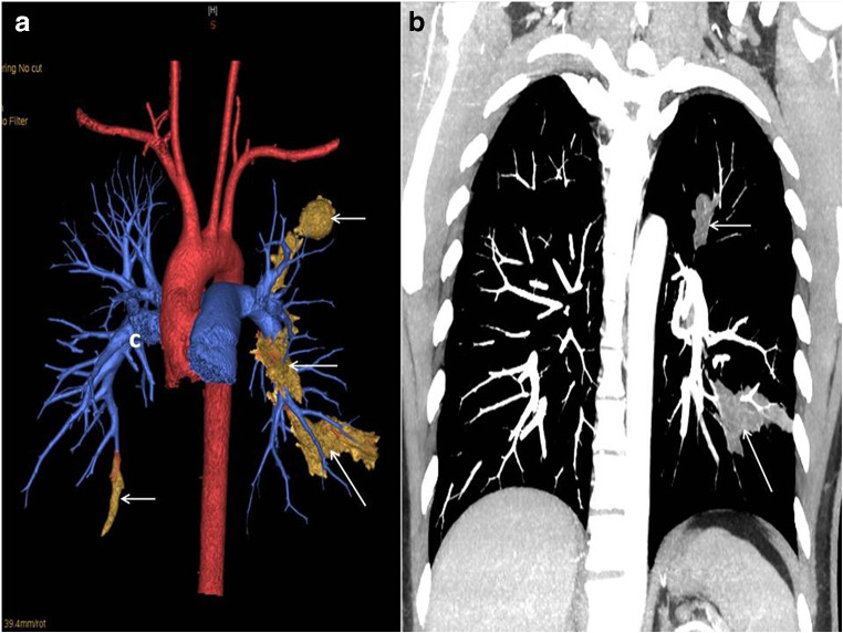
Metastatic hydatid disease of the lung may happen when a hydatid cyst (HC) anywhere in the body ruptures into a systemic vein, a right heart chamber or a pulmonary artery (PA), resulting in the embolisation of the cyst's contents into the lungs. We submit herewith, the images of embolised hydatids within the PA, in a patient who had surgery for HC involving the right ventricular (RV) wall in 2014. Despite adequate surgical and medical management, investigations in 2017 revealed multiple embolised cysts within PA branches. Further continued medical therapy resulted only in partial resolution of the disease, indicating probably the inadequacy of the currently available treatment strategies.

Keywords: Benzimidazole; Echinococcus granulosus; Hydatid cysts; Pulmonary embolism; Syncope.


Conflict of interest statement

Conflict of interestThe authors declare that there is no conflict of interest.

Figures

Fig. 1
Fig. 1
Images of 2014: Preoperative magnetic resonance images of the chest, a sagittal view and b coronal view, showing the RV wall hydatid compromising the RV chamber
Fig. 2
Fig. 2
Serial chest x-rays: a Preoperatively in 2014, showing an empty cyst in the LUL. b In 2017, showing recurrence in LUL and LLL. c In 2018, showing a persistent LUL cyst (single arrow) and areas of pulmonary infarction in LLL (double arrows). d In 2019, showing persisting LUL lesion and resolution of the LLL lesion
Fig. 3
Fig. 3
a Three-dimensional volume-rendered images in 2017, demonstrating the embolised hydatids as soft tissue densities and filling defects occluding multiple segmental and subsegmental PA. b Coronal maximum intensity projection image showing the same
Fig. 4
Fig. 4
Images of 2018. a Computerised tomogram images of the chest, axial view, showing embolised cysts in the anterior basal and subsegmental arteries of the LLL, and a wedge-shaped pulmonary infarct. b Coronal section image demonstrating the same

Similar articles

References

    1. Sarkar M, Pathania R, Jhobta A, Thakur BR, Chopra R. Cystic pulmonary hydatidosis. Lung India. 2016;33:179–191. doi: 10.4103/0970-2113.177449. - DOI - PMC - PubMed
    1. Shankar R, Thankachen R, Choudhary LK, Rao VM. Right ventricular hydatid cyst excision without the use of cardiopulmonary bypass. Indian J Thorac Cardiovasc Surg. 2016;32:163–164. doi: 10.1007/s12055-016-0419-4. - DOI
    1. Turak O, Özcan F, Sökmen E, İşleyen A, Ökten RS, Tüfekçioğlu O, Birincioğlu CL. Syncope as the primary manifestation of hydatid cyst. Report of two cases with different etiologies. Herz. 2014;39:287–290. doi: 10.1007/s00059-013-3827-x. - DOI - PubMed
    1. Namn Y, Maldjian PD. Hydatid cyst embolization to the pulmonary artery: CT and MR features. Emerg Radiol. 2013;20:565–568. doi: 10.1007/s10140-013-1130-z. - DOI - PubMed
    1. Akgun V, Battal B, Karaman B, Ors F, Deniz O, Daku A. Pulmonary artery embolism due to a ruptured hepatic hydatid cyst: clinical and radiologic imaging findings. Emerg Radiol. 2011;18:437–439. doi: 10.1007/s10140-011-0953-8. - DOI - PubMed

LinkOut - more resources