

Gingival squamous cell carcinoma masquerading as necrotizing ulcerative periodontitis
- PMID: 33642743
- PMCID: PMC7904020
- DOI: 10.4103/jisp.jisp_175_20
Gingival squamous cell carcinoma masquerading as necrotizing ulcerative periodontitis
Abstract
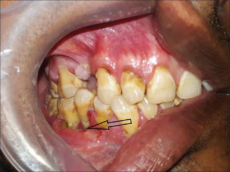
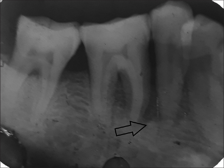
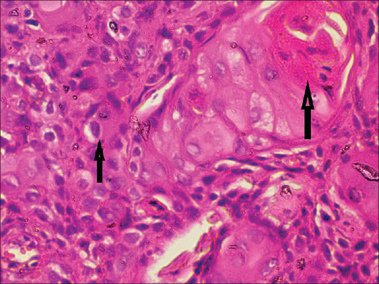
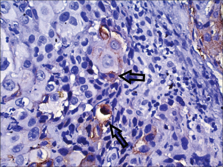
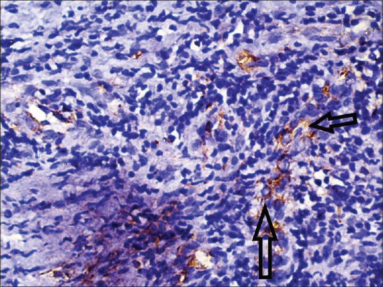
Necrotizing ulcerative periodontitis (NUP) is a painful and debilitating condition seen mostly in an immunocompromised state. Although squamous cell carcinoma (SCC) on gingiva is not uncommon, its presentation as a benign necrotizing lesion on gingiva is rare. Such presentations may lead to delayed diagnosis and poor prognosis. This report describes a case of a 34-year-old male presenting clinically with NUP around mandibular posterior teeth. Clinical features were misleading, but the histological findings established the diagnosis of well-differentiated SCC. Immunohistochemistry also showed features of epithelial-mesenchymal transition with decreased expression of E-cadherin and increased vimentin expression showing local invasion and metastasis. The patient was referred to the oncology department for evaluation of possible metastasis and further management of carcinoma.
Keywords: Gingiva; immunohistochemistry; periodontal disease; squamous cell carcinoma.
Copyright: © 2020 Journal of Indian Society of Periodontology.
Conflict of interest statement
There are no conflicts of interest.
Figures
Similar articles
-
Gingival squamous cell carcinoma mimicking as a desquamative lesion.J Indian Soc Periodontol. 2016 Jan-Feb;20(1):75-8. doi: 10.4103/0972-124X.164765. J Indian Soc Periodontol. 2016. PMID: 27041843 Free PMC article.
-
A case of squamous cell carcinoma presenting as localized severe periodontitis in the maxillary gingiva.J Periodontol. 2012 Jun;83(6):753-6. doi: 10.1902/jop.2011.110465. Epub 2011 Nov 3. J Periodontol. 2012. PMID: 22050549
-
Molecular changes in the gingival epithelium associated with necrotizing ulcerative periodontitis: a case report.Int J Periodontics Restorative Dent. 2006 Apr;26(2):191-6. Int J Periodontics Restorative Dent. 2006. PMID: 16642908
-
Two different protein expression profiles of oral squamous cell carcinoma analyzed by immunoprecipitation high-performance liquid chromatography.World J Surg Oncol. 2017 Aug 8;15(1):151. doi: 10.1186/s12957-017-1213-5. World J Surg Oncol. 2017. PMID: 28789700 Free PMC article. Review.
-
Necrotizing ulcerative periodontitis.Ann Periodontol. 1999 Dec;4(1):74-8. doi: 10.1902/annals.1999.4.1.74. Ann Periodontol. 1999. PMID: 10863377 Review.
References
-
- Soo KC, Spiro RH, King W, Harvey W, Strong EW. Squamous carcinoma of the gums. Am J Surg. 1988;156:281–5. - PubMed
-
- O'Sullivan B, Shah J. New TNM staging criteria for head and neck tumors. Semin Surg Oncol. 2003;21:30–42. - PubMed
-
- Glick M, Muzyka BC, Salkin LM, Lurie D. Necrotizing ulcerative periodontitis: A marker for immune deterioration and a predictor for the diagnosis of AIDS. J Periodontol. 1994;65:393–7. - PubMed
-
- Novak MJ. Necrotizing ulcerative periodontitis. Ann Periodontol. 1999;4:74–8. - PubMed
Publication types
LinkOut - more resources
Full Text Sources
Research Materials