

Dynamic Changes of Amplitude of Low-Frequency Fluctuations in Patients With Diabetic Retinopathy
- PMID: 33643197
- PMCID: PMC7905082
- DOI: 10.3389/fneur.2021.611702
Dynamic Changes of Amplitude of Low-Frequency Fluctuations in Patients With Diabetic Retinopathy
Abstract
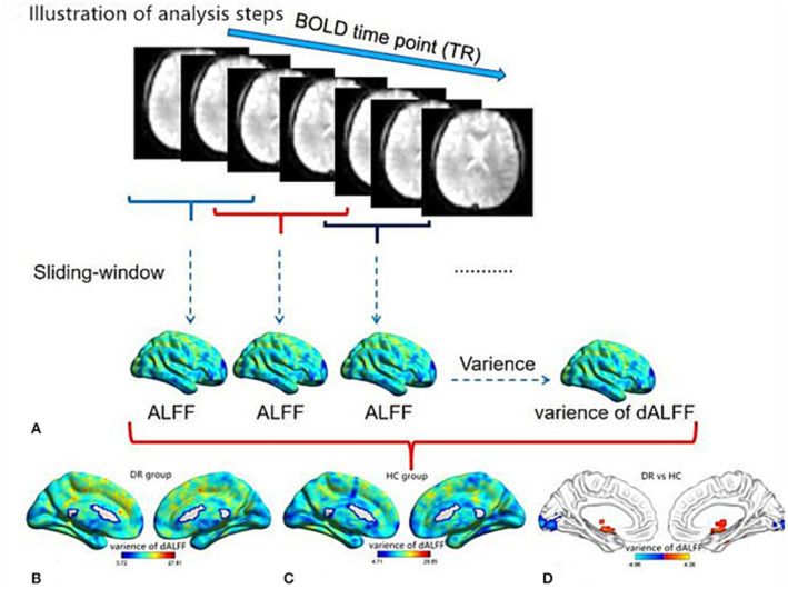
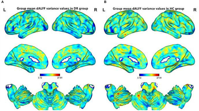
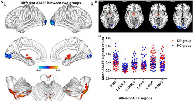
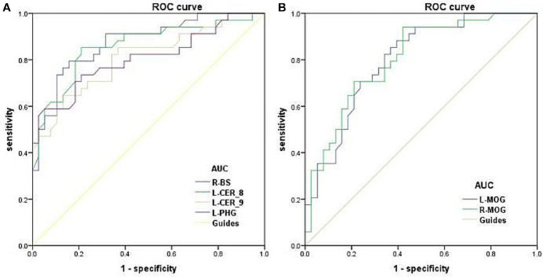
Background: Growing evidence demonstrate that diabetic retinopathy (DR) patients have a high risk of cognitive decline and exhibit abnormal brain activity. However, neuroimaging studies thus far have focused on static cerebral activity changes in DR patients. The characteristics of dynamic cerebral activity in patients with DR are poorly understood. Purpose: The purpose of the study was to investigate the dynamic cerebral activity changes in patients with DR using the dynamic amplitude of low-frequency fluctuation (dALFF) method. Materials and methods: Thirty-four DR patients (18 men and 16 women) and 38 healthy controls (HCs) (18 males and 20 females) closely matched in age, sex, and education were enrolled in this study. The dALFF method was used to investigate dynamic intrinsic brain activity differences between the DR and HC groups. Results: Compared with HCs, DR patients exhibited increased dALFF variability in the right brainstem, left cerebellum_8, left cerebellum_9, and left parahippocampal gyrus. In contrast, DR patients exhibited decreased dALFF variability in the left middle occipital gyrus and right middle occipital gyrus. Conclusion: Our study highlighted that DR patients showed abnormal variability of dALFF in the visual cortices, cerebellum, and parahippocampal gyrus. These findings suggest impaired visual and motor and memory function in DR individuals. Thus, abnormal dynamic spontaneous brain activity might be involved in the pathophysiology of DR.
Keywords: diabetic retinopathy; dynamic amplitude of low-frequency fluctuation; functional magnetic resonance imaging; functional network; network centrality.
Copyright © 2021 Huang, Wen, Qi, Tong and Shen.
Conflict of interest statement
The authors declare that the research was conducted in the absence of any commercial or financial relationships that could be construed as a potential conflict of interest.
Figures
Similar articles
-
Machine learning analysis reveals aberrant dynamic changes in amplitude of low-frequency fluctuations among patients with retinal detachment.Front Neurosci. 2023 Jul 20;17:1227081. doi: 10.3389/fnins.2023.1227081. eCollection 2023. Front Neurosci. 2023. PMID: 37547140 Free PMC article.
-
Changes in dynamic and static brain fluctuation distinguish minimal hepatic encephalopathy and cirrhosis patients and predict the severity of liver damage.Front Neurosci. 2023 Mar 28;17:1077808. doi: 10.3389/fnins.2023.1077808. eCollection 2023. Front Neurosci. 2023. PMID: 37056312 Free PMC article.
-
Dynamic changes of amplitude of low-frequency in systemic lupus erythematosus patients with cognitive impairment.Front Neurosci. 2022 Aug 23;16:929383. doi: 10.3389/fnins.2022.929383. eCollection 2022. Front Neurosci. 2022. PMID: 36081656 Free PMC article.
-
Aberrant spontaneous static and dynamic amplitude of low-frequency fluctuations in cerebral small vessel disease with or without mild cognitive impairment.Brain Behav. 2023 Dec;13(12):e3279. doi: 10.1002/brb3.3279. Epub 2023 Oct 10. Brain Behav. 2023. PMID: 37815202 Free PMC article.
-
Dynamic alterations of functional connectivity and amplitude of low-frequency fluctuations in patients with unilateral sudden sensorineural hearing loss.Neurosci Lett. 2022 Feb 16;772:136470. doi: 10.1016/j.neulet.2022.136470. Epub 2022 Jan 20. Neurosci Lett. 2022. PMID: 35066092
Cited by
-
Altered static and dynamic spontaneous brain activity in patients with dysthyroid optic neuropathy: a resting-state fMRI study.Front Neurosci. 2025 Jan 10;18:1530967. doi: 10.3389/fnins.2024.1530967. eCollection 2024. Front Neurosci. 2025. PMID: 39867455 Free PMC article.
-
Machine learning analysis reveals aberrant dynamic changes in amplitude of low-frequency fluctuations among patients with retinal detachment.Front Neurosci. 2023 Jul 20;17:1227081. doi: 10.3389/fnins.2023.1227081. eCollection 2023. Front Neurosci. 2023. PMID: 37547140 Free PMC article.
-
Genetic mechanisms of dynamic functional connectivity density in diabetic retinopathy brains: a combined transcriptomic and resting-state functional magnetic resonance imaging study.Front Cell Neurosci. 2025 Apr 10;19:1476038. doi: 10.3389/fncel.2025.1476038. eCollection 2025. Front Cell Neurosci. 2025. PMID: 40276708 Free PMC article.
-
Brain white matter microstructural alterations in patients with diabetic retinopathy: an automated fiber-tract quantification study.Quant Imaging Med Surg. 2025 May 1;15(5):3982-3992. doi: 10.21037/qims-24-1440. Epub 2025 Apr 8. Quant Imaging Med Surg. 2025. PMID: 40384690 Free PMC article.
-
Dynamic alterations in the amplitude of low-frequency fluctuation in patients with cerebral small vessel disease.Front Mol Neurosci. 2023 Sep 22;16:1200756. doi: 10.3389/fnmol.2023.1200756. eCollection 2023. Front Mol Neurosci. 2023. PMID: 37808469 Free PMC article.
References
-
- Liu Y, Yang J, Tao L, Lv H, Jiang X, Zhang M, et al. . Risk factors of diabetic retinopathy and sight-threatening diabetic retinopathy: a cross-sectional study of 13 473 patients with type 2 diabetes mellitus in mainland China. BMJ Open. (2017) 7:e016280. 10.1136/bmjopen-2017-016280 - DOI - PMC - PubMed
LinkOut - more resources
Full Text Sources
Other Literature Sources