

Physiological Ripples Associated With Sleep Spindles Can Be Identified in Patients With Refractory Epilepsy Beyond Mesio-Temporal Structures
- PMID: 33643198
- PMCID: PMC7902925
- DOI: 10.3389/fneur.2021.612293
Physiological Ripples Associated With Sleep Spindles Can Be Identified in Patients With Refractory Epilepsy Beyond Mesio-Temporal Structures
Abstract
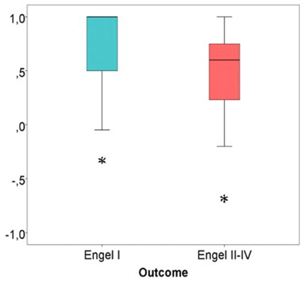
Introduction: High frequency oscillations (HFO) are promising biomarkers of epileptic tissue. While group analysis suggested a correlation between surgical removal of HFO generating tissue and seizure free outcome, HFO could not predict seizure outcome on an individual patient level. One possible explanation is the lack of differentiation between physiological and epileptic HFO. In the mesio-temporal lobe, a proportion of physiological ripples can be identified by their association with scalp sleep spindles. Spike associated ripples in contrast can be considered epileptic. This study investigated whether categorizing ripples by the co-occurrence with sleep spindles or spikes improves outcome prediction after surgery. Additionally, it aimed to investigate whether spindle-ripple association is limited to the mesio-temporal lobe structures or visible across the whole brain. Methods: We retrospectively analyzed EEG of 31 patients with chronic intracranial EEG. Sleep spindles in scalp EEG and ripples and epileptic spikes in iEEG were automatically detected. Three ripple subtypes were obtained: SpindleR, Non-SpindleR, and SpikeR. Rate ratios between removed and non-removed brain areas were calculated. We compared the distinct ripple subtypes and their rates in different brain regions, inside and outside seizure onset areas and between patients with good and poor seizure outcome. Results: SpindleR were found across all brain regions. SpikeR had significantly higher rates in the SOZ than in Non-SOZ channels. A significant positive correlation between removal of ripple-events and good outcome was found for the mixed ripple group (rs = 0.43, p = 0.017) and for ripples not associated with spindles (rs=0.40, p = 0.044). Also, a significantly high proportion of spikes associated with ripples were removed in seizure free patients (p = 0.036). Discussion: SpindleR are found in mesio-temporal and neocortical structures, indicating that ripple-spindle-coupling might have functional importance beyond mesio-temporal structures. Overall, the proportion of SpindleR was low and separating spindle and spike associated ripples did not improve outcome prediction in our patient group. SpindleR analysis therefore can be a tool to identify physiological events but needs to be used in combination with other methods to have clinical relevance.
Keywords: epileptic spikes; high frequency oscillations; post-surgical outcome; refractory epilepsy; ripples; sleep spindles.
Copyright © 2021 Bruder, Schmelzeisen, Lachner-Piza, Reinacher, Schulze-Bonhage and Jacobs.
Conflict of interest statement
The authors declare that the research was conducted in the absence of any commercial or financial relationships that could be construed as a potential conflict of interest.
Figures


Similar articles
-
Mesial-Temporal Epileptic Ripples Correlate With Verbal Memory Impairment.Front Neurol. 2022 Jun 3;13:876024. doi: 10.3389/fneur.2022.876024. eCollection 2022. Front Neurol. 2022. PMID: 35720106 Free PMC article.
-
Physiological Ripples Associated with Sleep Spindles Differ in Waveform Morphology from Epileptic Ripples.Int J Neural Syst. 2017 Nov;27(7):1750011. doi: 10.1142/S0129065717500113. Epub 2016 Nov 2. Int J Neural Syst. 2017. PMID: 28043201
-
High Frequency Oscillations in the Ripple Band (80-250 Hz) in Scalp EEG: Higher Density of Electrodes Allows for Better Localization of the Seizure Onset Zone.Brain Topogr. 2018 Nov;31(6):1059-1072. doi: 10.1007/s10548-018-0658-3. Epub 2018 Jul 6. Brain Topogr. 2018. PMID: 29980967
-
Strong relationship between NREM sleep, epilepsy and plastic functions - A conceptual review on the neurophysiology background.Epilepsy Res. 2019 Feb;150:95-105. doi: 10.1016/j.eplepsyres.2018.11.008. Epub 2019 Jan 31. Epilepsy Res. 2019. PMID: 30712997 Review.
-
Seizures and Sleep.In: Noebels JL, Avoli M, Rogawski MA, Vezzani A, Delgado-Escueta AV, editors. Jasper's Basic Mechanisms of the Epilepsies. 5th edition. New York: Oxford University Press; 2024. Chapter 14. In: Noebels JL, Avoli M, Rogawski MA, Vezzani A, Delgado-Escueta AV, editors. Jasper's Basic Mechanisms of the Epilepsies. 5th edition. New York: Oxford University Press; 2024. Chapter 14. PMID: 39637155 Free Books & Documents. Review.
Cited by
-
Pathological and Physiological High-frequency Oscillations on Electroencephalography in Patients with Epilepsy.Neurosci Bull. 2024 May;40(5):609-620. doi: 10.1007/s12264-023-01150-6. Epub 2023 Nov 24. Neurosci Bull. 2024. PMID: 37999861 Free PMC article. Review.
-
Association between Removal of High-Frequency Oscillations and the Effect of Epilepsy Surgery: A Meta-Analysis.J Neurol Surg A Cent Eur Neurosurg. 2024 May;85(3):294-301. doi: 10.1055/a-2202-9344. Epub 2023 Nov 2. J Neurol Surg A Cent Eur Neurosurg. 2024. PMID: 37918885 Free PMC article.
-
Fast activity chirp patterns in focal seizures from patients and animal models.Epilepsia. 2025 Mar;66(3):621-631. doi: 10.1111/epi.18245. Epub 2024 Dec 26. Epilepsia. 2025. PMID: 39723840 Free PMC article. Review.
-
Mesial-Temporal Epileptic Ripples Correlate With Verbal Memory Impairment.Front Neurol. 2022 Jun 3;13:876024. doi: 10.3389/fneur.2022.876024. eCollection 2022. Front Neurol. 2022. PMID: 35720106 Free PMC article.
References
LinkOut - more resources
Full Text Sources
Other Literature Sources