. 2021 Feb 12:11:626431.
doi: 10.3389/fimmu.2020.626431. eCollection 2020.

A Matter of Life or Death: Productively Infected and Bystander CD4 T Cells in Early HIV Infection

Affiliations

A Matter of Life or Death: Productively Infected and Bystander CD4 T Cells in Early HIV Infection

Dechao Cao et al. Front Immunol. .

Erratum in

Abstract

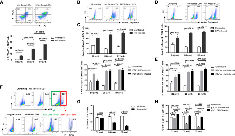
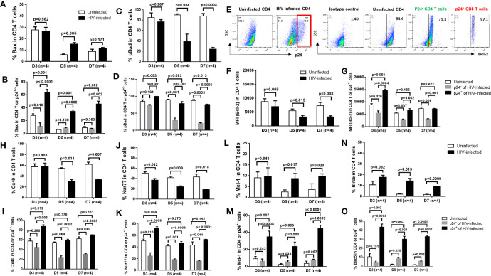
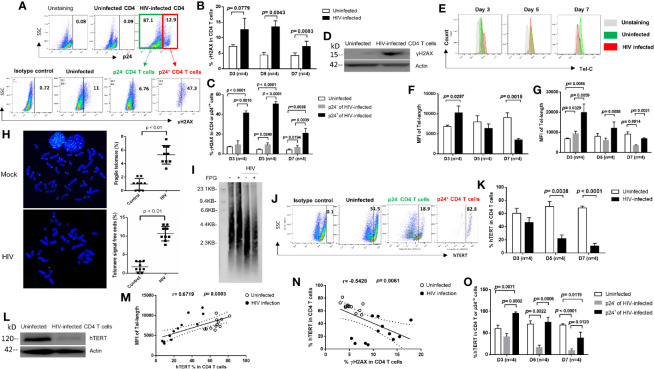
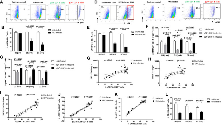
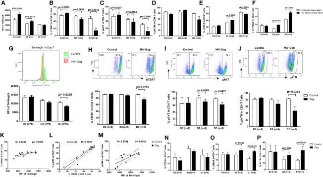
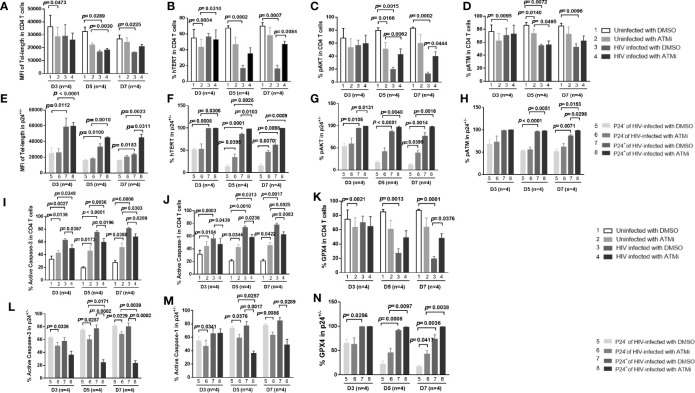
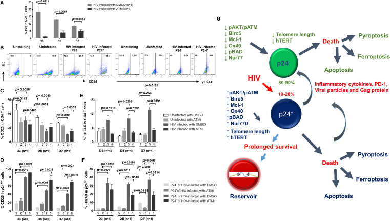
CD4 T cell death or survival following initial HIV infection is crucial for the development of viral reservoirs and latent infection, making its evaluation critical in devising strategies for HIV cure. Here we infected primary CD4 T cells with a wild-type HIV-1 and investigated the death and survival mechanisms in productively infected and bystander cells during early HIV infection. We found that HIV-infected cells exhibited increased programmed cell death, such as apoptosis, pyroptosis, and ferroptosis, than uninfected cells. However, productively infected (p24+) cells and bystander (p24-) cells displayed different patterns of cell death due to differential expression of pro-/anti-apoptotic proteins and signaling molecules. Cell death was triggered by an aberrant DNA damage response (DDR), as evidenced by increases in γH2AX levels, which inversely correlated with telomere length and telomerase levels during HIV infection. Mechanistically, HIV-infected cells exhibited a gradual shortening of telomeres following infection. Notably, p24+ cells had longer telomeres compared to p24- cells, and telomere length positively correlated with the telomerase, pAKT, and pATM expressions in HIV-infected CD4 T cells. Importantly, blockade of viral entry attenuated the HIV-induced inhibition of telomerase, pAKT, and pATM as well as the associated telomere erosion and cell death. Moreover, ATM inhibition promoted survival of HIV-infected CD4 T cells, especially p24+ cells, and rescued telomerase and AKT activities by inhibiting cell activation, HIV infection, and DDR. These results indicate that productively infected and bystander CD4 T cells employ different mechanisms for their survival and death, suggesting a possible pro-survival, pro-reservoir mechanism during early HIV infection.

Keywords: AKT; ATM; HIV; T cell death; survival; telomerase; telomere.


Conflict of interest statement

The authors declare that the research was conducted in the absence of any commercial or financial relationships that could be construed as a potential conflict of interest.

Figures

Figure 1
Figure 1
HIV induces CD4 T cell death in productively infected and bystander cells during early infection via different mechanisms. (A) Representative dot plots (day 7) and summary of the percentage (%) of Av and 7AAD levels in HIV-infected and uninfected CD4 T cells, as determined by flow cytometry. (B, C) Representative dot plots and summary data showing active caspase-3% in productively HIV-infected (p24+) and bystander (p24-) or uninfected cells at days 3, 5, and 7 in cultures. (D, E) Representative dot plots and summary data of active caspase-1% in HIV-infected (p24+) and bystander (p24-) or uninfected cells at day 3, day 5, and day 7. (F–H) Representative dot plots and summary data of GPX4% in HIV-infected (p24+) and bystander (p24-) or uninfected cells at days 3, 5, and 7.
Figure 2
Figure 2
Expression of pro- and anti-apoptotic proteins in productively infected cells and bystander cells during HIV infection. (A, B) Summary data of Bax % in HIV-infected (p24+ -) and bystander (p24-) or uninfected cells at days 3, 5, and 7, as determined by flow cytometry. (C, D) Summary data of pBad % in HIV-infected (p24+) and bystander (p24-) or uninfected cells at days 3, 5, and 7. (E–O) Representative dot plots and summary data of Bcl-2%, Ox40% (H, I), Nur77% (J, K), Mcl-1 (L, M), and Birc5% (N, O) in HIV-infected (p24+ vs. p24-) and uninfected cells at days 3, 5, and 7.
Figure 3
Figure 3
HIV causes CD4 T cell death by inducing DNA damage and disrupting telomeres via inhibition of telomerase during early viral infection. (A) Representative dot plots and gating strategy of γH2AX levels in p24+ and p24- CD4 T cells with HIV infection at day 7. Isotype staining and uninfected CD4 T cells serve as negative controls for flow cytometry. (B, C) Summary data of γH2AX % in HIV-infected (p24+ vs. p24-) and uninfected cells at days 3, 5, and 7. (D) Western blot analysis of γH2AX levels in HIV-infected versus uninfected CD4 T cells at day 5. (E–G) Representative overlaid histograms and summary data of the mean fluorescence intensity (MFI) of telomere length in HIV-infected (p24+ or p24-) and uninfected CD4 T cells at days 3, 5, and 7, as determined by Flow-FISH. (H) Representative imaging and summary data of meta-FISH analysis of the frequencies (%) of chromatin with fragile telomeres and telomere-free ends in CD4 T cells with or without HIV infection at day 5, measured by confocal microscopy. (I) Telomere length in CD4 T cells with or without HIV infection at day 5. Telomeric DNA was treated with or without FPG and analyzed by Southern blot. (J, K, O) Representative dot plots and summary data of the human telomerase (hTERT) % in HIV-infected (p24+ or p24-) and uninfected CD4 T cells at days 3, 5, and 7. (L) Western blot analysis of hTERT levels in HIV-infected versus uninfected CD4 T cells at day 5. (M) Correlation analysis between hTERT expression and telomere length. (N) Correlation analysis between hTERT expression and γH2AX level in HIV-infected and uninfected cells.
Figure 4
Figure 4
T cell receptor (TCR) signaling pathways are differentially regulated in productively HIV-infected and bystander cells. (A–C) Representative dot plots and summary data of the pAKT % in HIV-infected (p24+) and bystander (p24-) or uninfected D4 T cells at days 3, 5, and 7. (D–F) Representative dot blots and summary data of the pATM % in HIV-infected (p24+ and bystander (p24-) or uninfected CD4 T cells at days 3, 5, and 7, by flow cytometry. (G, H) Positive correlation between the MFI of telomere length and the % of pAKT or pATM expressions. (I–K) Positive correlation between the % of hTERT, pAKT, and pATM expressions in CD4 T cells with or without HIV infection. (L) Percentage of CD25 expression in CD4 T cells with or without HIV infection.
Figure 5
Figure 5
HIV Gag protein shortens telomere length by diminishing telomerase, AKT, and ATM activities in primary CD4 T cells. (A–F) Primary CD4 T cells were incubated with supernatants derived from 7-day culture of HIV-infected or uninfected cells in the presence of HIV-entry blockers (Enfuvirtide and Maraviroc) for 3, 5, and 7 days, followed by measuring telomere length, telomerase, pAKT, pATM, γH2AX, and Av/7AAD by flow cytometry. (G) Representative overlaid histograms and summary data of the median fluorescence intensity (MFI) of telomere length in Gag-treated and untreated CD4 T cells at days 3, 5, and 7, measured by Flow-FISH. (H–J) Representative dot plots and summary % of the hTERT, pAKT, and pATM expressions in Gag-treated and untreated CD4 T cells at days 3, 5, and 7. (K–M) Positive correlation between the MFI of telomere length and the % of hTERT, pATM, and hTERT, as well as pATM expression and telomere length in CD4 T cells with or without Gag treatment. (N–P) Summary data of the % of CD25+, γH2AX+, and Av+ cells in CD4 T cells treated with or without HIV Gag.
Figure 6
Figure 6
ATM blockade promotes HIV-infected cell survival via rescuing AKT and telomerase activities by inhibiting HIV infection and DNA damage response (DDR). (A–D) HIV-infected and uninfected CD4 T cells were treated with DMSO control or ATM inhibitor for 3, 5, and 7 days, followed by measuring telomere length, hTERT, pAKT, and pATM by flow cytometry. (E–H) Summary data of the MFI of telomere length, the % of hTERT, pAKT, and pATM expressions in p24+ and p24- cells following HIV infection with ATMi or DMSO treatment for 3, 5, and 7 days. (I–K) Summary data of the % of active caspase-3, active caspase-1, and GPX4 expressions in HIV-infected and uninfected CD4 T cells treated with DMSO or ATMi for 3, 5, and 7 days. (L–N) Summary data of the % of active caspase-3, active caspase-1, and GPX4 expressions in p24+ and p24- cells following HIV infection with ATMi or DMSO treatment for 3, 5, and 7 days.
Figure 7
Figure 7
Blockade of ATM promotes HIV-infected cell survival via inhibiting T cell activation, HIV infection, and DNA damage. (A) p24 expression in HIV-infected CD4 T cells treated with DMSO or ATM inhibitor (ATMi). (B–F) Summary data of the % of CD25 and γH2AX expressions in uninfected and HIV-infected (p24+ and p24-) CD4 T cells treated with DMSO or ATMi for 3, 5, and 7 days. (G) A schematic model of the fate of productively infected CD4 T cells and bystander cells during early HIV infection. HIV promotes CD4 T cell death through enhancing various programmed cell death pathways, including apoptosis and pyroptosis (especially in p24+ cells) or ferroptosis (especially in p24- cells) during early viral infection. Mechanistically, HIV infection promotes secretion of inflammatory cytokines as well as viral particles and proteins (such as Gag) that can differentially regulate the pro- and anti-apoptotic proteins, such as Bad, pBad, Bcl-2, OX40, Nur77, Mcl-1, and Birc5, in p24+ and p24- cells. HIV infection also dysregulates T cell receptor (TCR) signaling pathways, in particular PI3K (such as AKT/ATM) and telomerase hTERT activities, and thus affects telomere length and cell survival or death machineries in p24+ and p24- cells. While productively infected (p24+) cells experience more death than p24- bystander cells, p24+ cells appear to exhibit survival activity, thus favoring HIV reservoir formation and latency establishment. Specifically, productively infected CD4 T cells show prolonged telomeres, increased levels of telomerase, and more activated TCR signaling pathways compared to bystander CD4 T cells, indicating the involvement of cell survival and pro-reservoir machineries during HIV infection. Importantly, blocking the ATM pathway promotes uninfected cell death but enhances virus-infected cell survival, and blocking HIV infection enhances overall cell survival, both through regulating the signaling pathways involved in the maintenance of genomic telomere integrity. These results indicate that HIV-infected and uninfected cells employ different mechanisms for survival and death during HIV infection.

Similar articles

Cited by

References

    1. Okoye AA, Picker LJ. CD4(+) T-cell depletion in HIV infection: mechanisms of immunological failure. Immunol Rev (2013) 254:54–64. doi: 10.1111/imr.12066 - DOI - PMC - PubMed
    1. Vidya Vijayan KK, Karthigeyan KP, Tripathi SP, Hanna LE. Pathophysiology of CD4+ T-Cell Depletion in HIV-1 and HIV-2 Infections. Front Immunol (2017) 8:580. doi: 10.3389/fimmu.2017.00580 - DOI - PMC - PubMed
    1. Piconi S, Trabattoni D, Gori A, Parisotto S, Magni C, Meraviglia P, et al. . Immune activation, apoptosis, and Treg activity are associated with persistently reduced CD4+ T-cell counts during antiretroviral therapy. AIDS (2010) 24:1991–2000. doi: 10.1097/QAD.0b013e32833c93ce - DOI - PubMed
    1. Lederman MM, Calabrese L, Funderburg NT, Clagett B, Medvik K, Bonilla H, et al. . Immunologic failure despite suppressive antiretroviral therapy is related to activation and turnover of memory CD4 cells. J Infect Dis (2011) 204:1217–26. doi: 10.1093/infdis/jir507 - DOI - PMC - PubMed
    1. Tyagi M, Bukrinsky M. Human immunodeficiency virus (HIV) latency: the major hurdle in HIV eradication. Mol Med (2012) 18:1096–108. doi: 10.2119/molmed.2012.00194 - DOI - PMC - PubMed

Publication types