

Hip arthroscopy following slipped capital femoral epiphysis fixation: chondral damage and labral tears findings
- PMID: 33643455
- PMCID: PMC7907763
- DOI: 10.1302/1863-2548.15.200178
Hip arthroscopy following slipped capital femoral epiphysis fixation: chondral damage and labral tears findings
Abstract
Purpose: This study investigated the association between chondrolabral damage and time to arthroscopic surgery for slipped capital femoral epiphysis (SCFE).
Methods: This was a descriptive retrospective study that enrolled patients with SCFE who underwent hip arthroscopy for femoral osteochondroplasty after SCFE fixation. SCFE type, time from SCFE symptom onset or slip fixation surgery to hip arthroscopy and intraarticular arthroscopic findings were recorded. Acetabular chondrolabral damage was evaluated according to the Konan and Outerbridge classification systems. Nested analysis of variance and the chi-squared test were used for statistical analyses.
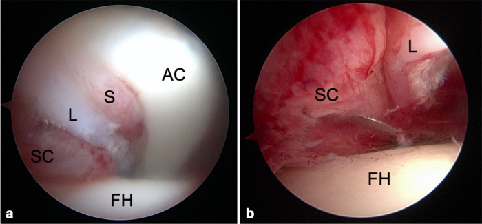
Results: We analyzed 22 cases of SCFE in 17 patients (five bilateral). The mean age at the time of hip arthroscopy was 13.6 years-old (8-20), and mean time from SCFE fixation to arthroscopy was 25.1 months (3 weeks to 8 years). Labral frying was present in 20 cases, labral tears in 16 and acetabular chondral damage in 17. The most frequent lesion was type 3 (41%) (Konan classification). Two cases had a grade III and 1 had a grade II acetabular chondral lesion (Outerbridge classification). Positive associations were observed between time from SCFE to hip arthroscopy and hip intraarticular lesions evaluated using Konan (p = 0.004) and Outerbridge (p = 0.000) classification systems. There was no association between SCFE severity (chi-squared = 0.315), stability (chi-squared = 0.558) or temporality (chi-squared = 0.145) type and hip intraarticular lesions.
Conclusion: A longer time from SCFE symptom onset and fixation to hip arthroscopy is associated with greater acetabular chondrolabral damage.
Level of evidence: IV.
Keywords: epiphysiolysis; femoroacetabular impingement; hip arthroscopy; hip preservation surgery; slipped capital femoral epiphysis; slipped upper femoral epiphysis.
Copyright © 2021, The author(s).
Figures


References
-
- Aronsson DD, Loder RT, Breur GJ, Weinstein SL. Slipped capital femoral epiphysis: current concepts. J Am Acad Orthop Surg 2006;14:666-679. - PubMed
-
- Purcell D, Varthi A, Lee MC. Slipped capital femoral epiphisis: current concepts review. Curr Orthop Pract 2011;22:81-88.
-
- Lehmann CL, Arons RR, Loder RT, Vitale MG. The epidemiology of slipped capital femoral epiphysis: an update. J Pediatr Orthop 2006;26:286-290. - PubMed
-
- Loder RT. The demographics of slipped capital femoral epiphysis. An international multicenter study. Clin Orthop Relat Res 1996;322:8-27. - PubMed
-
- Drehmann F. Drehmann’s sign. A clinical examination method in epiphysiolysis (slipping of the upper femoral epiphysis). Description of signs, aetiopathogenetic considerations, clinical experience (author’s transl). Z Orthop Ihre Grenzgeb 1979;117:333-344. - PubMed
LinkOut - more resources
Full Text Sources
Other Literature Sources
Research Materials
