

Paediatric hip ultrasound: uncertainties in examination and choice of treatment
- PMID: 33643457
- PMCID: PMC7907771
- DOI: 10.1302/1863-2548.15.200084
Paediatric hip ultrasound: uncertainties in examination and choice of treatment
Abstract
Purpose: In Germany, neonates undergo hip sonography examination using the Graf method during the routine U3 screening examination, performed by consultant physicians four to five weeks after birth, and are referred to specialized orthopaedic departments if there are any uncertainties. This study evaluated the quality of sonographic screening in the outpatient sector and the treatment requirements of referred children.
Methods: We performed a retrospective analysis of the patient data of 384 neonates collected in consultations performed between April 2016 and April 2019.
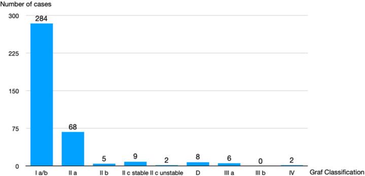
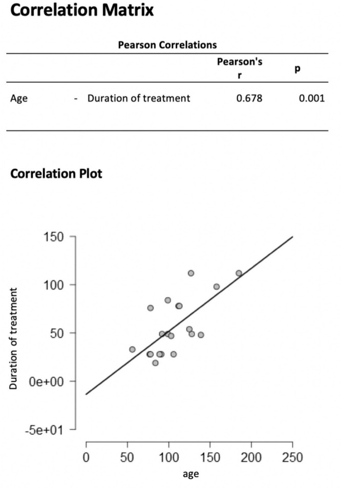
Results: In total, 74% (n = 284) of neonates presented a hip type Ia/b. Treatment (abduction brace or Fettweis cast) was required in 32% (n = 122) of cases. The treatment duration was significantly correlated with age at first presentation (Pearson's r = 0.678; p = 0.001). The treatment duration for patients aged > 200 days old at first presentation was twice as long as those aged 100 days at first presentation. Patients with public health insurance require referral by a consultant. Developmental dysplasia of the hip as referral diagnosis could not be confirmed in control examination in 64% (n = 132) of cases. Of the public health insured children, 97% (n = 200) were referred through a consultant paediatrician.
Conclusion: We identified deficits in performing and interpreting the Graf method of ultrasound examination. A total of 64% of referred pathological hips turned out to be physiological configurations in our control examination. The future goal should be to increase anatomical knowledge of the newborn hip and ensure the correct use of Graf ultrasound method. Advanced training courses are recommended and necessary.
Level of evidence: IV.
Keywords: developmental dysplasia of the hip; neonates; quality control; screening; ultrasound.
Copyright © 2021, The author(s).
Figures
References
-
- Rosendahl K, Markestad T, Lie RT. Developmental dysplasia of the hip: prevalence based on ultrasound diagnosis. Pediatr Radiol 1996;26:635-639. - PubMed
-
- Ihme N, Altenhofen L, von Kries R, et al. Hip ultrasound screening in Germany. Results and comparison with other screening procedures. Orthopade 2008;37:541-549. - PubMed
-
- Schmitz MR, Blumberg TJ, Nelson SE, Sees JP, Sankar WN. What’s new in pediatric hip? J Pediatr Orthop 2018;38:e300-e304. - PubMed
-
- Swarup I, Penny CL, Dodwell ER. Developmental dysplasia of the hip: an update on diagnosis and management from birth to 6 months. Curr Opin Pediatr 2018;30:84-92. - PubMed
LinkOut - more resources
Full Text Sources
Other Literature Sources
Research Materials
Miscellaneous
