

Imaging Characteristics of Brain SPECT, PET, and MRI in Neurosyphilis
- PMID: 33643490
- PMCID: PMC7881059
- DOI: 10.1007/s13139-021-00684-9
Imaging Characteristics of Brain SPECT, PET, and MRI in Neurosyphilis
Abstract
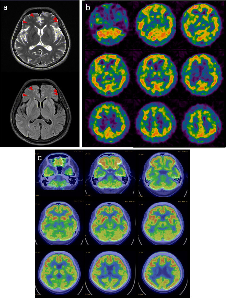
The incidence of neurosyphilis has declined since effective penicillin therapy against Treponema pallidum was introduced. However, the diagnosis of neurosyphilis early in the disease course is very important in order to select appropriate antibiotic therapy. We report brain MRI, SPECT with Tc-99m ECD, and PET with F-18 FDG findings before antibiotic therapy in a neurosyphilis patient with neurological symptoms. The cerebral cortices showed hypoperfusion with a patchy distribution on SPECT and foci with high signal intensity on MRI, suggesting ischemia. Brain PET showed areas with hypometabolism in the temporoparietal lobes bilaterally.
Keywords: Magnetic resonance imaging; Neurosyphilis; Positron-emission tomography; Tomography, emission-computed, single-photon.
© Korean Society of Nuclear Medicine 2021.
Conflict of interest statement
Conflict of InterestEun Kyoung Choi, Young Do Kim, Hyeonseok Jeong, Yong-An Chung, Jin Kyoung Oh, and In-Uk Song declare that they have no conflict of interest.
Figures

References
-
- Kitabayashi Y, Ueda H, Narumoto J, Nakamura K, Kita H, Tsuchida H, et al. Cerebral blood flow changes in general paresis following penicillin treatment: a longitudinal single photon emission computed tomography study. Psychiatry Clin Neurosci. 2002;56:65–70. doi: 10.1046/j.1440-1819.2002.00930.x. - DOI - PubMed
Publication types
LinkOut - more resources
Full Text Sources
Other Literature Sources