Review
. 2021 Feb 16:12:2041731421990536.
doi: 10.1177/2041731421990536. eCollection 2021 Jan-Dec.

Biomaterials for corneal endothelial cell culture and tissue engineering

Affiliations
Review

Biomaterials for corneal endothelial cell culture and tissue engineering

Mohit Parekh et al. J Tissue Eng. .

Abstract

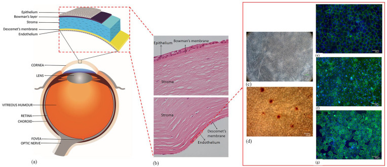
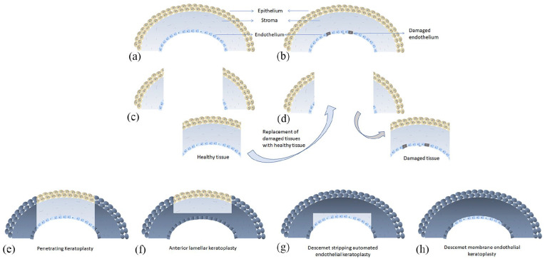
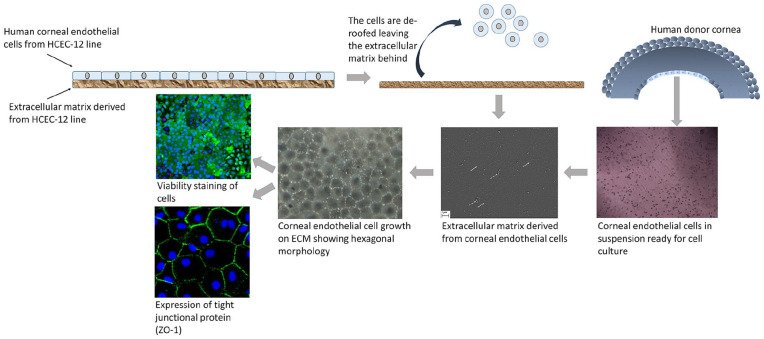
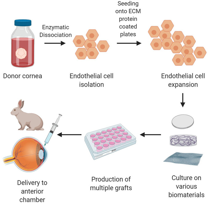
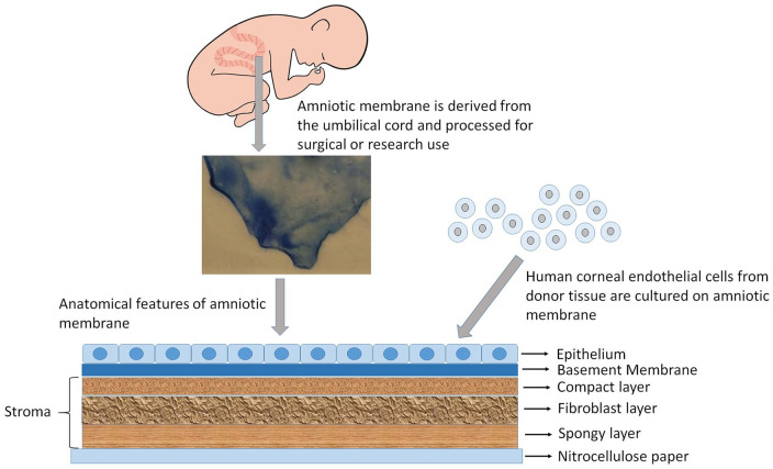
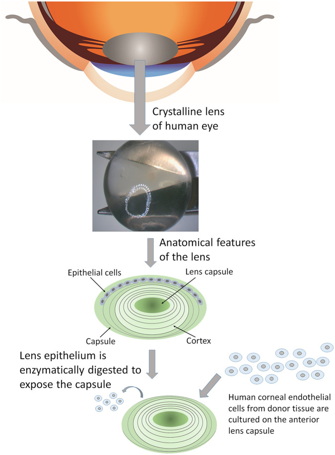
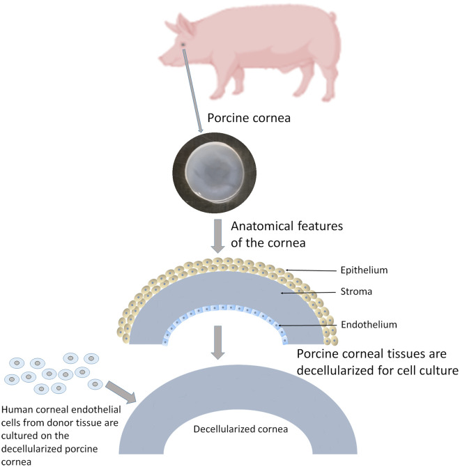
The corneal endothelium is the posterior monolayer of cells that are responsible for maintaining overall transparency of the avascular corneal tissue via pump function. These cells are non-regenerative in vivo and therefore, approximately 40% of corneal transplants undertaken worldwide are a result of damage or dysfunction of endothelial cells. The number of available corneal donor tissues is limited worldwide, hence, cultivation of human corneal endothelial cells (hCECs) in vitro has been attempted in order to produce tissue engineered corneal endothelial grafts. Researchers have attempted to recreate the current gold standard treatment of replacing the endothelial layer with accompanying Descemet's membrane or a small portion of stroma as support with tissue engineering strategies using various substrates of both biologically derived and synthetic origin. Here we review the potential biomaterials that are currently in development to support the transplantation of a cultured monolayer of hCECs.

Keywords: Cornea; biomaterials; cell culture; endothelial cells; tissue engineering.


Conflict of interest statement

Declaration of conflicting interests: The author(s) declared no potential conflicts of interest with respect to the research, authorship, and/or publication of this article.

Figures

Figure 1.
Figure 1.
Anatomy of the human eye and cornea: (a) The figure illustrates different parts of the eye and layers of the corneal tissue, (b) histological section of human cornea showing different layers of the tissue, (c) corneal endothelial cell viability using trypan blue staining observed under light microscope and (d) alizarin red staining showing the hexagonality of endothelial cells. Biomarkers such as (e) ZO-1, (f) Tag-1A3, and (g) Tag-2A12 to determine the presence of specific proteins like tight-junction and surface epitopes. These biomarkers are used on cultured corneal endothelial cells to characterize the cell type and for functional analysis.
Figure 2.
Figure 2.
Illustration of conventional corneal transplantation technique: (a) normal cornea, (b) cornea with diseased or dysfunctional endothelium, (c) part of a healthy donor tissue is replaced with (d) part of damaged recipient tissue. Representation of conventional full thickness (e) penetrating keratoplasty and selective modern transplantation procedures such as (f) anterior lamellar keratoplasty, (g) Descemet’s stripping automated endothelial keratoplasty, and (h) Descemet’s membrane endothelial keratoplasty.
Figure 3.
Figure 3.
Human corneal endothelial cell culture on HCEC-12 derived extracellular matrix (ECM). HCEC-12 cells are cultured on a culture plate. ECM is laid by the HCEC-12 cells naturally. Upon confluence, the cells are detached leaving behind the ECM. Corneal endothelial cells from human donor corneas are isolated and cultured on the ECM. White arrows show fiber-like collagen structures and dotted white arrow show cell debris. Cell morphology, viability and expression of tight-junction protein are checked to confirm the health and for end-stage characterization.
Figure 4.
Figure 4.
Tissue engineering of the cornea. Cadaveric donor corneas suitable for research are acquired. The hCECs are isolated as per optimized protocols and allowed to expand in number in culture. Once there are sufficient number of cells, they are transferred onto a scaffold which can be biologically derived, synthetic or semi-synthetic. Multiple grafts can then be produced as a confluent monolayer of cells on the biomaterials. These grafts can then be introduced into the anterior chamber of an animal for research purposes with the eventual goal of transplanting into humans. Figure created with BioRender.com.
Figure 5.
Figure 5.
Biologically derived material. Amniotic membrane is excised from the umbilical cord, processed and preserved on nitrocellulose paper. Amniotic membrane is used for culturing corneal endothelial cells.
Figure 6.
Figure 6.
Human crystalline lens is obtained from cadaveric eye donors. The lens epithelium is enzymatically isolated and the remaining capsule is used for culturing corneal endothelial cells in vitro.
Figure 7.
Figure 7.
Porcine cornea is excised from the eye globe. The tissue is decellularized to preserve the stromal layer. This layer is used for culturing corneal endothelial cells in vitro.
Figure 8.
Figure 8.
Preparation of Real Architecture for 3D Tissues (RAFT) scaffolds: (a) collagen mixture is pressurized using a plunger to form (b) RAFT scaffolds. The transparency is checked using (c) a transparent slide as a control and compared with (d) the RAFT scaffold. RAFT, as observed under light microscope (e) without cells, and (f) with cultured corneal endothelial cells in vitro.
Figure 9.
Figure 9.
Poly-ε-lysine peptide hydrogels: (a) the poly-ε-lysine backbone structure is cross-linked with diacid forming amide bonds (red circles) leaving free amine sites which can be functionalized (blue circles), (b) the hydrogels are highly transparent and (c) have mechanical strength to allow loading into an endothelial graft delivery device, and (d) porcine CECs attach and expand and form a monolayer on the surface of the gel.
Figure 10.
Figure 10.
CECs on electrospun PET membranes: (a) and (b) patterned plate used to collect eletrospun fibers, (c) wet membrane showing good transparency and conformity to the surface of a hemispherical contact lens, (d) optical coherence tomography images of (d) a contact lens alone, (e) a contact lens with the membrane on the anterior surface and (f) a side view showing the conformity of the membrane to the surface of the contact lens, (g) hematoxylin and eosin stained section of the membrane after 4 weeks of cell culture with HCEC-12 cells, and (h) DAPI and phalloidin staining of the HCEC-12 cells on the surface of the membrane showing a preference for the central region. White arrow indicates the start of the opaque frame of the membrane.
Figure 11.
Figure 11.
Illustration of 3D bioprinting. The cultured cells are expanded and mixed with the biomaterial to form the bioink. This bioink is then loaded into the printer. Computer program helps to design the final product, which is then printed in the 3D bioprinter on the desired scaffold. The final model is thus generated after cross-linking and transplanted.

References

    1. Zavala J, Jaime GL, Barrientos CR, et al. Corneal endothelium: developmental strategies for regeneration. Eye 2013; 27: 579–588. - PMC - PubMed
    1. Navaratnam J, Utheim TP, Rajasekhar VK, et al. Substrates for expansion of corneal endothelial cells towards bioengineering of human corneal endothelium. J Funct Biomater 2015; 6: 917–945. - PMC - PubMed
    1. Nishida T. Neurotrophic mediators and corneal wound healing. Ocul Surf 2005; 3: 194–202. - PubMed
    1. Van Horn DL, Sendele DD, Seideman S, et al. Regenerative capacity of the corneal endothelium in rabbit and cat. Invest Ophthalmol Vis Sci 1977; 16: 597–613. - PubMed
    1. Joyce NC. Proliferative capacity of the corneal endothelium. Prog Retin Eye Res 2002; 22: 359–389. - PubMed