. 2021 Feb 19:9:e10847.
doi: 10.7717/peerj.10847. eCollection 2021.

Retinoic Acid Induced Protein 14 (Rai14) is dispensable for mouse spermatogenesis

Affiliations

Retinoic Acid Induced Protein 14 (Rai14) is dispensable for mouse spermatogenesis

Yangyang Wu et al. PeerJ. .

Abstract

Background: Retinoic Acid Induced Protein 14 (Rai14) is an evolutionarily conserved gene that is highly expressed in the testis. Previous experiments have reported that small interfering RNA (siRNA)-mediated gene knockdown (KD) of Rai14 in rat testis disrupted spermatid polarity and transport. Of note, a gene knockout (KO) model is considered the "gold standard" for in vivo assessment of crucial gene functions. Herein, we used CRISPR/Cas9-based gene editing to investigate the in vivo role of Rai14 in mouse testis.

Methods: Sperm concentration and motility were assayed using a computer-assisted sperm analysis (CASA) system. Histological and immunofluorescence (IF) staining and transmission electron microscopy (TEM) were used to visualize the effects of Rai14 KO in the testes and epididymides. Terminal deoxynucleotidyl transferase-dUTP nick-end labeling (TUNEL) was used to determine apoptotic cells. Gene transcript levels were calculated by real-time quantitative PCR.

Results: Rai14 KO in mice depicted normal fertility and complete spermatogenesis, which is in sharp contrast with the results reported previously in a Rai14 KD rat model. Sperm parameters and cellular apoptosis did not appear to differ between wild-type (WT) and KO group. Mechanistically, in contrast to the well-known role of Rai14 in modulating the dynamics of F-actin at the ectoplasmic specialization (ES) junction in the testis, morphological changes of ES junction exhibited no differences between Rai14 KO and WT testes. Moreover, the F-actin surrounded at the ES junction was also comparable between the two groups.

Conclusion: In summary, our study demonstrates that Rai14 is dispensable for mouse spermatogenesis and fertility. Although the results of this study were negative, the phenotypic information obtained herein provide an enhanced understanding of the role of Rai14 in the testis, and researchers may refer to these results to avoid conducting redundant experiments.

Keywords: Ectoplasmic specialization; F-actin; Knockout; Rai14; Spermatogenesis.


Conflict of interest statement

The authors declare there are no competing interests.

Figures

Figure 1
Figure 1. Generation of Rai14−∕− mice.
(A) Schematic diagram of CRISPR/Cas9-mediated Rai14 editing; (B) PCR amplification of genomic DNA in Rai14+∕+,−∕− and+∕− mice; (C) Western blot analysis of RAI14 in Rai14+∕+ and −∕− testes; (D) Co-immunostaining of RAI14 and PNA in Rai14+∕+ and−∕− testes. The epithelial cycle is divided into 12 stages recognized by PNA-labeled acrosomes. RAI14 is specifically located in spermatids at steps 11–14. Rai14−∕− tubules are used as the negative control. Scale bar: 20 µm.
Figure 2
Figure 2. Rai14−∕− mice are fertile.
(A) Fertility test of Rai14+∕+,−∕− and +∕− males. For Rai14+∕+, n = 6; for Rai14+∕−, n = 7, for Rai14−∕−, n = 7 , P > 0.05; (B) Testes of Rai14+∕+ and −∕− mice; (C) Testis/body weight, n = 3, P > 0.05; (D) Sperm concentration in Rai14+∕+ and−∕− mice, n = 5, P > 0.05; (E) Sperm motility in Rai14 +∕+ and −∕− mice, n = 6, P > 0.05; (F) Sperm abnormality in Rai14+∕+ and −∕− mice, n = 4, P > 0.05; (G) HE staining of cauda epididymal sperm from Rai14+∕+ and −∕− mice. Scale bar: 50 µm; (H) Ultrastructural analysis of cauda epididymal sperm from Rai14+∕+ and −∕− mice. Note the normal head and axoneme with typical “9 + 2” microtubule structure (nine pairs of peripheral and two central microtubules, arrows) in Rai14+∕+ and −∕− mice. Nu, nucleus; Ac, acrosome.
Figure 3
Figure 3. Normal spermatogenesis in Rai14−∕− mice.
(A) Periodic Acid Schiff (PAS) staining of testicular sections from Rai14+∕+ and −∕− mice. The epithelial cycle is divided into 12 stages recognized by PAS, according to changes of the acrosome and nuclear morphology of spermatids. Scale bar: 20 µm; (B) HE staining of the cauda epididymis obtained from Rai14+∕+ and −∕− mice. Scale bar: 100 µm; (C) Immunostaining of Lin28 from Rai14+∕+ and −∕− testes; (D) Quantification of (C), n = 5, P > 0.05. Thirty tubules were counted per sample. Scale bar: 20 µm; (E) Immunostaining of γ H2AX from Rai14+∕+ and −∕− testes; (F) Quantification of (E), for Rai14+∕+, n = 5; for Rai14−∕−, n = 4; P > 0.05. Thirty tubules were counted per sample. Scale bar: 20 µm; (G) Immunostaining of Vimentin from Rai14+∕+ and −∕− testes; (H) Quantification of (G), n = 5, P > 0.05. Thirty tubules were counted per sample. Scale bar: 20 µm; (I) Immunostaining of HSD-3 β from Rai14+∕+ and −∕− testes; (J) Quantification of (I), n = 3, P > 0.05. Three slides were counted per sample. Scale bar: 50 µm; (K) TUNEL assay of Rai14+∕+ and −∕− testes; (L) Quantification of (K), n = 4, P > 0.05. Thirty tubules were counted per sample. Scale bar: 20 µm.
Figure 4
Figure 4. Expression and distribution of ES-associated genes/proteins.
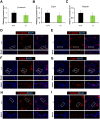
Real-time quantitative PCR analysis of β-catenin (A), Espin (B) and Palladin (C) from Rai14+∕+ and −∕− testes. n = 3, P > 0.05; Immunostaining of basal ES protein β-catenin (D, E) and Espin (F, G) from Rai14 +∕+ and −∕− testes. Scale bar: 20 µm; Immunostaining of apical ES protein Espin (F, G) and Palladin (H, I) from Rai14+∕+ and −∕− testes. Scale bar: 20 µm.
Figure 5
Figure 5. Rai14 is not required for F-actin organization.
(A) Phalloidin-labeled F-actin staining of Rai14+∕+ and −∕− spermatids at steps 13–14. Scale bar: 20 µm; (B) TEM analysis of the apical ES from Rai14+∕+ and −∕− spermatids at steps 13–14. ES synchronously stretches along with the acrosome, and is characterized by the presence of actin filament bundles (arrows). Nu, nucleus; Ac, acrosome; ER, endoplasmic reticulum.

References

    1. Alsemeh AE, Samak MA, El-Fatah SSA. Therapeutic prospects of hydroxytyrosol on experimentally induced diabetic testicular damage: potential interplay with AMPK expression. Cell and Tissue Research. 2020;380:173–189. doi: 10.1007/s00441-019-03143-2. - DOI - PubMed
    1. Bonilla E, Xu EY. Identification and characterization of novel mammalian spermatogenic genes conserved from fly to human. Molecular Human Reproduction. 2008;14:137–142. doi: 10.1093/molehr/gan002. - DOI - PubMed
    1. Cen C, Chen M, Zhou J, Zhang L, Duo S, Jiang L, Hou X, Gao F. Inactivation of Wt1 causes pre-granulosa cell to steroidogenic cell transformation and defect of ovary developmentdagger. Biology of Reproduction. 2020;103:60–69. doi: 10.1093/biolre/ioaa042. - DOI - PubMed
    1. Cheng CY, Mruk DD. A local autocrine axis in the testes that regulates spermatogenesis. Nature Reviews Endocrinology. 2010;6:380–395. doi: 10.1038/nrendo.2010.71. - DOI - PMC - PubMed
    1. Clement TM, Anway MD, Uzumcu M, Skinner MK. Regulation of the gonadal transcriptome during sex determination and testis morphogenesis: comparative candidate genes. Reproduction. 2007;134:455–472. doi: 10.1530/REP-06-0341. - DOI - PMC - PubMed

LinkOut - more resources