. 2021 Feb 10:9:615218.
doi: 10.3389/fbioe.2021.615218. eCollection 2021.

All-Polymer Printed Low-Cost Regenerative Nerve Cuff Electrodes

Affiliations

All-Polymer Printed Low-Cost Regenerative Nerve Cuff Electrodes

Laura M Ferrari et al. Front Bioeng Biotechnol. .

Abstract

Neural regeneration after lesions is still limited by several factors and new technologies are developed to address this issue. Here, we present and test in animal models a new regenerative nerve cuff electrode (RnCE). It is based on a novel low-cost fabrication strategy, called "Print and Shrink", which combines the inkjet printing of a conducting polymer with a heat-shrinkable polymer substrate for the development of a bioelectronic interface. This method allows to produce miniaturized regenerative cuff electrodes without the use of cleanroom facilities and vacuum based deposition methods, thus highly reducing the production costs. To fully proof the electrodes performance in vivo we assessed functional recovery and adequacy to support axonal regeneration after section of rat sciatic nerves and repair with RnCE. We investigated the possibility to stimulate the nerve to activate different muscles, both in acute and chronic scenarios. Three months after implantation, RnCEs were able to stimulate regenerated motor axons and induce a muscular response. The capability to produce fully-transparent nerve interfaces provided with polymeric microelectrodes through a cost-effective manufacturing process is an unexplored approach in neuroprosthesis field. Our findings pave the way to the development of new and more usable technologies for nerve regeneration and neuromodulation.

Keywords: PEDOT:PSS; inkjet printing; low-cost fabrication; organic bioelectronics; peripheral nerve interfaces; regenerative cuff electrodes; wrinkling.


Conflict of interest statement

The authors declare that the research was conducted in the absence of any commercial or financial relationships that could be construed as a potential conflict of interest.

Figures
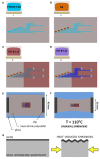
Figure 1
Figure 1
The Print and Shrink fabrication process. (A) Inkjet printing of the conductive ink based on PEDOT:PSS. (B) Inkjet printing of the Ag ink. (C) Inkjet printing of the first SU-8 10% layer. (D) Inkjet printing of the SU-8 29% layer. (E) PO film clamped onto the glass slide. (F) Thermally-induced uniaxial shrinking. (G) Schematics of surface wrinkling induced by heat-shrinking.
Figure 2
Figure 2
Regenerative nerve Cuff Electrodes (RnCEs). (A) Planar view of the final device, with the design of the tube lumen and geometrical references before the rolling up. (B) Picture of the final RnCE. (C) Rendering of the device with an inset of the custom PCB.
Figure 3
Figure 3
PEDOT:PSS and SU-8 wrinkles. (A,B) SEM images of PEDOT:PSS wrinkled active site. Scale bar: 10 and 2 μm. (C) AFM image of wrinkled PEDOT:PSS (scale bar: 5 μm), and extracted x-z profile (gray line scan). (D,E) SEM images of SU-8 10% wrinkled micro-grooves. Scale bar: 20 and 2 μm. (F) AFM image of wrinkled SU-8 10% (scale bar: 5 μm), and extracted x-z profile (gray line scan).
Figure 4
Figure 4
The implanted regenerative cuff electrode. Representative images of: (A) silicone tube, (B) PEDOT:PSS coated PO tube (PPP tube), and (C) Regenerative nerve Cuff Electrode (RnCE) at the time of implant (0 dpi) bridging a 8 mm gap in the rat sciatic nerve, and of (D) silicone tube, (E) PPP tube and (F) RnCE at 90 dpi, showing the regenerated nerve cable inside. Yellow arrows point proximal and distal sciatic stumps sutured to each device. White arrows point the PCB. Scale bar: 5 mm.
Figure 5
Figure 5
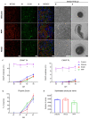
Histological and electrophysiological evaluation of nerve regeneration. (A–E) Representative images of cross sectioned nerves repaired with silicone, PEDOT:PSS coated PO tube (PPP) and Regenerative nerve Cuff Electrode (RnCE) at 90 dpi. (A) NF200 (red) for axons; (B) S100 (green) for Schwann cells; (C) Merge of A and B with nuclear staining (DAPI, blue); (D,E) Brightfield images of regenerated nerves stained with toluidine blue labeling myelin sheaths at high (D) and low (E) magnification. Scale bar: 50 μm in (A–D), 200 μm in (E). (F) Amplitude of the CMAP of tibialis anterior (TA) and plantar interossei (PL) muscles along the 90 days follow-up after sciatic nerve section and repair. (G) Skin paw reinnervation tests by pinprick test. (H) Quantification of myelinated fibers in the sciatic nerve distal to the tube in the three tested groups. *p < 0.05 vs. other groups.
Figure 6
Figure 6
Muscle recruitment curves. (A,B) Representative muscle recruitment curves from selected active sites at (A) 0 dpi and (B) 90 dpi. The increase of the electrical charge delivered through the device elicited recruitment of more muscular fibers thus increasing the CMAP amplitude. (C) Values of the electrical charge needed to elicit 5, 30, and 95% of the maximum CMAP amplitude of gastrocnemius (GM), tibialis anterior (TA) and plantar interossei muscles (PL). (D) Maximal selectivity index (SImax) for GM, TA, and PL muscles over time. *p < 0.05 vs. time.

References

    1. Allodi I., Mecollari V., González-Pérez F., Eggers R., Hoyng S., Verhaagen J., et al. . (2014). Schwann cells transduced with a lentiviral vector encoding Fgf-2 promote motor neuron regeneration following sciatic nerve injury. Glia 62, 1736–1746. 10.1002/glia.22712 - DOI - PubMed
    1. Altuna A., Bellistri E., Cid E., Aivar P., Gal B., Berganzo J., et al. . (2013). SU-8 based microprobes for simultaneous neural depth recording and drug delivery in the brain. Lab Chip. 13, 1422–1430. 10.1039/c3lc41364k - DOI - PubMed
    1. Asensio-Pinilla E., Udina E., Jaramillo J., Navarro X. (2009). Electrical stimulation combined with exercise increase axonal regeneration after peripheral nerve injury. Exp. Neurol. 219, 258–265. 10.1016/j.expneurol.2009.05.034 - DOI - PubMed
    1. Asplund M., Nyberg T., Inganäs O. (2010). Electroactive polymers for neural interfaces. Polym. Chem. 1, 1374–1391. 10.1039/c0py00077a - DOI
    1. Badia J., Boretius T., Andreu D., Azevedo-Coste C., Stieglitz T., Navarro X. (2011). Comparative analysis of transverse intrafascicular multichannel, longitudinal intrafascicular and multipolar cuff electrodes for the selective stimulation of nerve fascicles. J. Neural Eng. 8:036023. 10.1088/1741-2560/8/3/036023 - DOI - PubMed

LinkOut - more resources