Review
. 2021 Feb 12:9:621748.
doi: 10.3389/fbioe.2021.621748. eCollection 2021.

Increased Mesenchymal Stem Cell Functionalization in Three-Dimensional Manufacturing Settings for Enhanced Therapeutic Applications

Affiliations
Review

Increased Mesenchymal Stem Cell Functionalization in Three-Dimensional Manufacturing Settings for Enhanced Therapeutic Applications

Dimitrios Kouroupis et al. Front Bioeng Biotechnol. .

Abstract

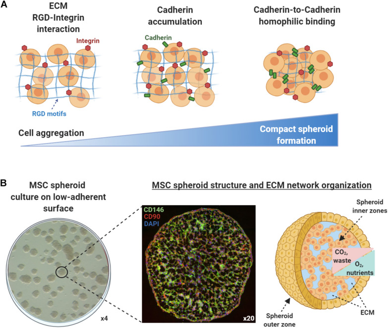
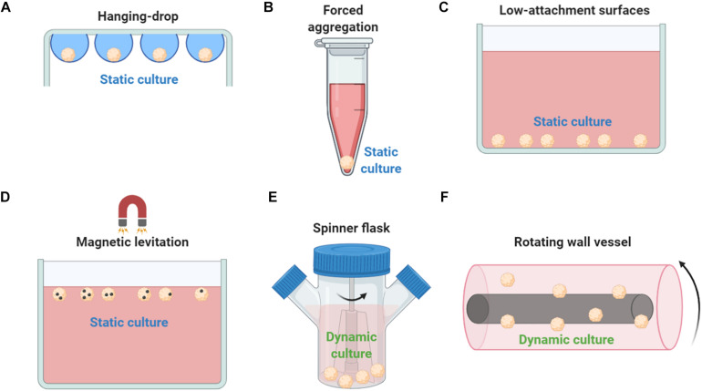
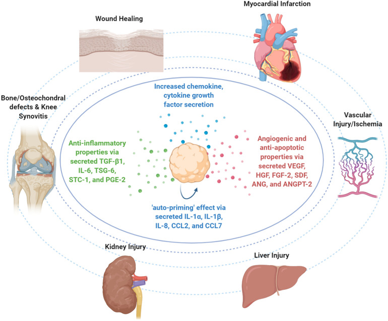
Mesenchymal stem/stromal cell (MSC) exist within their in vivo niches as part of heterogeneous cell populations, exhibiting variable stemness potential and supportive functionalities. Conventional extensive 2D in vitro MSC expansion, aimed at obtaining clinically relevant therapeutic cell numbers, results in detrimental effects on both cellular characteristics (e.g., phenotypic changes and senescence) and functions (e.g., differentiation capacity and immunomodulatory effects). These deleterious effects, added to the inherent inter-donor variability, negatively affect the standardization and reproducibility of MSC therapeutic potential. The resulting manufacturing challenges that drive the qualitative variability of MSC-based products is evident in various clinical trials where MSC therapeutic efficacy is moderate or, in some cases, totally insufficient. To circumvent these limitations, various in vitro/ex vivo techniques have been applied to manufacturing protocols to induce specific features, attributes, and functions in expanding cells. Exposure to inflammatory cues (cell priming) is one of them, however, with untoward effects such as transient expression of HLA-DR preventing allogeneic therapeutic schemes. MSC functionalization can be also achieved by in vitro 3D culturing techniques, in an effort to more closely recapitulate the in vivo MSC niche. The resulting spheroid structures provide spatial cell organization with increased cell-cell interactions, stable, or even enhanced phenotypic profiles, and increased trophic and immunomodulatory functionalities. In that context, MSC 3D spheroids have shown enhanced "medicinal signaling" activities and increased homing and survival capacities upon transplantation in vivo. Importantly, MSC spheroids have been applied in various preclinical animal models including wound healing, bone and osteochondral defects, and cardiovascular diseases showing safety and efficacy in vivo. Therefore, the incorporation of 3D MSC culturing approach into cell-based therapy would significantly impact the field, as more reproducible clinical outcomes may be achieved without requiring ex vivo stimulatory regimes. In the present review, we discuss the MSC functionalization in 3D settings and how this strategy can contribute to an improved MSC-based product for safer and more effective therapeutic applications.

Keywords: MSC anti-inflammatory properties; MSC functionalization; MSC spheroids; MSC therapeutic properties; mesenchymal stem cell manufacturing.


Conflict of interest statement

The authors declare that the research was conducted in the absence of any commercial or financial relationships that could be construed as a potential conflict of interest.

Figures

FIGURE 1
FIGURE 1
Mesenchymal stem/stromal cell (MSC) spheroids formation process and structure. (A) Cell aggregation and spheroid formation involving three phases. Initially, cells form loose aggregates via the tight binding of extracellular matrix arginine–glycine–aspartate (RGD) motifs with membrane-bound integrin. Due to increased cell–cell interactions, cadherin gene expression levels are upregulated and cadherin protein is accumulated on the cell membrane. In the later phase, homophilic cadherin-to-cadherin binding induce the formation of compact cell spheroids from cell aggregates. (B) Methylcellulose-based technique can be used to generate viable MSC spheroids on low-attachment gas-permeable plates (left panel). Generated MSC spheroids show stable immunophenotypic profile by expressing high levels of the pericytic marker CD146 (green) and MSC-related marker CD90 (red) (middle panel) (unpublished data). Structurally, based on their size and abundance of nutrients and oxygen in vitro, MSC spheroids can be divided into zones (outer and inner). The nutrients, oxygen, and waste concentration gradients within the spheroids should be always taken into consideration when selecting the optimal technique to generate spheroids in vitro in order to achieve increased spheroid functionality in in vivo settings (right panel).
FIGURE 2
FIGURE 2
Methods used to generate MSC spheroids ex vivo. (A) In hanging drop technique, MSCs are aggregated by gravitational force. (B) Forced aggregation technique (or pellet culture) is used to generate MSC aggregates by gravitational force that are further induced toward 3D differentiation protocols. (C) Low-attachment surfaces allow the self-organization of cells into suspended spheroids. (D) Magnetic levitation diminish gravitational force, promotes cell–cell contact and induces cell aggregation in vitro. (E) In a spinner flask bioreactor system, cells are continuously mixed by stirring. (F) Rotating wall vessel technique simulates microgravity by constant circular rotation and, therefore, cells are continuously in suspension. Both dynamic culturing techniques (spinner flask and rotating wall vessel) form viable compact MSC spheroids but with altered cell size, altered phenotypic and molecular profiles, and enhanced differentiation potential compared to 2D conventional MSC cultures.
FIGURE 3
FIGURE 3
Therapeutic properties of MSC spheroids in vivo. Upon infusion in vivo, MSC spheroid “medicinal signaling” activities are exerted by the paracrine secretion of modulatory mediators that possess immunomodulatory and trophic (i.e., angiogenic, anti-fibrotic, anti-apoptotic, and mitogenic) actions. MSC spheroids have been safely and effectively applied in various preclinical animal models for the treatment of skin wounds, myocardial infarction, vascular injury/ischemia, liver injury, kidney injury, bone and osteochondral defects, and knee synovitis.

Similar articles

Cited by

References

    1. Agarwal S. K., Brenner M. B. (2006). Role of adhesion molecules in synovial inflammation. Curr. Opin. Rheumatol. 18 268–276. 10.1097/01.bor.0000218948.42730.39 - DOI - PubMed
    1. Aggarwal S., Pittenger M. F. (2005). Human mesenchymal stem cells modulate allogeneic immune cell responses. Blood 105 1815–1822. - PubMed
    1. Alimperti S., Andreadis S. T. (2015). CDH2 and CDH11 act as regulators of stem cell fate decisions. Stem Cell Res. 14 270–282. 10.1016/j.scr.2015.02.002 - DOI - PMC - PubMed
    1. Alvarez-Pérez J., Ballesteros P., Cerdán S. (2005). Microscopic images of intraspheroidal pH by 1H magnetic resonance chemical shift imaging of pH sensitive indicators. Magma 18 293–301. 10.1007/s10334-005-0013-z - DOI - PubMed
    1. Amos P. J., Kapur S. K., Stapor P. C., Shang H., Bekiranov S., Khurgel M., et al. (2009). Human adipose-derived stromal cells accelerate diabetic wound healing: impact of cell formulation and delivery. Tissue Eng. Part A 16 1595–1606. 10.1089/ten.tea.2009.0616 - DOI - PMC - PubMed