

Case Reports
doi: 10.1016/j.case.2020.09.006.
eCollection 2021 Feb.
Nonvisualization of the Left Atrial Appendage and Role of Multimodality Imaging
Affiliations
- PMID: 33644505
- PMCID: PMC7887443
- DOI: 10.1016/j.case.2020.09.006
Item in Clipboard
Case Reports
Nonvisualization of the Left Atrial Appendage and Role of Multimodality Imaging
CASE (Phila).
.
No abstract available
Keywords: Cardiac computed tomographic angiography; Left atrial appendage; Transesophageal echocardiography.
Figures


Congenital LAA absence. (A, B) Multiplanar reconstruction axial image from CCTA demonstrates nonvisualization of the LAA adjacent to the left superior pulmonary vein (LSPV). (C) Three-dimensional TEE shows the surgeon's view and congenital absence of the LAA. (D) Transesophageal echocardiographic view at 60° confirms congenital absence of the LAA. AO, Aorta; AV, aortic valve; LA, left atrium; LV, left ventricle; MV, mitral valve.
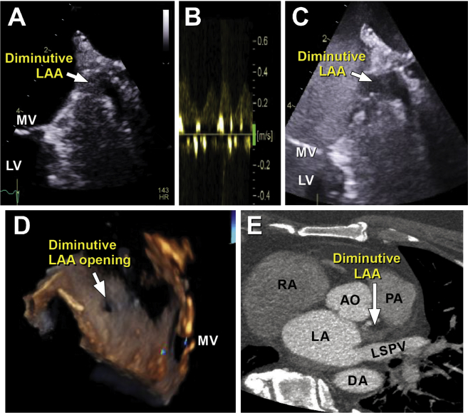
Diminutive LAA. (A) A small LAA sliver with minimal blood flow is seen on TEE in the 60° view. (B) Pulsed-wave Doppler evaluation shows low-velocity blood flow. (C) Definity contrast image shows minimal late filling of contrast in the LAA sliver. (D) The LAA orifice is seen on three-dimensional TEE. (E) CCTA shows a rudimentary appendage that did not opacify with contrast. AO, Aorta; AV, aortic valve; DA, descending aorta; LA, left atrium; LSPV, left superior pulmonary vein; LV, left ventricle; MV, mitral valve; PA, pulmonary artery; RA, right atrium.
References
-
- Odell J.A., Blackshear J.L., Davies E., Byrne W.J., Kollmorgen C.F., Edwards W.D. Thoracoscopic obliteration of the left atrial appendage: potential for stroke reduction? Ann Thorac Surg. 1996;61:565–569. - PubMed
-
- Di Biase L., Santangeli P., Anselmino M., Mohanty P., Salvetti I., Gili S. Does the left atrial appendage morphology correlate with the risk of stroke in patients with atrial fibrillation? Results from a multicenter study. J Am Coll Cardiol. 2012;60:531–538. - PubMed
-
- Di Biase L., Burkhardt J.D., Mohanty P., Sanchez J., Mohanty S., Horton R. Left atrial appendage: an underrecognized trigger site of atrial fibrillation. Circulation. 2010;122:109–118. - PubMed
-
- Veinot J.P., Harrity P.J., Gentile F., Khandheria B.K., Bailey K.R., Eickholt J.T. Anatomy of the normal left atrial appendage: a quantitative study of age-related changes in 500 autopsy hearts: implications for echocardiographic examination. Circulation. 1997;96:3112–3115. - PubMed
-
- Yamamoto M., Seo Y., Kawamatsu N., Sato K., Sugano A., Machino-Ohtsuka T. Complex left atrial appendage morphology and left atrial appendage thrombus formation in patients with atrial fibrillation. Circ Cardiovasc Imaging. 2014;7:337–343. - PubMed
Publication types
LinkOut - more resources
Full Text Sources
Other Literature Sources
