

Case Reports
doi: 10.1016/j.case.2020.09.007.
eCollection 2021 Feb.
Vital Role of Transesophageal Echocardiographic Surveillance of a Left Atrial Appendage Perforation Complicating Attempted Percutaneous Appendage Occlusion
Affiliations
- PMID: 33644506
- PMCID: PMC7887453
- DOI: 10.1016/j.case.2020.09.007
Item in Clipboard
Case Reports
Vital Role of Transesophageal Echocardiographic Surveillance of a Left Atrial Appendage Perforation Complicating Attempted Percutaneous Appendage Occlusion
CASE (Phila).
.
No abstract available
Keywords: Atrial fibrillation; Cardiac tamponade; Complication; Pericardial effusion; Watchman.
Figures

Midesophageal TEE image of guiding catheter in the LAA, which has a chicken-wing morphology. LA, Left atrium.

Midesophageal TEE image of the Watchman device malpositioned and incompletely expanded upon attempted deployment in the LAA. LA, Left atrium.

Midesophageal TEE image of the developing pericardial effusion (red arrow) noted following the third attempt of Watchman device placement. LA, Left atrium; LV, left ventricle; RA, right atrium; RV, right ventricle.

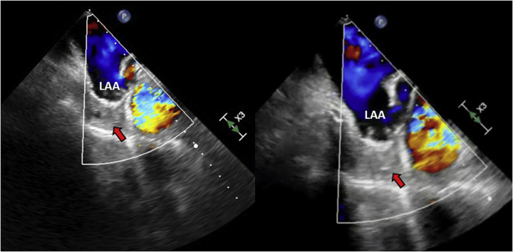
TEE images of LAA perforation visualized by color Doppler flow from the LAA into the pericardial space (red arrows), seen in two midesophageal imaging planes, 129° and 68°. LV, Left ventricle.

Midesophageal TEE images of LAA perforation resolving, evidenced by decreased flow on color Doppler (red arrow) as well as the presence of a small thrombus (orange arrow) visualized at site of perforation.

Midesophageal TEE color Doppler images demonstrating the cessation of flow through the LAA perforation as well as the presence of thrombus (red arrow) in the pericardial space.
References
-
- Schwartz R.S., Holmes D.R., Van Tassel R.A., Hauser R., Henry T.D., Mooney M. Left atrial appendage obliteration: mechanisms of healing and intracardiac integration. JACC Cardiovasc Interv. 2010;3:870–877. - PubMed
-
- Holmes D.R., Kar S., Price M.J., Whisenant B., Sievert H., Doshi S.K. Prospective randomized evaluation of the Watchman left atrial appendage closure device in patients with atrial fibrillation versus long-term warfarin therapy: the PREVAIL trial. J Am Coll Cardiol. 2014;64:1–12. - PubMed
-
- Reddy V.Y., Gibson D.N., Kar S., O'Neill W., Doshi S.K., Horton R.P. Post-approval U.S. experience with left atrial appendage closure for stroke prevention in atrial fibrillation. J Am Coll Cardiol. 2017;69:253–261. - PubMed
-
- Steinbeck G., Sinner M.F., Lutz M., Müller-Nurasyid M., Kääb S., Reinecke H. Incidence of complications related to catheter ablation of atrial fibrillation and atrial flutter: a nationwide in-hospital analysis of administrative data for Germany in 2014. Eur Heart J. 2018;39:4020–4029. - PMC - PubMed
Publication types
LinkOut - more resources
Full Text Sources
Other Literature Sources
