

Case Reports
doi: 10.1016/j.case.2020.10.002.
eCollection 2021 Feb.
Echocardiographic Features of Cardiac Echinococcal Infection
Affiliations
- PMID: 33644510
- PMCID: PMC7887517
- DOI: 10.1016/j.case.2020.10.002
Item in Clipboard
Case Reports
Echocardiographic Features of Cardiac Echinococcal Infection
CASE (Phila).
.
No abstract available
Keywords: Cardiac mass; Echinococcosis; Echocardiography; Hydatid cyst; Ultrasound enhancing agent.
Figures
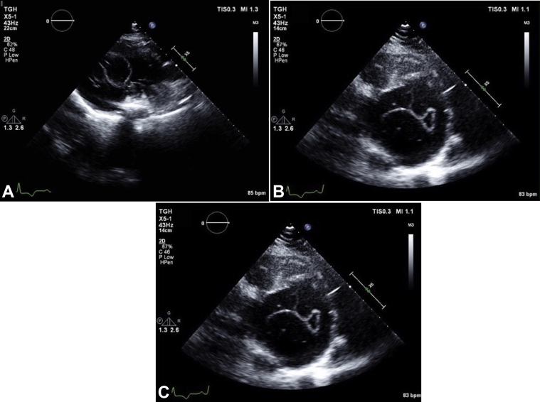
Transthoracic echocardiographic parasternal long-axis view (A), parasternal short-axis view (B), and apical four-chamber view (C) demonstrate a large mass occupying almost the entire mid to distal LV cavity. An echo-bright layer appears to encompass a more echo-lucent center, with small linear densities within the structure.
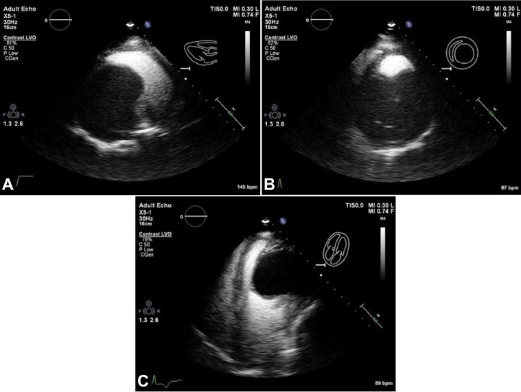
Transthoracic echocardiographic parasternal long-axis view (A), parasternal short-axis view (B), and apical four-chamber view (C) with ultrasound enhancing agent. The large mass is seen along the mid-distal inferolateral, anterolateral, and apical walls without contrast enhancement, suggesting that the lesion is not vascular as per the American Society of Echocardiography 2018 guidelines on ultrasound enhancing agents in echocardiography.
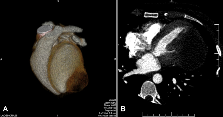
Cardiac computed tomographic three-dimensional volume-rendered image (A) demonstrating the mass along the LV apical lateral wall. Contrast-enhanced axial computed tomographic image (B) shows a large cystic appearing lesion likely embedded within the inferolateral LV wall.
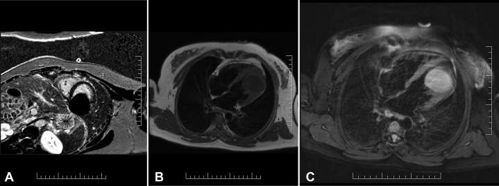
Cardiac MRI short axis phase-sensitive inversion recovery delayed enhancement images (A). The cyst appears fully nulled, suggesting that it is fluid filled. Horizontal long-axis T1 weighted turbo-spin echo (B) showing a hypointense lesion. Horizontal long-axis, T2 spectral attenuated inversion recovery image (C) showing hyperintense signal from the mass, suggesting that it contains fluid. There is a hypointense peripheral ring, which likely represents a pericyst.
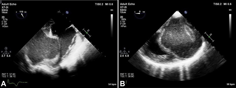
Intraoperative transesophageal echocardiographic four-chamber view (A) and short-axis view (B) demonstrate a large mass occupying the entire mid to distal LV cavity.
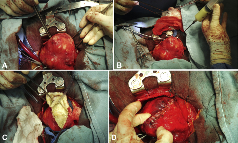
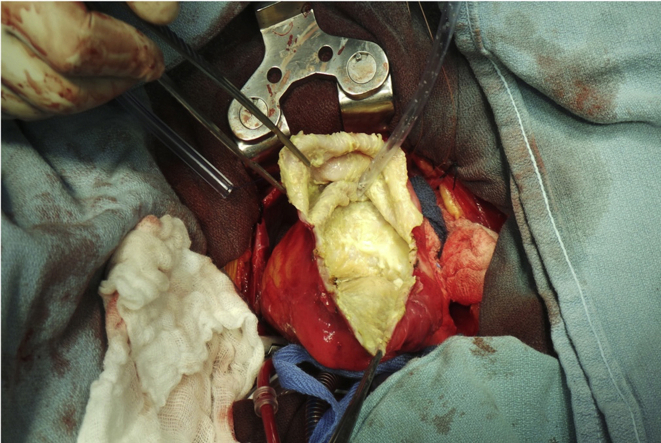
Gross intraoperative surgical inspection identified a large lesion adhered to the pericardium (A) containing a green- and yellow-colored liquid (B). The inner wall of the cyst (C) was dissected and resected before being sutured shut from the base of the heart to the apex of the heart (D).
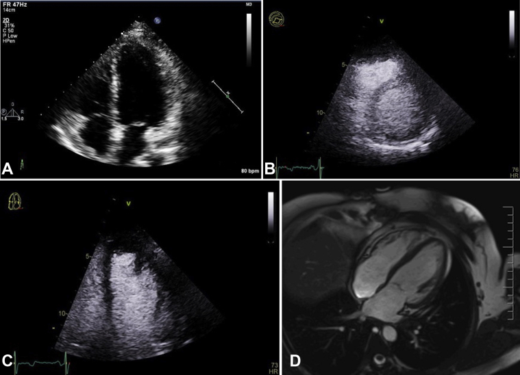
Postoperative transthoracic echocardiographic apical four-chamber view (A), parasternal short-axis view with ultrasound enhancing agent (B), apical four-chamber with ultrasound enhancing agent (C), and cardiac MRI horizontal long-axis cine (D) showing normal LV systolic function after mass resection. There is no evidence of residual or recurrent cyst.
References
-
- Basso C., Rizzo S., Valente M., Thiene G. Cardiac masses and tumours. BMJ Heart. 2016;102:1230–1245. - PubMed
-
- Peters P.J., Reinhardt S. The echocardiographic evaluation of intracardiac masses: a review. J Am Soc Echocardiogr. 2006;19:230–240. - PubMed
-
- Ben-Hamda K., Maatouk F., Ben-Farhat M., Betbout F., Gamra H., Addad F. Eighteen-year experience with echinococcosus of the heart: clinical and echocardiographic features in 14 patients. Int J Cardiol. 2003;91:145–151. - PubMed
-
- Moro P., Schantz P.M. Echinococcosis: a review. Int J Infect Dis. 2009;13:125–133. - PubMed
-
- Brunetti E., Kern P., Vuitton D.A., Writing Panel for the WHO-IWGE Expert consensus for the diagnosis and treatment of cystic and alveolar echinococcosis in humans. Acta Trop. 2010;114:1–16. - PubMed
Publication types
LinkOut - more resources
Full Text Sources
Other Literature Sources
