Review
. 2021 Apr 1;40(7):e106922.
doi: 10.15252/embj.2020106922. Epub 2021 Mar 1.

Sealing holes in cellular membranes

Affiliations
Review

Sealing holes in cellular membranes

Yan Zhen et al. EMBO J. .

Abstract

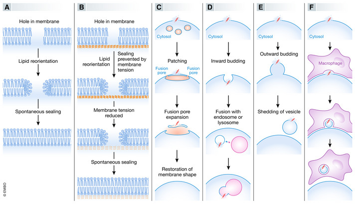
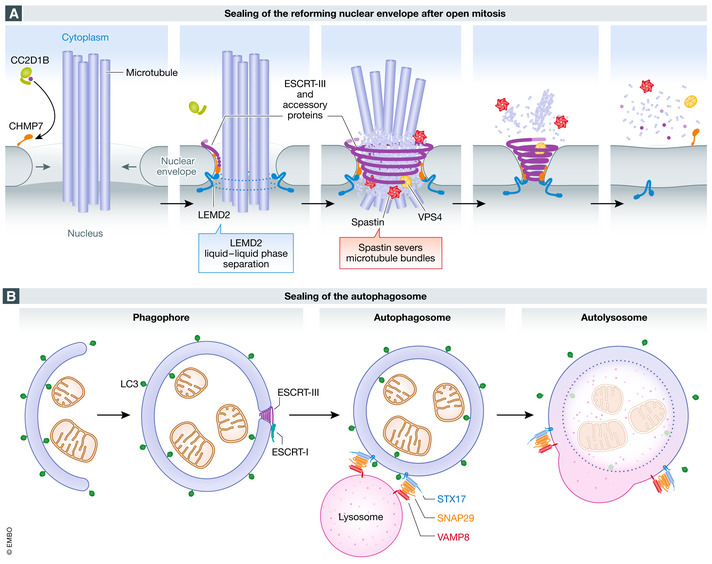
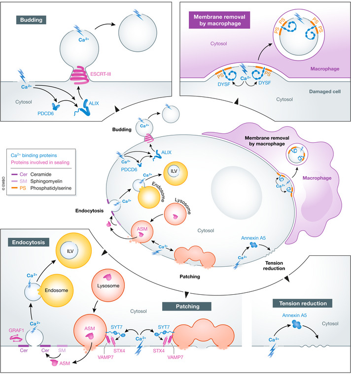
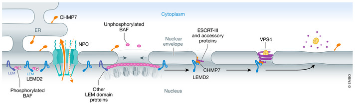
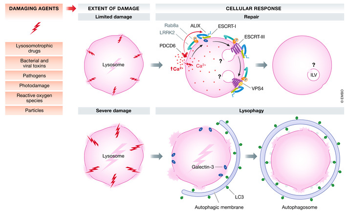
The compartmentalization of eukaryotic cells, which is essential for their viability and functions, is ensured by single or double bilayer membranes that separate the cell from the exterior and form boundaries between the cell's organelles and the cytosol. Nascent nuclear envelopes and autophagosomes, which both are enveloped by double membranes, need to be sealed during the late stage of their biogenesis. On the other hand, the integrity of cellular membranes such as the plasma membrane, lysosomes and the nuclear envelope can be compromised by pathogens, chemicals, radiation, inflammatory responses and mechanical stress. There are cellular programmes that restore membrane integrity after injury. Here, we review cellular mechanisms that have evolved to maintain membrane integrity during organelle biogenesis and after injury, including membrane scission mediated by the endosomal sorting complex required for transport (ESCRT), vesicle patching and endocytosis.

Keywords: ESCRT; autophagy; endocytosis; lysosome; membrane repair.


Conflict of interest statement

The authors declare that they have no conflict of interest.

Figures

Figure 1
Figure 1. Mechanisms of membrane sealing
Holes in lipid bilayers can be sealed by several alternative mechanisms, including membrane self‐sealing (A), reduction in membrane tension to promote self‐sealing (B), patching of hole by vertex fusion of large intracellular vesicles (C), inward budding of hole‐containing membrane area (D), outward budding of hole‐containing membrane area (E), or removal of hole‐containing membrane area by adjacent cell (F).
Figure 2
Figure 2. Sealing of holes during biogenesis of double‐membrane organelles
(A) sealing of the reforming nuclear envelope after open mitosis. During anaphase, when the reforming nuclear envelope meets microtubule bundles still connected to chromatin, the inner nuclear membrane protein LEMD2 undergoes liquid‐phase separation and activates the ESCRT‐III specific nuclear envelope recruitment factor CHMP7, which drives ESCRT‐III polymerization. Timing of ESCRT‐III recruitment is also regulated by CC2D1B, which prevents its premature localization to the reforming membrane. Spastin recruitment by ESCRT‐III is required for severing of mitotic spindle microtubules, while VPS4 modelling of ESCRT‐III filaments promotes membrane constriction and sealing. (B) sealing of the autophagosome. During autophagy, the double‐membrane phagophore expands to sequester cytoplasmic material for degradation. When the resulting LC3‐positive autophagosome is complete, a small hole remains. This hole is sealed by ESCRT‐III, which is recruited by ESCRT‐I. Sealing is followed by recruitment to the autophagosome membrane of the SNARE protein STX17, which forms a complex with the cytosolic SNARE SNAP29 and the lysosomal SNARE VAMP8, and this mediates fusion of the autophagosome with the lysosome to form an autolysosome.
Figure 3
Figure 3. Plasma membrane repair
Holes in the plasma membrane can be sealed by patching, endocytosis, budding, macrophage‐mediated membrane removal or reduction in membrane tension. Ca2+ influx triggers all these processes. Ca2+ binding proteins are in green font, other proteins involved in sealing are in red font. Cer, ceramide; ILV, intraluminal vesicle; PS, phosphatidylserine.
Figure 4
Figure 4. Nuclear envelope repair
Under unperturbed conditions, the inner nuclear membrane protein LEMD2 and the endoplasmic reticulum‐associated protein CHMP7 are spatially separated because CHMP7 is actively exported out of the nucleus through nuclear pore complexes (NPCs). Following rupture, unphosphorylated cytosolic BAF coats the exposed chromatin. Interaction of BAF with integral membrane LEM‐domain proteins facilitates recruitment of nuclear membrane and decreases the size of the rupture. Subsequently, the CHMP7 and LEMD2 interaction promotes nucleation and polymerization of ESCRT‐III, which together with the regulatory ATPase VPS4 further constricts the rupture and promotes sealing.
Figure 5
Figure 5. Lysosome repair
Damaging agents such as lysosomotropic drugs, photodamage, pathogens etc. may inflict varying extent of lysosomal damage, leading to Ca2+ efflux from damaged lysosomes. The ESCRT machinery orchestrates repair of the limited lysosomal damage through multiple mechanisms in a coordinated manner. The increase in cytosolic Ca2+ is probably sensed by PDCD6 which in turn recruits the ESCRT‐III binding protein ALIX. The ESCRT‐I protein TSG101 and ALIX further recruit the ESCRT‐III machinery together with VPS4 to seal the membrane lesions. Although Ca2+ efflux might provide an immediate signal for ESCRT‐III recruitment, the β‐galactoside sensor GAL3, which interacts with ALIX, is required for efficient recruitment of ALIX and ESCRT‐III to the damaged lysosomes. This sealing might be accompanied by formation of ILVs containing the damage, similar to the process of endosomal ILV biogenesis. In macrophages, phagolysosome or lysosome damage triggers activation of the kinase LRRK2. Once activated, LRRK2 phosphorylates the small GTPase Rab8A, and ESCRT‐III is recruited to mediate endolysosomal membrane repair in Ca2+‐dependent fashion. Severely damaged lysosomes are engulfed and degraded via lysophagy, initiated by β‐galactoside sensors such as GAL3, which recruits LC3‐containing autophagic membranes.

References

    1. Anderson DJ, Hetzer MW (2008) The life cycle of the metazoan nuclear envelope. Curr Opin Cell Biol 20: 386–392 - PMC - PubMed
    1. Andrews NW, Almeida PE, Corrotte M (2014) Damage control: cellular mechanisms of plasma membrane repair. Trends Cell Biol 24: 734–742 - PMC - PubMed
    1. von Appen A, LaJoie D, Johnson IE, Trnka MJ, Pick SM, Burlingame AL, Ullman KS, Frost A (2020) LEM2 phase separation promotes ESCRT‐mediated nuclear envelope reformation. Nature 582: 115–118 - PMC - PubMed
    1. Bendix PM, Simonsen AC, Florentsen CD, Hager SC, Mularski A, Zanjani AAH, Moreno‐Pescador G, Klenow MB, Sonder SL, Danielsen HM et al (2020) Interdisciplinary synergy to reveal mechanisms of annexin‐mediated plasma membrane shaping and repair. Cells 9: 1029 - PMC - PubMed
    1. Berg TO, Fengsrud M, Stromhaug PE, Berg T, Seglen PO (1998) Isolation and characterization of rat liver amphisomes ‐ Evidence for fusion of autophagosomes with both early and late endosomes. J Biol Chem 273: 21883–21892 - PubMed

Publication types

MeSH terms

Substances