. 2021 May;10(10):e2002152.
doi: 10.1002/adhm.202002152. Epub 2021 Feb 28.

In Vivo Printing of Nanoenabled Scaffolds for the Treatment of Skeletal Muscle Injuries

Affiliations

In Vivo Printing of Nanoenabled Scaffolds for the Treatment of Skeletal Muscle Injuries

Jacob P Quint et al. Adv Healthc Mater. 2021 May.

Abstract

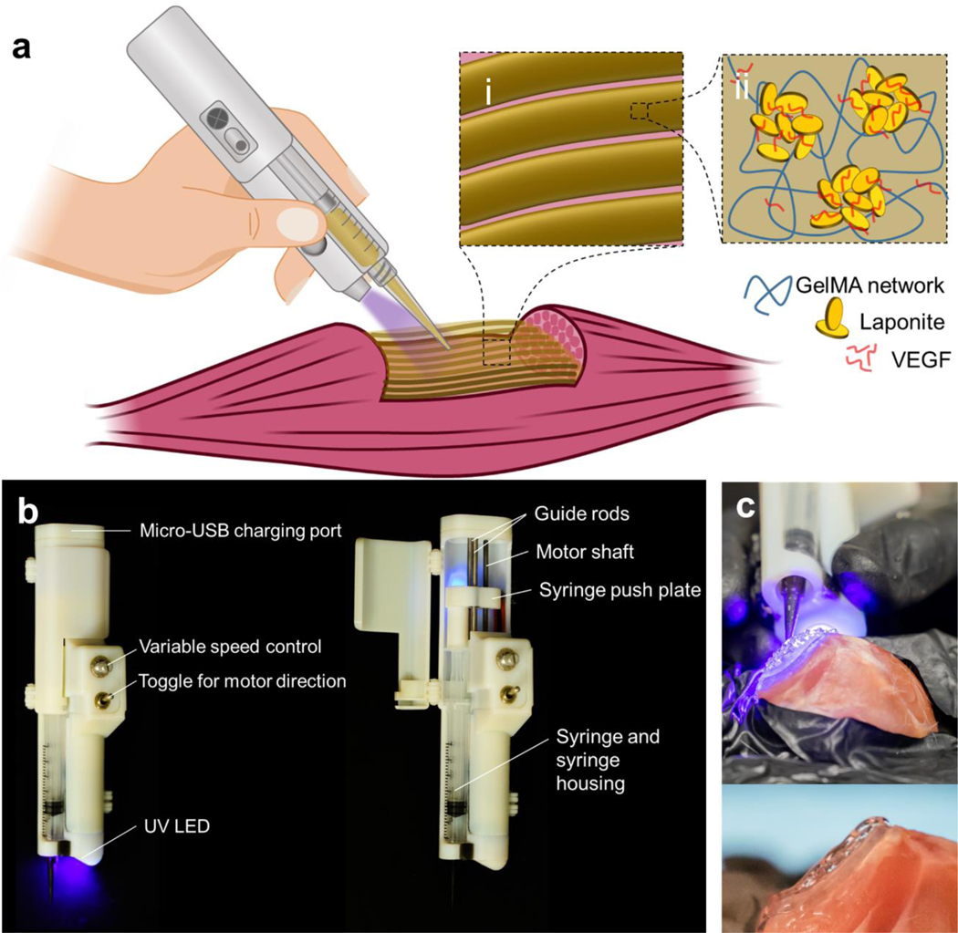
Extremity skeletal muscle injuries result in substantial disability. Current treatments fail to recoup muscle function, but properly designed and implemented tissue engineering and regenerative medicine techniques can overcome this challenge. In this study, a nanoengineered, growth factor-eluting bioink that utilizes Laponite nanoclay for the controlled release of vascular endothelial growth factor (VEGF) and a GelMA hydrogel for a supportive and adhesive scaffold that can be crosslinked in vivo is presented. The bioink is delivered with a partially automated handheld printer for the in vivo formation of an adhesive and 3D scaffold. The effect of the controlled delivery of VEGF alone or paired with adhesive, supportive, and fibrilar architecture has not been studied in volumetric muscle loss (VML) injuries. Upon direct in vivo printing, the constructs are adherent to skeletal muscle and sustained release of VEGF. The in vivo printing of muscle ink in a murine model of VML injury promotes functional muscle recovery, reduced fibrosis, and increased anabolic response compared to untreated mice. The in vivo construction of a therapeutic-eluting 3D scaffold paves the way for the immediate treatment of a variety of soft tissue traumas.

Keywords: functional recovery; handheld printers; hydrogel scaffolds; in vivo printing; soft tissue injuries; volumetric muscle loss.


Figures

Figure 1.
Figure 1.
In vivo handheld printing of therapeutic eluting scaffolds. a) Conceptual diagram of organized scaffold deposition directly into a wound using a handheld printer. Schematics of the composite bioink (Muscle Ink) (i) printed into filament scaffold and (ii) its components including VEGF bound to Laponite® nanodisks and supported by a GelMA hydrogel network. b) Callouts of the features and components of the handheld printer. The force to compress the plunger of a syringe that is fixed within the printer housing is transferred via the push plate from the rotational shaft of an electric motor. Photopolymerizable materials are crosslinked through the embedded ultraviolet (UV) and blue light. c) The handheld printer is able to reconstruct the geometry of volumetric muscle loss (top) and adhere to local tissue (bottom) through the designed Muscle Ink composite hydrogel.
Figure 2.
Figure 2.
Protein interaction and release kinetics from Laponite® in physiological buffers and mechanical characterization of Muscle Ink composite hydrogel. a) Diagram of a single Laponite® nanodisk with a diameter ranging from 20–50 nm and a thickness of 1–2 nm. The uneven distribution of negative face charges and positive edge charges enable the electrostatic binding of proteins. Reproduced with permission (pending).[21] b) The zeta potential of Laponite® becomes less negative after binding with BSA, the model protein. c) Binding efficiency of Laponite® was near 100% binding was achieved at 5 mg/mL. d) Cumulative model protein release of VEGF over a period of 22 days. SEM images of e) 7% (w/v) GelMA and f) Muscle Ink at 5,000× (scale bars 10 μm) and at 20,000× (scale bars 5 μm). There is no difference between the pore size of GelMA and Muscle Ink hydrogels. g) Hydrogel disks were compressed (left) to determine the compressive modulus (right). h) Muscle Ink and GelMA were printed onto skeletal muscle to determine ultimate normal adhesion strength.
Figure 3.
Figure 3.
VEGF eluting Muscle Ink induced angiogenic potential in HUVECs. a) A schematic representing the scratch assay. (i) A confluent layer of HUVECs was scratched to generate (ii) a gap in the cells. The cells were exposed to (iii) a positive control with VEGF in the media and (iv) VEGF+ Muscle Ink crosslinked into porous cell culture inserts to assess the angiogenic potential of released VEGF. The VEGF+ Muscle Ink was prepared fresh to model scaffolds at the time of implantation or presoaked in DPBS to replicate scaffolds 24 h after implantation. b) Micrographs of the scratch assay to assess the induced migratory effects of VEGF+ Muscle Ink (scale bar 500 μm). c) The relative scratch closure area was quantified over 4 h. d) A tube formation assay was performed to analyze the tubulogenesis of HUVECs that were exposed to VEGF+ Muscle Ink (scale bar 1 mm). e) The number of closed-loop structures shows the networking ability of the tube formation assay. f) Total tube length demonstrates the effect of eluted VEGF on tubulogenesis.
Figure 4.
Figure 4.
In situ printing and an in vivo murine model demonstrated the feasibility of in vivo printing and functional improvement with the treatment of VEGF+ Muscle Ink. a) In situ printing into an induced VML wound: uninjured quadriceps with a yellow dotted line indicating the location of muscle resection (top left), quadriceps after muscle resection to simulate VML injury (top right), in situ printing of Muscle Ink (bottom left), and reconstructed of Muscle Ink scaffold that replicates the geometry of the native muscle (bottom right). b) The functional performance of the VML animal model after treatment was assessed using a treadmill. Functional treadmill data shows improvement in c) maximum running speed and d) maximum running distance of VEGF+ Muscle Ink treated animals compared to other injured groups. There was no statistical significance in the maximum running speed between VEGF+ Muscle Ink treated group and the uninjured group. e) Trichrome staining was used to investigate fibrosis (blue) formation (scale bar 5 mm). (f) Fibrosis was quantified, and the Muscle Ink VEGF+ treated group showed less fibrosis compared to the VML injured animals. g) Average fiber area (μm2) in quadricep muscles of the uninjured, injured and untreated, injured and treated with printed VEGF- or VEGF+ Muscle Ink scaffolds. h) The distribution of fiber cross-sectional area (μm2) in the tested groups and controls.

Similar articles

Cited by

References

    1. Cezar CA, Roche ET, Vandenburgh HH, Duda GN, Walsh CJ, Mooney DJ, Proceedings of the National Academy of Sciences 2016, 113, 1534. - PMC - PubMed
    1. Almada AE, Wagers AJ, Nat Rev Mol Cell Biol 2016, 17, 267. - PMC - PubMed
    1. Nuutila K, Sakthivel D, Kruse C, Tran P, Giatsidis G, Sinha I, Wound repair and regeneration : official publication of the Wound Healing Society [and] the European Tissue Repair Society 2017, 25, 408. - PubMed
    1. Grogan BF, Hsu JR, The Journal of the American Academy of Orthopaedic Surgeons 2011, 19 Suppl 1, S35. - PubMed
    1. Garg K, Corona BT, Walters TJ, Front Pharmacol 2015, 6, 87. - PMC - PubMed

Publication types

Substances