. 2021 Jan-Dec;13(1):1-18.
doi: 10.1080/19490976.2021.1887720.

Gut microbiota shape the inflammatory response in mice with an epithelial defect

Affiliations

Gut microbiota shape the inflammatory response in mice with an epithelial defect

Ran Wang et al. Gut Microbes. 2021 Jan-Dec.

Abstract

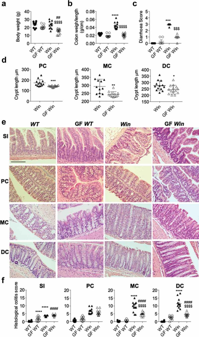
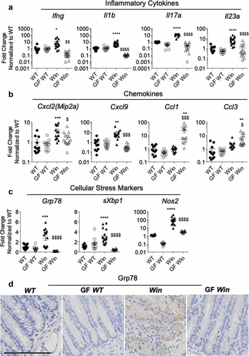
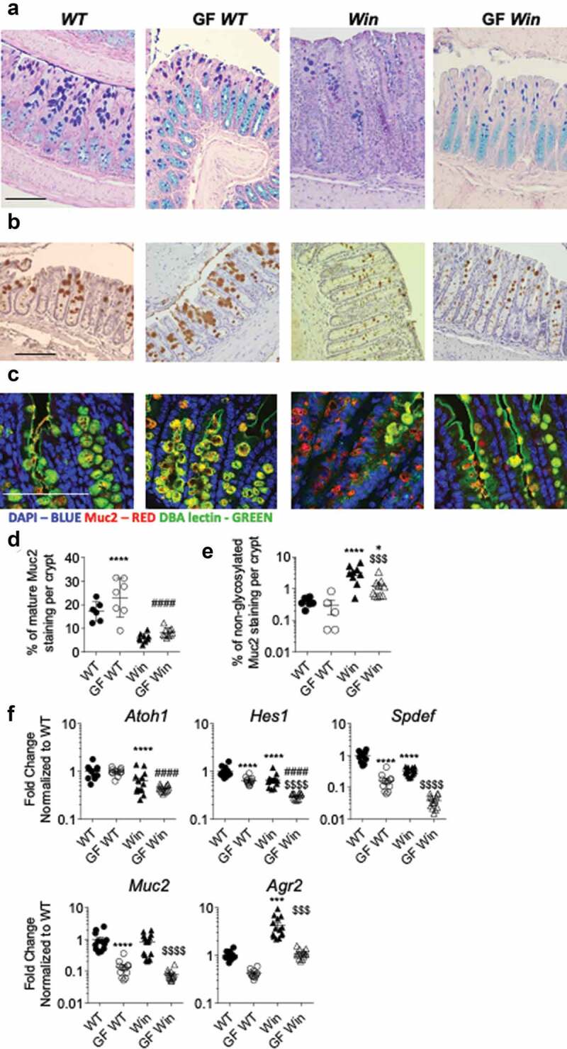
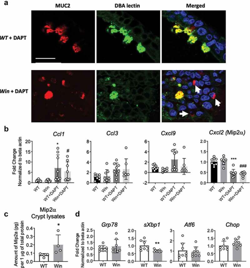
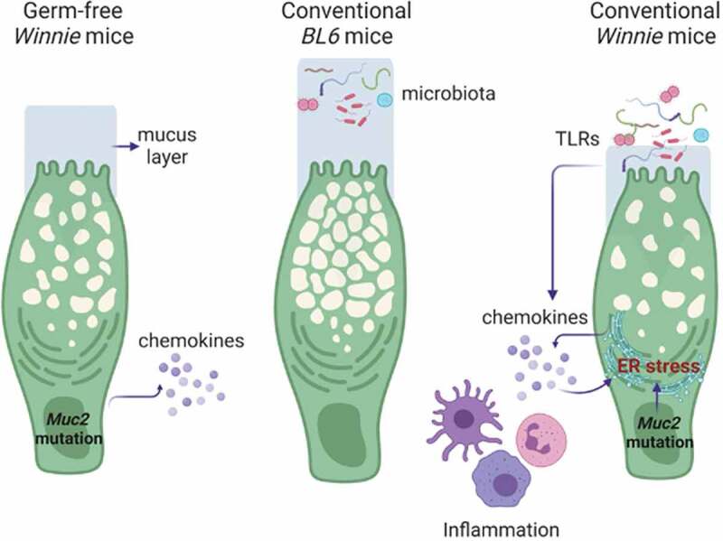
Intestinal epithelial cell endoplasmic reticulum (ER) stress has been implicated in intestinal inflammation. It remains unclear whether ER stress is an initiator of or a response to inflammation. Winnie mice, carrying a Muc2 gene mutation resulting in intestinal goblet cell ER stress, develop spontaneous colitis with a depleted mucus barrier and increased bacterial translocation. This study aims to determine whether the microbiota was required for the development of Winnie colitis, and whether protein misfolding itself can initiate inflammation directly in absence of the microbiota. To assess the role of microbiota in driving Winnie colitis, WT and Winnie mice on the same background were rederived into the germ-free facility and housed in the Trexler-type soft-sided isolators. The colitis phenotype of these mice was assessed and compared to WT and Winnie mice housed within a specific pathogen-free facility. We found that Winnie colitis was substantially reduced but not abolished under germ-free conditions. Expression of inflammatory cytokine genes was reduced but several chemokines remained elevated in absence of microbiota. Concomitantly, ER stress was also diminished, although mucin misfolding persisted. RNA-Seq revealed that Winnie differentiated colon organoids have decreased expression of the negative regulators of the inflammatory response compared to WT. This data along with the increase in Mip2a chemokine expression, suggests that the epithelial cells in the Winnie mice are more responsive to stimuli. Moreover, the data demonstrate that intestinal epithelial intrinsic protein misfolding can prime an inflammatory response without initiating the unfolded protein response in the absence of the microbiota. However, the microbiota is necessary for the amplification of colitis in Winnie mice. Genetic predisposition to mucin misfolding in secretory cells initiates mild inflammatory signals. However, the inflammatory signal sets a forward-feeding cycle establishing progressive inflammation in the presence of microbiota.Abbreviations: Endoplasmic Reticulum: ER; Mucin-2: Muc-2; GF: Germ-Free; Inflammatory Bowel Disease: IBD.

Keywords: Germ free; Inflammation; Microbiota; epithelial cells; mucin.


Figures

Figure 1.
Figure 1.
Absence of microbiota alleviated colitis in Winnie mice. WT, GF WT, Winnie and GF Winnie mice were sacrificed at 6–8 weeks of age. (a), Body weight. (b), Colon weight to length ratio. (c), Diarrhea score. (d), Crypt length in all mice was measured microscopically and presented as average crypt length per mouse in proximal (PC), middle (MC) and distal colon (DC) regions. (e), Representative H&E-stained small intestine and colon sections. (f), Blind assessment of histological colitis of mice in abased on H&E stained sections (maximum score = 20). Values are expressed as mean ± SEM and individual data points, n = 10 to 14. a, b, cand f, One-way ANOVA with Bonferroni’s multiple comparison test; *compared to WT control, #compared to GF WT, and $compared to Win (**P < .01; ***P < .001; ****P < .0001). Scale bar = 200 µm
Figure 2.
Figure 2.
Inflammation and ER stress were reduced in the distal colon of GF Winnie. (a-c): Distal colonic mRNA expression of genes encoding inflammatory cytokines (a), intestinal epithelial-specific chemokines (b), and cellular stress markers (c) in mice described in Figure 1. (d), Representative immunohistochemistry with Grp78 antibody reflective of ER stress in distal colon sections. (a-c): Data presented as fold change corrected to b actin and normalized to WT mice. Values are expressed as mean ± SEM and individual data points, n = 10 to 14. One-way ANOVA with Bonferroni’s multiple comparison test; *compared to WT control, #compared to GF WT, and $compared to Win (*P < .05; **P < .01; ***P < .001; ****P < .0001)
Figure 3.
Figure 3.
Muc2 misfolding and goblet cell mucin storage were partially improved in the distal colon of GF Winnie mice. (a), Representative sections of Alcian blue and periodic-acid Schiff stain, (b) immunohistochemistry with mature Muc2 antibody showing goblet cell morphology and (c) immunofluorescent staining of Muc2 core peptide (Red) and DBA lectin O-glycan staining (Green) showing accumulation of non-glycosylated misfolded Muc2 precursor in the distal part of the colon of mice described in Figure 1. D, Quantification of total Muc2 staining in the distal colon. (e), Quantification of non-glycosylated Muc2 staining (misfolded Muc2 in Red) in the distal colon. (f), mRNA expression of Atoh1(Math1), Hes1, Spdef, Muc2 and Agr2 in the distal colon of mice in Figure 1. Values are expressed as mean ± SEM and individual data points, n = 10 to 14, mixed gender. One-way ANOVA with Bonferroni’s multiple comparison test; *compared to WT control, #compared to GF WT, and $compared to Win (***P < .001; ****P < .0001). Scale bar = 200 µm
Figure 4.
Figure 4.
RNA-sequencing reveals lower expression of negative regulators of inflammation in differentiated Winnie compare to WT organoids. (a), DAVID gene ontology analysis reveals top five pathways/keywords associated with the 119 mostly differentially expressed genes between differentiated Winnie and WT organoids. (b), Heatmap showing expression levels of genes in “Negative regulation of inflammatory response”. (c), mRNA expression of Slpi, Nt5e, Enpp3, Nov, Cxcl11, Cma1 and Calcrl in a separate set of colon organoids isolated from WT and Winnie mice and differentiated with DAPT for 48 h. n = 8 in C per group, each dot represents organoid isolated from one mouse. Mann-Whitney test (*P < .05)
Figure 5.
Figure 5.
Muc2 misfolding persists in in vitro differentiated colonic organoids resulting in altered chemokine expression in epithelial cells. (a), Immunofluorescent staining of Muc2 core peptide (Red) and DBA lectin O-glycan staining (Green) showing Muc2 precursor accumulating in the non-glycosylated form in the DAPT differentiated Winnie organoids compared to WT. (b), mRNA expression of chemokines Ccl1, Ccl3,Cxcl9 and Mip2a in the organoids in (a). (c), Levels of Mip2a protein in colonic crypt isolated from whole colon of 8 weeks old WT and Winnie mice raised in conventional condition. (d), mRNA expression of ER stress and UPR markers in organoids in (a). For b and d, each dot indicates organoid isolated from one mouse, n = 8 per group, values are expressed as mean ± SD and individual data points: For C, each dot indicates one mouse, n = 4 per group. B, One-way ANOVA with Bonferroni’s multiple comparison test; *compared to WT control, #compared to Winnie (*P < .05; ** P < .01; ***P < .001). C and D, Mann-Whitney test, ns – not significant. Scale bar in A = 20 µm
Figure 6.
Figure 6.
Complex relationship of epithelium defect, microbiome translocation and mucosal inflammation. Under a germ-free environment, Muc2 misfolding in epithelial cells initiate inflammatory signals directly, but the signal is only significantly amplified by the presence of gut microbiota and immune cells in a conventional environment. In turn, the inflammation and microbiota translocation (or TLR leakage) can exacerbate Muc2 misfolding and ER stress resulting in mucus layer depletion and increased bacteria translocation. This sets a feed-forward cycle for progressive intestinal inflammation in Winnie mice. Figure created using BioRender

Similar articles

Cited by

References

    1. Kim YS, Ho SB.. Intestinal goblet cells and mucins in health and disease: recent insights and progress. Curr Gastroenterol Rep. 2010;12(5):319–18. doi:10.1007/s11894-010-0131-2. - DOI - PMC - PubMed
    1. Velcich A, Yang W, Heyer J, Fragale A, Nicholas C, Viani S, Kucherlapati R, Lipkin M, Yang K, Augenlicht L. Colorectal cancer in mice genetically deficient in the mucin Muc2. Science. 2002;295(5560):1726–1729. doi:10.1126/science.1069094. - DOI - PubMed
    1. Van Der Sluis M, De Koning BAE, De Bruijn ACJM, Velcich A, Meijerink JPP, Van Goudoever JB, Büller HA, Dekker J, Van Seuningen I, Renes IB, et al. Muc2-deficient mice spontaneously develop colitis, indicating that MUC2 is critical for colonic protection. Gastroenterology. 2006;131(1):117–129. doi:10.1053/j.gastro.2006.04.020. - DOI - PubMed
    1. An G, Wei B, Xia B, McDaniel JM, Ju T, Cummings RD, Braun J, Xia L. Increased susceptibility to colitis and colorectal tumors in mice lacking core 3-derived O-glycans. J Exp Med. 2007;204(6):1417–1429. doi:10.1084/jem.20061929. - DOI - PMC - PubMed
    1. Tobisawa Y, Imai Y, Fukuda M, Kawashima H. Sulfation of colonic mucins by N-acetylglucosamine 6-O-sulfotransferase-2 and its protective function in experimental colitis in mice. J Biol Chem. 2010;285(9):6750–6760. doi:10.1074/jbc.M109.067082. - DOI - PMC - PubMed

Publication types

MeSH terms

LinkOut - more resources