. 2021 Feb 11:(168):10.3791/62172.
doi: 10.3791/62172.

In vivo Calcium Imaging of Mouse Geniculate Ganglion Neuron Responses to Taste Stimuli

Affiliations

In vivo Calcium Imaging of Mouse Geniculate Ganglion Neuron Responses to Taste Stimuli

Bryan E Fowler et al. J Vis Exp. .

Abstract

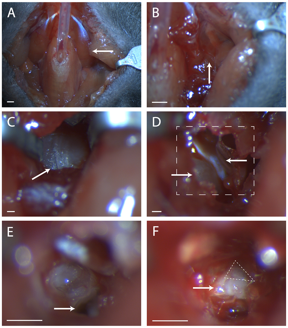
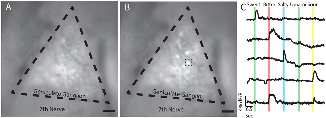
Within the last ten years, advances in genetically encoded calcium indicators (GECIs) have promoted a revolution in in vivo functional imaging. Using calcium as a proxy for neuronal activity, these techniques provide a way to monitor the responses of individual cells within large neuronal ensembles to a variety of stimuli in real time. We, and others, have applied these techniques to image the responses of individual geniculate ganglion neurons to taste stimuli applied to the tongues of live anesthetized mice. The geniculate ganglion is comprised of the cell bodies of gustatory neurons innervating the anterior tongue and palate as well as some somatosensory neurons innervating the pinna of the ear. Imaging the taste-evoked responses of individual geniculate ganglion neurons with GCaMP has provided important information about the tuning profiles of these neurons in wild-type mice as well as a way to detect peripheral taste miswiring phenotypes in genetically manipulated mice. Here we demonstrate the surgical procedure to expose the geniculate ganglion, GCaMP fluorescence image acquisition, initial steps for data analysis, and troubleshooting. This technique can be used with transgenically encoded GCaMP, or with AAV-mediated GCaMP expression, and can be modified to image particular genetic subsets of interest (i.e., Cre-mediated GCaMP expression). Overall, in vivo calcium imaging of geniculate ganglion neurons is a powerful technique for monitoring the activity of peripheral gustatory neurons and provides complementary information to more traditional whole-nerve chorda tympani recordings or taste behavior assays.


Conflict of interest statement

The authors have no conflict of interest to report.

Figures

Figure 1:
Figure 1:. Surgical exposure of the geniculate ganglion
(A) Image of the mouse neck cavity post tracheotomy. Arrow is pointing to the digastric muscle lying over the surgical area explored in the rest of the figure. (B) Image of region under the previously indicated digastric muscle. Arrow indicates the seam in musculature for blunt dissection. (C) Image of the Tympanic Bulla. Arrow indicates seam in the bone to break with a surgical probe. (D) Image of surgical area after opening the bulla. Lower left arrow indicates the cochlea, upper arrow points to the tensor tympani. Boxed line indicates area in (E) and (F). (E) Image of surgical area after cochlea has been broken and the contents removed. White arrow indicates where to place surgical probe referenced in protocol step 5.6. (F) An image of the exposed geniculate ganglion. Arrow indicates body of the seventh nerve, dashed triangle surrounds the geniculate ganglion. Panels A-B, Scale = 5 mm. Panels C-F, Scale = 1 mm.
Figure 2:
Figure 2:. Responses of geniculate ganglion neurons to tastants using in vivo GCaMP6s imaging.
(A) Epifluorescent image of the geniculate ganglion of a Snap25-GCaMP6s transgenic mouse during baseline as water is perfused over the tongue. Dashed lines indicate the approximate boundaries of the geniculate ganglion. The seventh cranial nerve is labelled as such. (B) Snapshot of the same ganglion in (A) as a sweet tastant (AceK 30 mM) is applied to the tongue of the mouse. Notice several individual neurons increase in fluorescence intensity. Dashed line box indicates sweet responding cell used in (C). end(C) Traces from five neurons indicating the amplitude of their GCaMP6s mediated fluorescence in response to a panel of tastants comprising sweet (30 mM acesulfame K), bitter (5 mM quinine); salty (60 mM NaCl); umami (50 mM monopotassium glutamate and 1mM inosine monophosphate); and sour (50 mM citric acid). Colored bars show the placement and duration (2 s) of the stimulus over the time course of the experiment. These representative data do not include a response to umami. Individual neurons commonly respond to both bitter and sour stimuli (bottom trace),,. Panels A-B, Scale = 5 mm. Panel C, horizontal scale bar indicates 6.5 seconds, vertical scale bar indicates threshold of 4% dF/F.

References

    1. Krimm RF Factors that regulate embryonic gustatory development. BMC Neurosci. 8 Suppl 3 S4, (2007). - PMC - PubMed
    1. Taruno A, Matsumoto I, Ma Z, Marambaud P & Foskett JK How do taste cells lacking synapses mediate neurotransmission? CALHM1, a voltage-gated ATP channel. Bioessays. 10.1002/bies.201300077 (35), 1111–1118, (2013). - DOI - PMC - PubMed
    1. Taruno A et al. Taste transduction and channel synapses in taste buds. Pflugers Arch. 10.1007/s00424-020-02464-4, (2020). - DOI - PMC - PubMed
    1. Kinnamon SC & Finger TE A taste for ATP: neurotransmission in taste buds. Front Cell Neurosci. 7 264, (2013). - PMC - PubMed
    1. Chandrashekar J, Hoon MA, Ryba NJ & Zuker CS The receptors and cells for mammalian taste. Nature. 444 (7117), 288–294, (2006). - PubMed

Publication types

LinkOut - more resources