. 2021 Feb 10:(168):10.3791/61848.
doi: 10.3791/61848.

A Murine Tail Lymphedema Model

Affiliations

A Murine Tail Lymphedema Model

Aladdin H Hassanein et al. J Vis Exp. .

Abstract

Lymphedema is extremity swelling caused by lymphatic dysfunction. The affected limb enlarges because of accumulation of fluid, adipose, and fibrosis. There is no cure for this disease. A mouse tail model that uses a focal full thickness skin excision near the base of the tail, resulting in tail swelling, has been used to study lymphedema. However, this model may result in vascular comprise and consequent tail necrosis and early tail swelling resolution, limiting its clinical translatability. The chronic murine tail lymphedema model induces sustained lymphedema over 15 weeks and a reliable perfusion to the tail. Enhancements of the traditional murine tail lymphedema model include 1) precise full thickness excision and lymphatic clipping using a surgical microscope, 2) confirmation of post-operative arterial and venous perfusion using high resolution laser speckle, and 3) functional assessment using indocyanine green near infrared laser lymphangiography. We also use tissue nanotransfection technology (TNT) for novel non-viral, transcutaneous, focal delivery of genetic cargo to the mouse tail vasculature.


Conflict of interest statement

Disclosures

The authors have no competing conflicts of interest.

Figures

Figure 1:
Figure 1:. Mouse tail model for sustained lymphedema.
(A) A 3 mm wide full-thickness skin excision is performed on a murine tail 20 mm from the base under the surgical micrscope. Care is taken to preserve the vasculature. (B) A schematic of the cross-section of the mouse tail. DV=dorsal vein, LV=lateral veins, A=ventral caudal artery, CV=caudal vertebra, T=tendon and muscle, yellow arrows shows the lymphatics. (C) Following administration of isosulfan blue into the tail tip to localize the lymphatics, the lymphatics (yellow arrow) exhibit blue color. The lymphatics are disrupted while preserving the adjacent lateral veins (white arrow).
Figure 2:
Figure 2:. Progressive swelling of the mouse tail lymphedema model.
(A) Following full-thickness skin excision and lymphatic transection, the mouse tail exhibits progressive swelling that is sustained over 15 weeks. The bracket denotes 20 mm from the base of the tail to the start of the surgical full thickness skin excision. (B-C) Quantification of the change in tail volume over 15 weeks represented as (B) bar graphs, each dot representing an animal, n=15, or as (C) line graph. Data represented as ± SEM.
Figure 3:
Figure 3:. High resolution laser speckle contrast imaging to confirm mouse tail perfusion in the lymphedema mouse tail model.
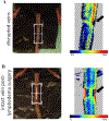
Laser speckle is used to assess mouse tail vasculature postoperatively to validate swelling of lymphatic etiology and minimize tail necrosis. (A) A mouse tail with injured lateral veins (black arrow) detected by laser speckle. (B) Intact lateral tail vein (black arrow) post-lymphedema surgery detected by laser speckle. (n=5) resolution 0.02 mm; Color coded bar indicates perfusion (blue: low, red: high) as measured in arbitrary relative units.
Figure 4:
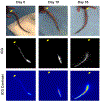
Figure 4:. Assessment of lymphatic function using near infrared laser lymphangioraphy in the mouse tail model.
Indocyanine green (ICG) injected into the tip of the mouse tail localizes to the lymphatics. Preoperatively, the lymphatics are intact along the mouse tail. Postoperatively, there is no ICG transit beyond the surgical site, confirming that swelling is caused by lymphatic dysfunction. Yellow arrow indicates the base of the tail.
Figure 5:
Figure 5:. Focal delivery of genetic cargo using tissue nanotransfection technology (TNT).
(A) Illustration of TNT delivery. (B) Plasmids are loaded into the TNT2.0 reservoir. The positive and negative electrical probes are attached and a brief, square wave pulse electric stimulation is delivered (10 × 10 ms pulses, 250 V, 10 mA), facilitating focal, non-viral, transcutaneous transfection. (C) Efficiency of genetic cargo delivery using TNT2.0 as observed through fluorescein amidite (FAM) labeled DNA delivery to the murine tail. Mouse tails were sectioned two days after TNT treatment and assessed through fluorescence microscopy. White dotted lines indicate the epithelium of the skin of murine tail. White arrows indicate the FAM labelled DNA.

References

    1. Kataru RP et al. Fibrosis and secondary lymphedema: chicken or egg? Translation Research. 209, 68–76 (2019). - PMC - PubMed
    1. Brayton KM et al. Lymphedema prevalence and treatment benefits in cancer: impact of a therapeutic intervention on health outcomes and costs. PLoS One. 9 (12), e114597 (2014). - PMC - PubMed
    1. Mendoza N, Li A, Gill A, Tyring S Filariasis: diagnosis and treatment. Dermatology and Therapy. 22 (6), 475–490 (2009). - PubMed
    1. Rockson SG, Rivera KK Estimating the population burden of lymphedema. Annals of the New York Academy of Sciences. 1131, 147–154 (2008). - PubMed
    1. Soran A et al. Breast cancer-related lymphedema--what are the significant predictors and how they affect the severity of lymphedema? Breast Journal. 12 (6), 536–543 (2006). - PubMed

Publication types