. 2021 Jun;53(3):351-363.
doi: 10.1007/s10863-021-09882-8. Epub 2021 Mar 1.

Supramolecular associations between atypical oxidative phosphorylation complexes of Euglena gracilis

Affiliations

Supramolecular associations between atypical oxidative phosphorylation complexes of Euglena gracilis

H V Miranda-Astudillo et al. J Bioenerg Biomembr. 2021 Jun.

Erratum in

Abstract

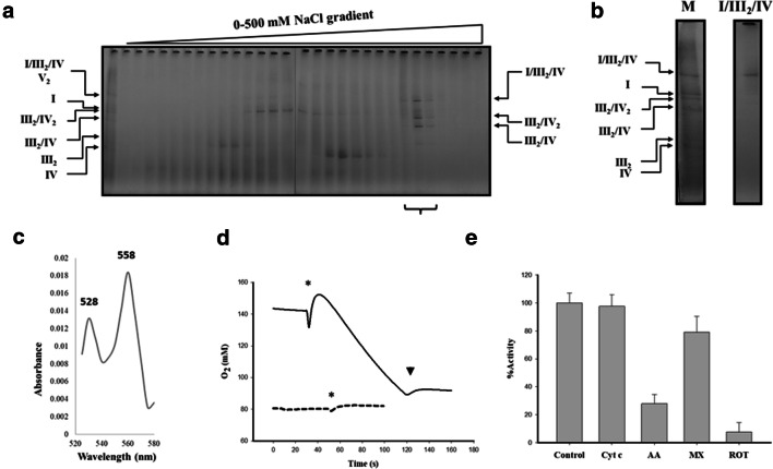
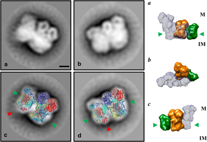
In vivo associations of respiratory complexes forming higher supramolecular structures are generally accepted nowadays. Supercomplexes (SC) built by complexes I, III and IV and the so-called respirasome (I/III2/IV) have been described in mitochondria from several model organisms (yeasts, mammals and green plants), but information is scarce in other lineages. Here we studied the supramolecular associations between the complexes I, III, IV and V from the secondary photosynthetic flagellate Euglena gracilis with an approach that involves the extraction with several mild detergents followed by native electrophoresis. Despite the presence of atypical subunit composition and additional structural domains described in Euglena complexes I, IV and V, canonical associations into III2/IV, III2/IV2 SCs and I/III2/IV respirasome were observed together with two oligomeric forms of the ATP synthase (V2 and V4). Among them, III2/IV SC could be observed by electron microscopy. The respirasome was further purified by two-step liquid chromatography and showed in-vitro oxygen consumption independent of the addition of external cytochrome c.

Keywords: Euglena gracilis; F1FO ATP synthase; Mitochondrial supercomplexes; Oligomeric complex V; Oxidative phosphorylation; Respirasome.


Conflict of interest statement

The authors declare no competing interests.

Figures

Fig. 1
Fig. 1
ATPase oligomers and respiratory supercomplexes in E. gracilis. Isolated mitochondria were solubilized with the indicated detergent: n-dodecyl-β-D-maltoside (DDM) at 4.0 g/ g protein or digitonin (Dig) at 8.0 g/g protein, after removing the insoluble material, each sample was resolved by BN-PAGE in a 3–10% polyacrylamide gradient gel. a Coomassie-stained gel showing the main bands with DDM- and digitonin-solubilization. b In-gel NADH-dehydrogenase activity; the BN-gel was incubated in the presence of NADH and Nitro blue tetrazolium chloride (NBT). c Detection of in-gel ATPase activity. The gel was incubated with ATP, MgSO4 and Pb(NO3)2. The determined molecular mass (kDa) of each isolated complex or supercomplexes is indicated. Nomenclature used: I, III2 and IV for the corresponding mitochondrial complexes, V2 and V4 for the dimeric and tetrameric ATP synthase respectively. Supercomplexes were: III2/IV, III2/IV2, the so-called “respirasome” association I/III2/IV and the putative Vx/I association, their stoichiometries are indicated as subindexes
Fig. 2
Fig. 2
Two-dimensional resolution of OXPHOS complexes and supercomplexes in E. gracilis mitochondria. a Upper panels: The OXPHOS complexes and supercomplexes from Euglena mitochondria were solubilized using digitonin (Dig) and separated by BN-PAGE followed by in-gel NADH-dehydrogenase and ATPase activities. Lower panel: NADH-dehydrogenase activity stain of two-dimensional gel from digitonin-extracted complexes and supercomplexes, the isolated spots show the complexes present in each supercomplex. III2/IV and III2/IV2 (red arrowheads), the respirasome (I/III2/IV, purple arrowhead), V4 oligomer (yellow arrowhead) and the Vx/I association (green arrowhead), isolated complexes were used as molecular mass markers (left lane). b The OXPHOS complexes and supercomplexes from Euglena mitochondria were solubilized using digitonin (upper lane) and separated by BN-PAGE. Lower panel: Two-dimensional SDS-tricine gel from digitonin-extracted complexes and supercomplexes. Representative subunits of each complex NDUFS3/NDUFA6/NDUFA9/NDUFA13/NDTB12/GapC3 for CI (purple arrows), QCR1/QCR2/QCR7 for CIII (red arrows), COX1/COX3/COXTB4/COXTB5/COX6b for CIV (green arrows) and ATPTB1/ATPTB2/Alpha-C/Beta for CV (blue arrows) are indicated. Molecular masses from the molecular mass marker are indicated on left side
Fig. 3
Fig. 3
Purification of the Euglenoid respirasome (I/III2/IV) by ion exchange/size exclusion chromatography and in-vitro oxygen consumption. Thirty milligrams of mitochondria were solubilized with glyco-diosgenin (GDN101) and loaded into an anion exchange column, then eluted with a 0–500 mM NaCl linear gradient. a BN- PAGE from the eluted 0.5 mL fractions, fractions containing III2/IV, III2/IV2 and I/III2/IV SCs (lower bracket) were concentrated and subjected to size exclusion column. b BN-PAGE from the pure respirasome (I/III2/IV) and the sample load onto the column (M), the identities of the SCs are indicated. c Differential redox absorption spectrum (520–580 nm) of purified respirasome was obtained as the sodium dithionite reduced spectrum minus the potassium ferricyanide oxidized spectrum. Alpha (558 nm) and beta (528 nm) peaks are signaled. d Oxygen consumption of the purified respirasome. The purified respirasome was incubated in the presence of NADH as electron donor (segmented line). External oxidized 2,3-Dimethoxy-5-methyl-p-benzoquinone was added (continuous line). The asterisk indicates the addition of the protein sample and the arrowhead indicates the addition of the complex I inhibitor rotenone. The lines were moved along the y axis for clarity. e Effect of external cytochrome c and inhibitory effect of antimycin A, myxothiazol and rotenone over the purified Euglena respirasome. The values represent the mean of three independent experiments and the bars represent the standard deviation
Fig. 4
Fig. 4
2D Projection maps of III2/IV supercomplex from E. gracilis obtained by single particle averaging. Left panels: A fraction containing both complexes was obtained after a two-step chromatographic procedure in presence of β-dodecyl-n-maltoside and analyzed by TEM (a and b). Overlap of the coupled model (see material and methods point 2.6 for further details) built with chicken dimeric complex III (pdb: 4U3F (Hao et al. 2015)) and the monomeric bovine complex together with the cytochrome c (pdb: 5IY5 (Shimada et al. 2017)) over the TEM images was performed (c and d). The membrane region is indicated by the green arrowheads, the cytochrome c binding site is indicated with red arrowheads. Right panels (a-c): model showing the position of the III2/IV supercomplex inside the mammalian respirasome map (EMD: 9539 (Wu et al. 2016)). Orange: dimeric complex III, green monomeric complex IV, light grey: monomeric complex one. The membrane region is signaled (green arrowheads), topology of this supercomplex in the mitochondrial inner membrane is signaled (M: matrix, IM: intermembrane space). The scale bar is 10 nm
Fig. 5
Fig. 5
Putative tetrameric structure of the E. gracilis ATP synthase. Putative tetrameric structure of the E. gracilis ATP synthase based in the electron cryotomography images from intact mitochondria membranes (Mühleip et al. 2017). The externally located peripheral stalks are shown in purple, the inter membrane space density below the c-ring is shown in orange, the F1/central rotor sector is shown in cyan, the membrane region is signalled (green arrowheads) and the position of the euglonoid specific subunit p18 is indicated (red arrowheads), the structure of the Euglena ATP synthase dimer (PDB: 6TDU (Mühleip et al. 2019)) is fitted inside the electron density for one dimer. The scale bar is 10 nm
Fig. 6
Fig. 6
Assembly pathway of mitochondrial respirasome in E. gracilis. Schematic representation of the associations of complexes into supercomplexes. The species observed by BN-PAGE (I, III2, IV, V2, III2/IV, III2/IV2, I/III2/IV, I/III2/IV2) and TEM analysis (I, III2, IV, and III2/IV) from Euglena mitochondria ((Yadav et al. ; Miranda-Astudillo et al. 2018b), this work) are shown in green and dark blue boxes, respectively

Similar articles

Cited by

References

    1. Acin-Perez R, Enriquez JA. The function of the respiratory supercomplexes: the plasticity model. Biochim Biophys Acta Bioenerg. 2014;1837:444–450. doi: 10.1016/j.bbabio.2013.12.009. - DOI - PubMed
    1. Allegretti M, Klusch N, Mills DJ, Vonck J, Kühlbrandt W, Davies KM. Horizontal membrane-intrinsic α-helices in the stator a-subunit of an F-type ATP synthase. Nature. 2015;521:237–240. doi: 10.1038/nature14185. - DOI - PubMed
    1. Allen RD, Schroeder CC, Fok AK. An investigation of mitochondrial inner membranes by rapid-freeze deep-etch techniques. J Cell Biol. 1989;108:2233–2240. doi: 10.1083/jcb.108.6.2233. - DOI - PMC - PubMed
    1. Atteia A, Van Lis R, Mendoza-Hernández G, et al. Bifunctional aldehyde/alcohol dehydrogenase (ADHE) in chlorophyte algal mitochondria. Plant Mol Biol. 2003;53:175–188. doi: 10.1023/B:PLAN.0000009274.19340.36. - DOI - PubMed
    1. Balabaskaran Nina P, Dudkina NV, Kane LA, van Eyk JE, Boekema EJ, Mather MW, Vaidya AB. Highly divergent mitochondrial ATP synthase complexes in Tetrahymena thermophila. PLoS Biol. 2010;8:3–6. doi: 10.1371/journal.pbio.1000418. - DOI - PMC - PubMed

Publication types

LinkOut - more resources