Review
. 2021 Apr 15;40(8):e105789.
doi: 10.15252/embj.2020105789. Epub 2021 Mar 1.

Peering into tunneling nanotubes-The path forward

Affiliations
Review

Peering into tunneling nanotubes-The path forward

Diégo Cordero Cervantes et al. EMBO J. .

Abstract

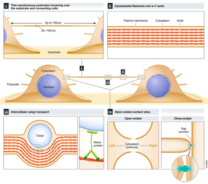
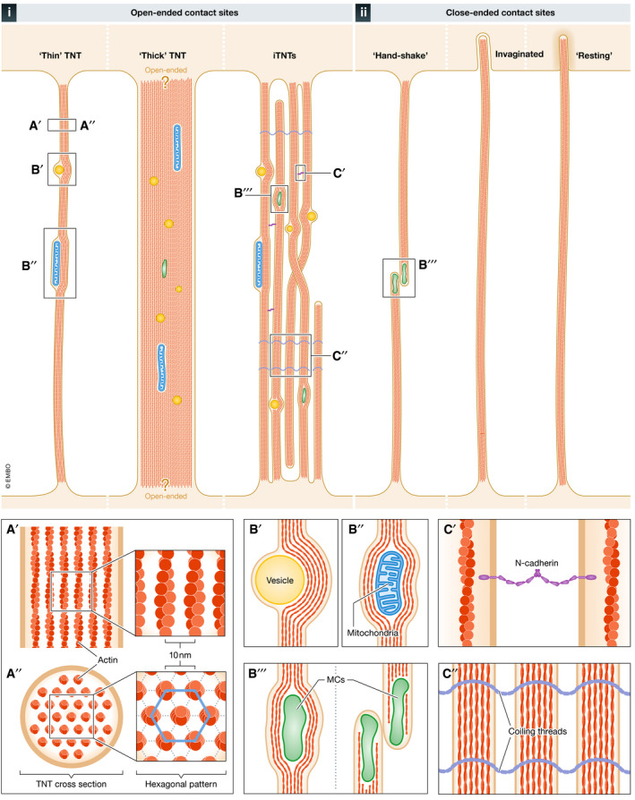
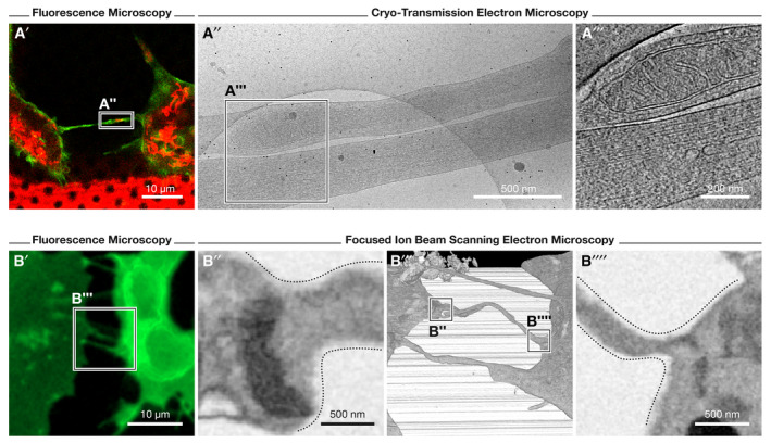
The identification of Tunneling Nanotubes (TNTs) and TNT-like structures signified a critical turning point in the field of cell-cell communication. With hypothesized roles in development and disease progression, TNTs' ability to transport biological cargo between distant cells has elevated these structures to a unique and privileged position among other mechanisms of intercellular communication. However, the field faces numerous challenges-some of the most pressing issues being the demonstration of TNTs in vivo and understanding how they form and function. Another stumbling block is represented by the vast disparity in structures classified as TNTs. In order to address this ambiguity, we propose a clear nomenclature and provide a comprehensive overview of the existing knowledge concerning TNTs. We also discuss their structure, formation-related pathways, biological function, as well as their proposed role in disease. Furthermore, we pinpoint gaps and dichotomies found across the field and highlight unexplored research avenues. Lastly, we review the methods employed to date and suggest the application of new technologies to better understand these elusive biological structures.

Keywords: actin protrusions; cell communication; cell signaling; tunneling nanotubes.


Conflict of interest statement

The authors declare that they have no conflict of interest.

Figures

Figure 1
Figure 1. Distinctive properties of TNTs
TNTs are (I) thin (20–700 nm) and long reaching (up to 100 µm in thin TNTs; < 700 nm in diameter) membranous protrusions that hover over the substrate and connect distant cells. Thick TNTs (diameter of > 700 nm) (not shown) can extend up to 250 µm in length. TNTs are (II) rich in F‐actin cytoskeletal filaments, which enable (III) internal intercellular cargo transport using motor proteins. Bona fide TNTs feature (IV) open‐ended contact sites at each end allowing for cytoplasmic continuity. A subset of TNTs contains one close‐ended contact site that facilitates electrical coupling between cells through gap junctions (GJ) at the tip of the protrusion and recipient cell.
Figure 2
Figure 2. Structural features of open‐ and close‐ended TNTs
Schematic representation of the 6 main types of open‐ and close‐ended TNTs observed in vitro using electron microscopy (EM) approaches. i) Open‐ended: “Thin” TNTs, observed in photoreceptor and stem cells via SEM and in neuronal cells via cryo‐TEM/ET; “Thick” (> 700 nm) TNTs, observed in epithelial and neuronal cells with contact sites displaying questions marks to illustrate they were not demonstrated to be open; and a bundle of individual TNTs (iTNTs), a bundle of thin parallel nanotubes that get twisted together or go over and under each other, together forming one group of intertwined tubes that cannot be resolved using fluorescence microscopy; observed in neuronal and stromal cells using cryo‐TEM/ET. ii) Close‐ended (presumably facilitating cell–cell communication through gap junctions, which are not visible by EM approaches): “hand‐shake” TNTs observed in lymphocytes via ssTEM; invaginated TNTs, observed in lymphocytes via ssTEM and in neuronal cells via FIB‐SEM; and “resting” TNTs, observed in neuronal cells via FIB‐SEM. A) Top‐ (A′) and cross‐sectional (A′′) views of a TNT showing its internal actin arrangement: hexagonal actin arrays with a 10 nm distance between the center of adjacent actin filaments. B) Three types of cargoes observed within TNTs by EM: vesicles (B′), mitochondria (B′′), and membranous components (MCs) (B′′′), which could represent unidentified organelles. (C) Types of iTNT‐iTNT interlinkage processes identified by immunogold labeling and cryo‐TEM/ET: N‐Cadherin adhesion molecules between iTNTs (C′) and coiling threads (C′′), whose protein and/or lipid composition are currently known.
Figure 3
Figure 3. Observation of TNTs by various electron microscopy‐based approaches
(A) Cryo‐correlative electron microscopy (cryo‐CLEM) and focused ion beam scanning EM (FIB‐SEM) approaches employed to study TNTs of SH‐SY5Y cells; images modified from (Sartori‐Rupp et al, 2019). (A′) Identification of TNT‐connected cells is first performed through fluorescence microscopy of WGA (green)‐ and mitochondria (red)‐labeled cells (seeded on EM finder grids). After vitrification of cells, the recorded position of TNTs acquired by fluorescence microscopy is imaged by cryo‐transmission EM (cryo‐TEM) and electron tomography (cryo‐ET) at medium‐ and high‐magnification (A″ and A‴, respectively). A mitochondrion can be observed within an iTNT in A‴. (B) Correlative, FIB‐SEM method employed to visualize TNTs using fluorescence microscopy (cells labeled with WGA, green) and FIB‐SEM to assess open‐ended contact sites (B″ and B′′′′, respectively) through high‐resolution 3D, volume imaging (B‴).

Similar articles

Cited by

References

    1. Abounit S, Bousset L, Loria F, Zhu S, de Chaumont F, Pieri L, Olivo‐Marin J‐C, Melki R, Zurzolo C (2016a) Tunneling nanotubes spread fibrillar α‐synuclein by intercellular trafficking of lysosomes. EMBO J 35: 2120–2138 - PMC - PubMed
    1. Abounit S, Delage E, Zurzolo C (2015) Identification and characterization of tunneling nanotubes for intercellular trafficking. Curr Protoc Cell Biol 67: 12.10.1‐21 - PubMed
    1. Abounit S, Wu JW, Duff K, Victoria GS, Zurzolo C (2016b) Tunneling nanotubes: a possible highway in the spreading of tau and other prion‐like proteins in neurodegenerative diseases. Prion 10: 344–351 - PMC - PubMed
    1. Abounit S, Zurzolo C (2012) Wiring through tunneling nanotubes–from electrical signals to organelle transfer. J Cell Sci 125: 1089–1098 - PubMed
    1. Ady J, Thayanithy V, Mojica K, Wong P, Carson J, Rao P, Fong Y, Lou E (2016) Tunneling nanotubes: an alternate route for propagation of the bystander effect following oncolytic viral infection. Mol Ther Oncolytics 3: 16029 - PMC - PubMed

Publication types