. 2021 Apr 26:39:719-757.
doi: 10.1146/annurev-immunol-093019-125918. Epub 2021 Mar 1.

Eosinophil Knockout Humans: Uncovering the Role of Eosinophils Through Eosinophil-Directed Biological Therapies

Affiliations

Eosinophil Knockout Humans: Uncovering the Role of Eosinophils Through Eosinophil-Directed Biological Therapies

Elizabeth A Jacobsen et al. Annu Rev Immunol. .

Abstract

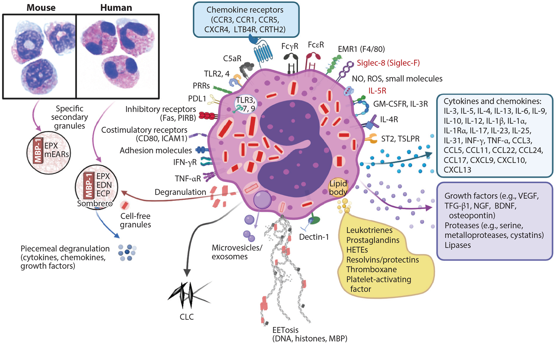
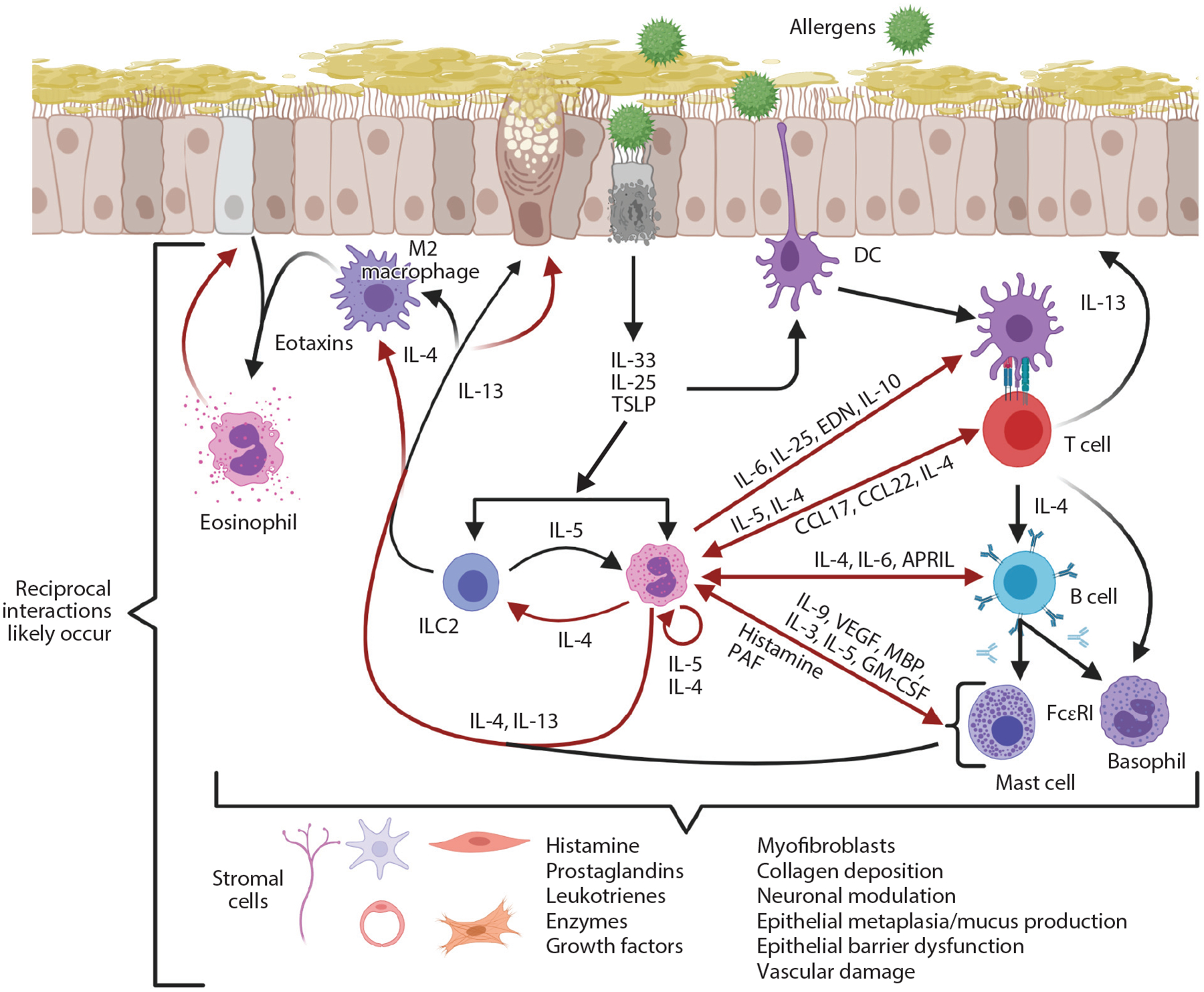
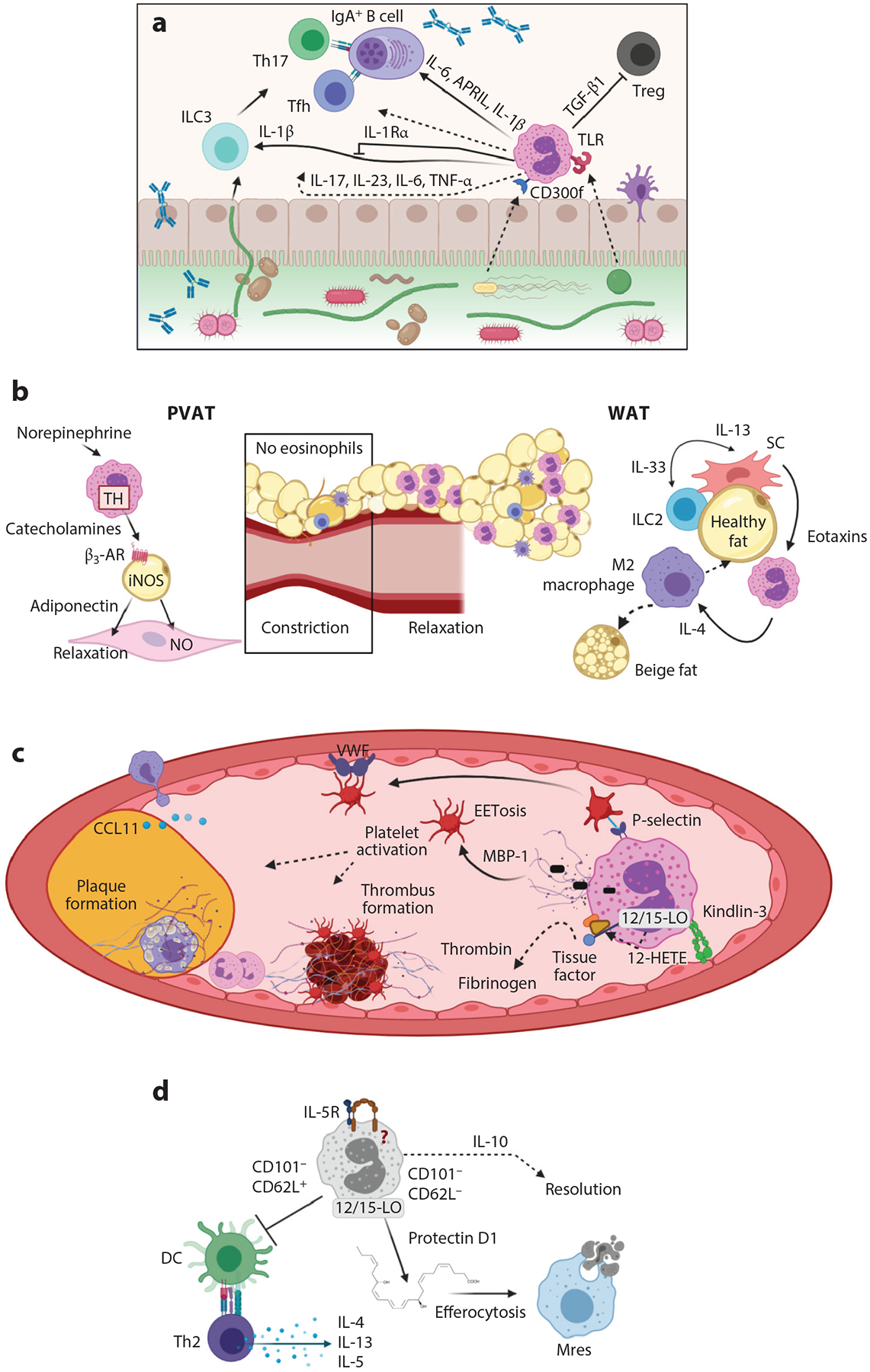
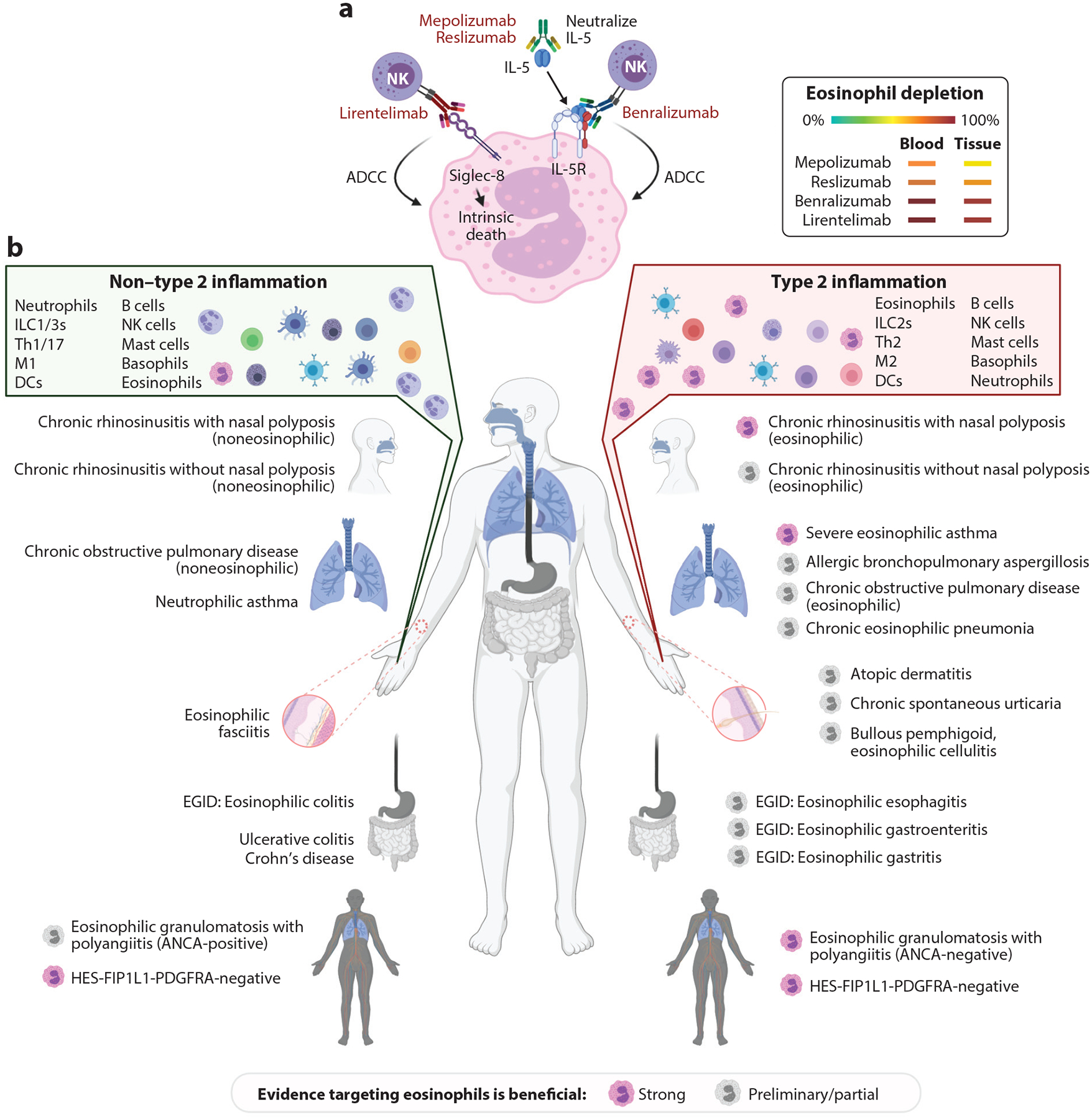
The enigmatic eosinophil has emerged as an exciting component of the immune system, involved in a plethora of homeostatic and inflammatory responses. Substantial progress has been achieved through experimental systems manipulating eosinophils in vivo, initially in mice and more recently in humans. Researchers using eosinophil knockout mice have identified a contributory role for eosinophils in basal and inflammatory processes and protective immunity. Primarily fueled by the purported proinflammatory role of eosinophils in eosinophil-associated diseases, a series of anti-eosinophil therapeutics have emerged as a new class of drugs. These agents, which dramatically deplete eosinophils, provide a valuable opportunity to characterize the consequences of eosinophil knockout humans. Herein, we comparatively describe mouse and human eosinophil knockouts. We put forth the view that human eosinophils negatively contribute to a variety of diseases and, unlike mouse eosinophils, do not yet have an identified role in physiological health; thus, clarifying all roles of eosinophils remains an ongoing pursuit.

Keywords: IL-5; biologic agents; eosinophil; eosinophil knockout; eosinophil-associated diseases; eosinophil-deficient mice.


Figures

Figure 1
Figure 1
Eosinophil biology. Mouse and human peripheral blood eosinophils show similarities in eosin staining (pink) and differences in nuclei (blue), with mouse nuclei in a circular or figure-eight shape and human nuclei being bilobed, as seen by standard light microscopy. Secondary granules in both contain MBP-1, which creates a crystalline core visible by electron microscopy, and the matrix contains EPX. Human eosinophils have ECP and EDN, whereas mouse eosinophils have only divergent mEARs. Eosinophils undergo several forms of degranulation, including classical exocytosis with release of entire granule contents or piecemeal degranulation, which may occur through formation of sombrero vesicles or exosomes, resulting in the differential release of cytokines, chemokines, or growth factors. Finally, eosinophils may undergo cytolysis, which includes release of cell-free granules and may also involve EETosis whereby DNA, histones, and granule proteins are released, forming extracellular nets. Mediators from lipid bodies are shown, as well as the many cytokines, chemokines, and growth factors. Siglec-8, IL-5R (receptors in red), and IL-5 are targets for monoclonal antibodies to deplete eosinophils. This figure shows some key representative eosinophil molecules and not a complete list. Abbreviations: BDNF, brain-derived neurotrophic factor; CCL, C-C motif chemokine ligand; CCR, C-C motif chemokine receptor; CLC, Charcot–Leyden crystal protein; CRTH2, chemoattractant receptor-homologous molecule expressed on Th2 cells; CXCL, C-X-C motif chemokine ligand; CXCR, C-X-C motif chemokine receptor; ECP, eosinophil cationic protein; EDN, eosinophil-derived neurotoxin; EPX, eosinophil peroxidase; FcεR, Fc-epsilon receptor; FcγR, Fc-gamma receptor; GM-CSFR, granulocyte-macrophage colony-stimulating factor receptor; HETE, hydroxyeicosatetraenoic acid; ICAM1, intercellular adhesion molecule 1; LTB4R, leukotriene B4 receptor; MBP-1, major basic protein 1; mEAR, mouse eosinophil-associated ribonuclease; NGF, nerve growth factor; NO, nitric oxide; PIRB, paired immunoglobulin-like receptor B; PDLI, programmed death ligand 1; PRR, pathogen recognition receptor; ROS, reactive oxygen species; Siglec, sialic acid–binding immunoglobulin-like lectin; ST2, suppressor of tumor 2 [also known as IL-1RL1 (interleukin-1 receptor-like 1)]; TGF-β1, transforming growth factor beta 1; TLR, Toll-like receptor; TSLPR, thymic stromal lymphopoietin receptor; VEGF, vascular endothelial growth factor. Figure adapted from images created with BioRender.com.
Figure 2
Figure 2
Type 2 immune responses in allergy. Allergens induce epithelial responses that lead to production of IL-33, IL-25, and TSLP, which are immediate cytokine signals released upon epithelial activation or damage. A wide variety of cells, including ILC2s, eosinophils, DCs, eosinophils, M2 macrophages, mast cells, basophils, and Th2 cells, respond to these cytokines. In principle, this can result in reciprocal interactions between these cells to release additional mediators. IL-13 enhances expression of eotaxins to promote eosinophil recruitment and leads to goblet cell metaplasia/mucus production. Red arrows represent products derived from eosinophils. Abbreviations: APRIL, a proliferation-inducing ligand; CCL, C-C motif chemokine ligand; DC, dendritic cell; EDN, eosinophil-derived neurotoxin; FcεRI, Fc-epsilon receptor I; GM-CSF, granulocyte-macrophage colony-stimulating factor; ILC2, group 2 innate lymphoid cell; MBP, major basic protein; PAF, platelet-activating factor; TSLP, thymic stromal lymphopoietin; VEGF, vascular endothelial growth factor. Figure adapted from images created with BioRender.com.
Figure 3
Figure 3
Eosinophil activities in homeostasis in mice. (a) Eosinophil activities with the microbiome, B cells, and Th17 responses in the gastrointestinal tract. Eosinophils express receptors such as TLRs and CD300f that recognize bacterial components to signal for immune responses. Eosinophils release the cytokines IL-6, APRIL, and IL-1β to influence B cell IgA production. This may happen through Tfh cells or Th17 cells, which express IL-17. Eosinophil release of IL-1β increases the activity of ILC3s; TGF-β1, which inhibits Tregs; and IL-17, IL-23, IL-6, and TNF-α, which may directly participate in the above responses. (b) Eosinophil functions in metabolic and vascular health. Eosinophils induce relaxation of blood vessels in PVAT through use of TH to release catecholamines that bind β3-AR, which stimulates iNOS in adipocytes to make NO that results in smooth muscle cell relaxation. In WAT, ILC2s produce IL-13 in response to the IL-33 produced by SCs. The IL-13 induces production of eotaxins that recruit eosinophils into fat. These eosinophils release IL-4 to polarize macrophages into alternatively activated M2 macrophages that contribute to transformation of white to beige fat cells, which are thermogenic cells that aid in metabolic health. (c) Eosinophils have multiple functions in vascular health. Eosinophils release a 12-hydroxyeicosatetraenoic acid (HETE) product that is generated from 12/15-LO and ultimately aids in tissue factor activation and development of thrombi through thrombin and fibrinogen activation. Eosinophils activate platelets through P-selectin promoting platelet binding to VWF in endothelial cells, which also aids in clot and plaque formation. Through integrin kindlin-3 binding of the endothelium, eosinophils are activated to undergo EETosis, releasing MBP-1 that further activates platelets and promotes atherosclerotic inflammation. (d) Eosinophils as lung-resident cells. Resident lung eosinophils are different from inflammatory recruited eosinophils due to their differential expression of CD101. CD101 cells that are CD62L+ suppress DC activation, which in turn inhibits Th2 cell release of cytokines IL-4, IL-13, and IL-5. CD101 eosinophils that are CD62L are reported as important in resolution of inflammation in that they produce protectin D1 through 12/15-LO activity. Protectin D1 promotes efferocytosis, whereby Mres phagocytose dead neutrophils. Eosinophil-derived IL-10 is also important in resolution functions of eosinophils in the lung. Dashed arrows indicate either less-defined or indirect pathways of eosinophils in the roles presented. Abbreviations: APRIL, a proliferation-inducing ligand; β3-AR, beta 3 adrenergic receptor; CCL, C-C motif chemokine ligand; DC, dendritic cell; ILC2, group 2 innate lymphoid cell; iNOS, inducible nitric oxide synthase; MBP-1, major basic protein 1; Mres, resolution macrophage; NO, nitric oxide; PVAT, perivascular adipose tissue; SC, stromal vascular cell; Tfh, T follicular helper; TGF-β1, transforming growth factor beta 1; Th, T helper; TH, tyrosine hydroxylase; TLR, Toll-like receptor; TNF-α, tumor necrosis factor alpha; Treg, regulatory T cell; 12/15-LO, 12/15 lipoxygenase; VWF, Von Willebrand factor; WAT, white adipose tissue. Figure adapted from images created with BioRender.com.
Figure 4
Figure 4
Monoclonal antibodies that target eosinophils and eosinophil-associated diseases. (a) Biologics that target eosinophils for depletion. Mepolizumab and reslizumab are monoclonal antibodies that neutralize IL-5, leading to inhibition of eosinophilopoiesis and impaired eosinophil activation and survival. Benralizumab is a monoclonal antibody that targets the IL-5Rα component. Benralizumab induces Fc-mediated killing by NK cells that mediate ADCC. Lirentelimab binds Siglec-8. This antibody induces intrinsic cell death and mediates ADCC. Eosinophil depletion that occurs in the blood or tissues is shown, with green representing no depletion and red representing complete depletion. (b) Eosinophil-associated diseases and their diversity. Eosinophils classically participate in type 2 diseases and also in non–type 2 diseases. Pathological responses may be eosinophil dependent or independent; in the latter case, other effector cells direct the disease phenotype. Pink eosinophils denote that substantial clinical studies demonstrate clear evidence of some benefit by antibodies targeting eosinophils. Gray eosinophils denote that preliminary clinical studies or a portion of the clinical study outcomes indicate that targeting eosinophils leads to improvement. Please note that studies indicating lack of response to anti-eosinophil therapy are not reflected in this figure. Please also note that HES is on both sides of the figure, as both type 2 and non–type 2 mechanisms are involved and are amenable to anti-eosinophil therapy. Abbreviations: ADCC, antibody-dependent cellular cytotoxicity; ANCA, antineutrophil cytoplasmic antibody; DC, dendritic cell; EGID, eosinophilic gastrointestinal disease; HES, hypereosinophilic syndrome; ILC2, group 2 innate lymphoid cell; M1 macrophage, classically activated macrophage; M2 macrophage, alternatively activated macrophage; NK, natural killer; Siglec-8, sialic acid–binding immunoglobulin-like lectin 8; Th1, T helper 1. Figure adapted from images created with BioRender.com.

References

    1. Acharya KR, Ackerman SJ. 2014. Eosinophil granule proteins: form and function. J. Biol. Chem 289:17406–15 - PMC - PubMed
    1. Klion AD, Ackerman SJ, Bochner BS. 2020. Contributions of eosinophils to human health and disease. Annu. Rev. Pathol. Mech. Dis 15:179–209 - PMC - PubMed
    1. Lee JJ, Jacobsen EA, McGarry MP, Schleimer RP, Lee NA. 2010. Eosinophils in health and disease: the LIAR hypothesis. Clin. Exp. Allergy 40:563–75 - PMC - PubMed
    1. Weller PF, Spencer LA. 2017. Functions of tissue-resident eosinophils. Nat. Rev. Immunol 17:746–60 - PMC - PubMed
    1. Rothenberg ME, Hogan SP. 2006. The eosinophil. Annu. Rev. Immunol 24:147–74 - PubMed

Publication types

Substances

LinkOut - more resources