. 2021 Mar 1;17(3):e1009368.
doi: 10.1371/journal.ppat.1009368. eCollection 2021 Mar.

The allelic rice immune receptor Pikh confers extended resistance to strains of the blast fungus through a single polymorphism in the effector binding interface

Affiliations

The allelic rice immune receptor Pikh confers extended resistance to strains of the blast fungus through a single polymorphism in the effector binding interface

Juan Carlos De la Concepcion et al. PLoS Pathog. .

Abstract

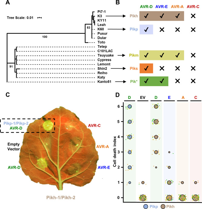
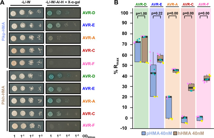
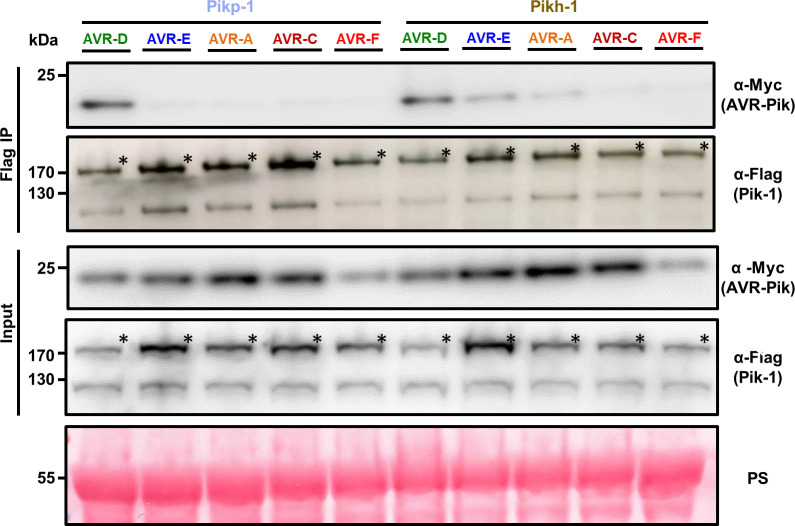
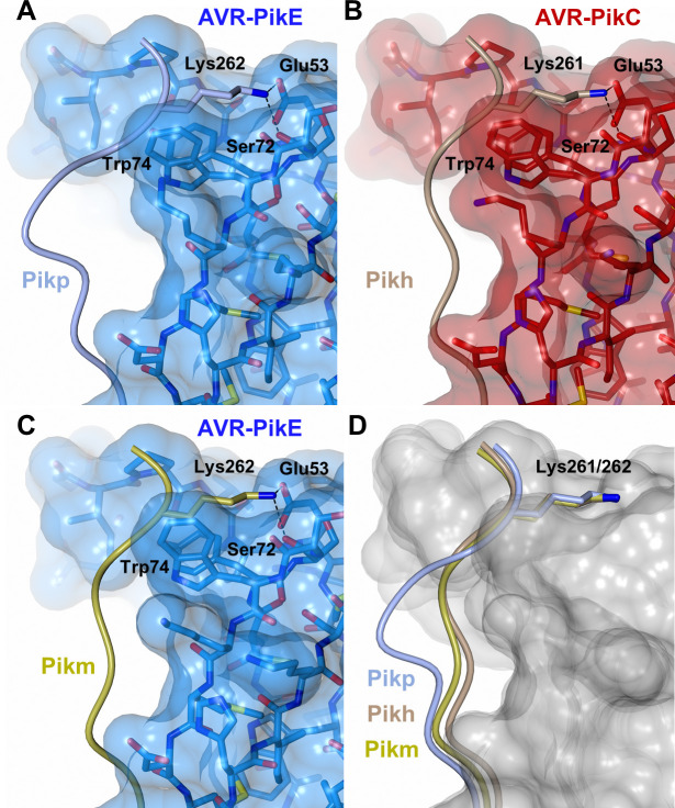
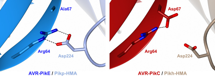
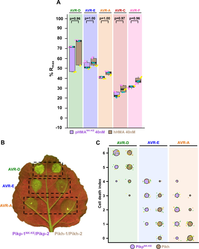
Arms race co-evolution drives rapid adaptive changes in pathogens and in the immune systems of their hosts. Plant intracellular NLR immune receptors detect effectors delivered by pathogens to promote susceptibility, activating an immune response that halts colonization. As a consequence, pathogen effectors evolve to escape immune recognition and are highly variable. In turn, NLR receptors are one of the most diverse protein families in plants, and this variability underpins differential recognition of effector variants. The molecular mechanisms underlying natural variation in effector recognition by NLRs are starting to be elucidated. The rice NLR pair Pik-1/Pik-2 recognizes AVR-Pik effectors from the blast fungus Magnaporthe oryzae, triggering immune responses that limit rice blast infection. Allelic variation in a heavy metal associated (HMA) domain integrated in the receptor Pik-1 confers differential binding to AVR-Pik variants, determining resistance specificity. Previous mechanistic studies uncovered how a Pik allele, Pikm, has extended recognition to effector variants through a specialized HMA/AVR-Pik binding interface. Here, we reveal the mechanistic basis of extended recognition specificity conferred by another Pik allele, Pikh. A single residue in Pikh-HMA increases binding to AVR-Pik variants, leading to an extended effector response in planta. The crystal structure of Pikh-HMA in complex with an AVR-Pik variant confirmed that Pikh and Pikm use a similar molecular mechanism to extend their pathogen recognition profile. This study shows how different NLR receptor alleles functionally converge to extend recognition specificity to pathogen effectors.


Conflict of interest statement

The authors have declared that no competing interests exist.

Figures

Fig 1
Fig 1. Pikh responses to AVR-Pik variants in N. benthamiana.
(A) Maximum Likelihood Phylogenetic tree of coding sequences of rice Pik-1 HMA domains. The tree was prepared using Interactive Tree Of Life (iTOL) v4 [76]. Cultivar names are placed next to their corresponding branch. Significant bootstrap values (>75) are indicated. (B) Schematic representations of immune response profiles of rice cultivars K3 (Pikh), K60 (Pikp), Tsuyuake (Pikm), Shin2 (Piks) and Kanto51 (Pik*) as reported in [43]. (C) Representative leaf image showing Pikh-mediated response to AVR-Pik variants as autofluorescence under UV light. Pikp-mediated response with AVR-PikD is included as a positive control (surrounded by a dashed square), and a spot inoculated with empty vector instead of AVR-Pik effector is included a negative control. (D) In planta response scoring represented as dot plots. Fluorescence intensity is scored as previously described in [20,21]. Pikh-mediated responses are coloured in brown while the Pikp control is coloured in blue. For each sample, all the data points are represented as dots with a distinct colour for each of the three biological replicates; these dots are jittered about the cell death score for visualisation purposes. The size of the centre dot at each value is directly proportional to the number of replicates in the sample with that score. The total number of repeats was 60.
Fig 2
Fig 2. Pikh-HMA has increased binding to AVR-Pik effector alleles in vivo and in vitro.
(A) Yeast two-hybrid assay of Pikp-HMA and Pikh-HMA with AVR-Pik variants. For each combination of HMA/AVR-Pik, 5μl of yeast were spotted and incubated for ~60 h in double dropout plate for yeast growth control (left) and quadruple dropout media supplemented with X-α-gal (right). Growth, and development of blue colouration, in the selection plate are both indicative of protein:protein interaction. HMA domains were fused to the GAL4 DNA binding domain, and AVR-Pik alleles to the GAL4 activator domain. Each experiment was repeated a minimum of three times, with similar results. (B) Measurement of Pikp-HMA and Pikh-HMA binding to AVR-Pik effector variants by surface plasmon resonance. The binding is expressed as %Rmax at an HMA concentration of 40 nM. Pikp-HMA and Pikh-HMA are represented by blue and brown boxes, respectively. For each experiment, three biological replicates with three internal repeats each were performed, and the data are presented as box plots. The centre line represents the median, the box limits are the upper and lower quartiles, the whiskers extend to the largest value within Q1-1.5× the interquartile range (IQR) and the smallest value within Q3 + 1.5× IQR. All the data points are represented as dots with distinct colours for each biological replicate. “p” is the p-value obtained from statistical analysis and Tukey’s HSD. For results of experiments with 4 and 100 nM HMA protein concentrations, see S5 Fig.
Fig 3
Fig 3. The Asn261Lys polymorphism in Pikh-1 extends association to AVR-PikE and AVR-PikA in planta.
Co-immunoprecipitation of full length Pikp-1 and Pikh-1 with AVR-Pik variants. N-terminally 4xMyc tagged AVR-Pik effectors were transiently co-expressed with Pikp-1:6xHis3xFLAG (left) or Pikh-1:6xHis3xFLAG (right) in N. benthamiana. Immunoprecipitates (IPs) obtained with M2 anti-FLAG resin and total protein extracts were probed with appropriate antisera. Each experiment was repeated at least three times, with similar results. The asterisks mark the Pik-1 band. Total protein extracts were coloured with Ponceau Staining (PS).
Fig 4
Fig 4. The Pikh-HMA domain adopts a favourable conformation at the effector binding interface.
Schematic representation of the conformations adopted by Pikp-HMA (PDB: 6G11), Pikm-HMA (PDB: 6FUB) and Pikh-HMA at interface 3 in complex with AVR-PikE or AVR-PikC. In each panel, the effector is represented in cylinders, with the molecular surface also shown and coloured as labelled. Pik-HMA residues are coloured as labelled and shown as the Cα-worm. For clarity, only the Lys-261/262 side chain is shown. Hydrogen bonds between Lys-261/262 and the effector are represented by dashed black lines. (A) Pikp-HMA bound to AVR-PikE, (B) Pikh-HMA bound to AVR-PikC, (C) Pikm-HMA bound to AVR-PikE. (D) Superposition of HMA chains bound to AVR-Pik. For clarity, only the Lys-261/262 side chain is shown. Two different effector alleles, AVR-PikE and AVR-PikC, are represented by their molecular surface coloured in grey.
Fig 5
Fig 5. The polymorphic Asp67 in AVR-PikC disrupts hydrogen bonding between the effector and the HMA domain.
Close-up views of the position and interactions of Asp224 of the HMA domain in complex with either AVR-PikE (Pikp-HMA (PDB: 6G11), left) or AVR-PikC (Pikh-HMA, right). HMA domains are presented as cartoon ribbons with the side chain of Asp224 displayed as a cylinder; Pikh-HMA and Pikp-HMA are coloured in brown and ice blue, respectively. The effectors are shown in cartoon ribbon representation, with the side chains of Arg64 and Asp67/Ala67 as cylinders. AVR-PikC and AVR-PikE are coloured in crimson and bright blue, respectively. Hydrogen bonds/salt bridges are shown as black dashed lines. For clarity, the N-terminal residues 32 to 52 of the AVR-Pik effector are hidden from the foreground in both structures.
Fig 6
Fig 6. Pikh and PikpNK-KE display similar binding affinity for AVR-Pik effectors but Pikh shows a reduced response in planta.
(A) Pikp-HMANK-KE and Pikh-HMA binding to AVR-Pik effector variants determined by surface plasmon resonance. The binding is expressed as %Rmax at an HMA concentration of 40 nM. Pikp-HMANK-KE and Pikh-HMA are represented by purple and brown boxes, respectively. For each experiment, three biological replicates with three internal repeats each were performed and the data are presented as box plots. The centre line represents the median, the box limits are the upper and lower quartiles, the whiskers extend to the largest value within Q1–1.5× the interquartile range (IQR) and the smallest value within Q3 + 1.5× IQR. All the data points are represented as dots with distinct colours for each biological replicate. “p” is the p-value obtained from statistical analysis and Tukey’s HSD. Data for Pikh-HMA is also presented in Fig 2B and were collected side-by-side at the same time. For results of experiments with 4 and 100 nM HMA protein concentration see S7 Fig. (B) Representative leaf image showing a side-by-side responses for Pikp-NK-KE and Pikh with AVR-PikD, AVR-PikE and AVR-PikA. (C) In planta response scoring represented as dot plots. Fluorescence intensity is scored as previously described in [20,21]. Responses mediated by PikpNK-KE and Pikh are coloured in purple and brown, respectively. For each sample, all the data points are represented as dots with a distinct colour for each of the three biological replicates; these dots are jittered about the cell death score for visualisation purposes. The size of the centre dot at each value is directly proportional to the number of replicates in the sample with that score. The total number of repeats was 57. For statistical analysis of the differences between the responses mediated by PikpNK-KE and Pikh see S8 Fig.

Similar articles

Cited by

References

    1. Savary S, Willocquet L, Pethybridge SJ, Esker P, McRoberts N, Nelson A. The global burden of pathogens and pests on major food crops. Nat Ecol Evol. 2019;3(3):430–9. Epub 2019/02/06. 10.1038/s41559-018-0793-y . - DOI - PubMed
    1. Win J, Chaparro-Garcia A, Belhaj K, Saunders DG, Yoshida K, Dong S, et al.. Effector biology of plant-associated organisms: concepts and perspectives. Cold Spring Harb Symp Quant Biol. 2012;77:235–47. Epub 2012/12/12. 10.1101/sqb.2012.77.015933 . - DOI - PubMed
    1. Jones JDG, Vance RE, Dangl JL. Intracellular innate immune surveillance devices in plants and animals. Science. 2016;354(6316):aaf6395. 10.1126/science.aaf6395 - DOI - PubMed
    1. Bentham AR, De la Concepcion JC, Mukhi N, Zdrzałek R, Draeger M, Gorenkin D, et al.. A molecular roadmap to the plant immune system. J Biol Chem. 2020. Epub 2020/08/21. 10.1074/jbc.REV120.010852 . - DOI - PMC - PubMed
    1. Jones JDG, Dangl JL. The plant immune system. Nature 2006;444(7117):323–9. Epub 2006/11/17. 10.1038/nature05286 . - DOI - PubMed

Publication types

MeSH terms

Supplementary concepts