. 2021 Jul 15:414:125514.
doi: 10.1016/j.jhazmat.2021.125514. Epub 2021 Feb 24.

Low doses of zeolitic imidazolate framework-8 nanoparticles alter the actin organization and contractility of vascular smooth muscle cells

Affiliations

Low doses of zeolitic imidazolate framework-8 nanoparticles alter the actin organization and contractility of vascular smooth muscle cells

Divya Kota et al. J Hazard Mater. .

Abstract

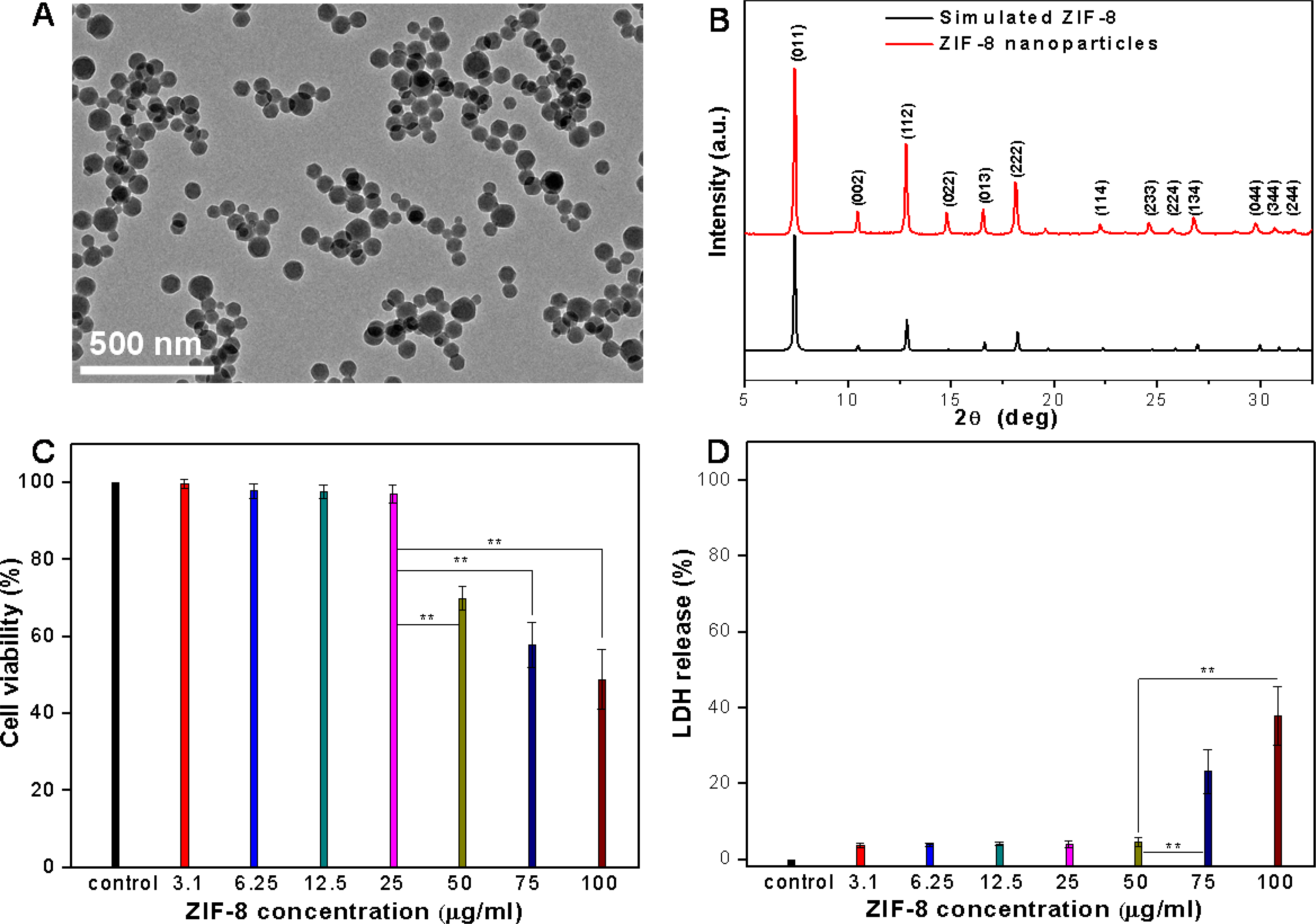
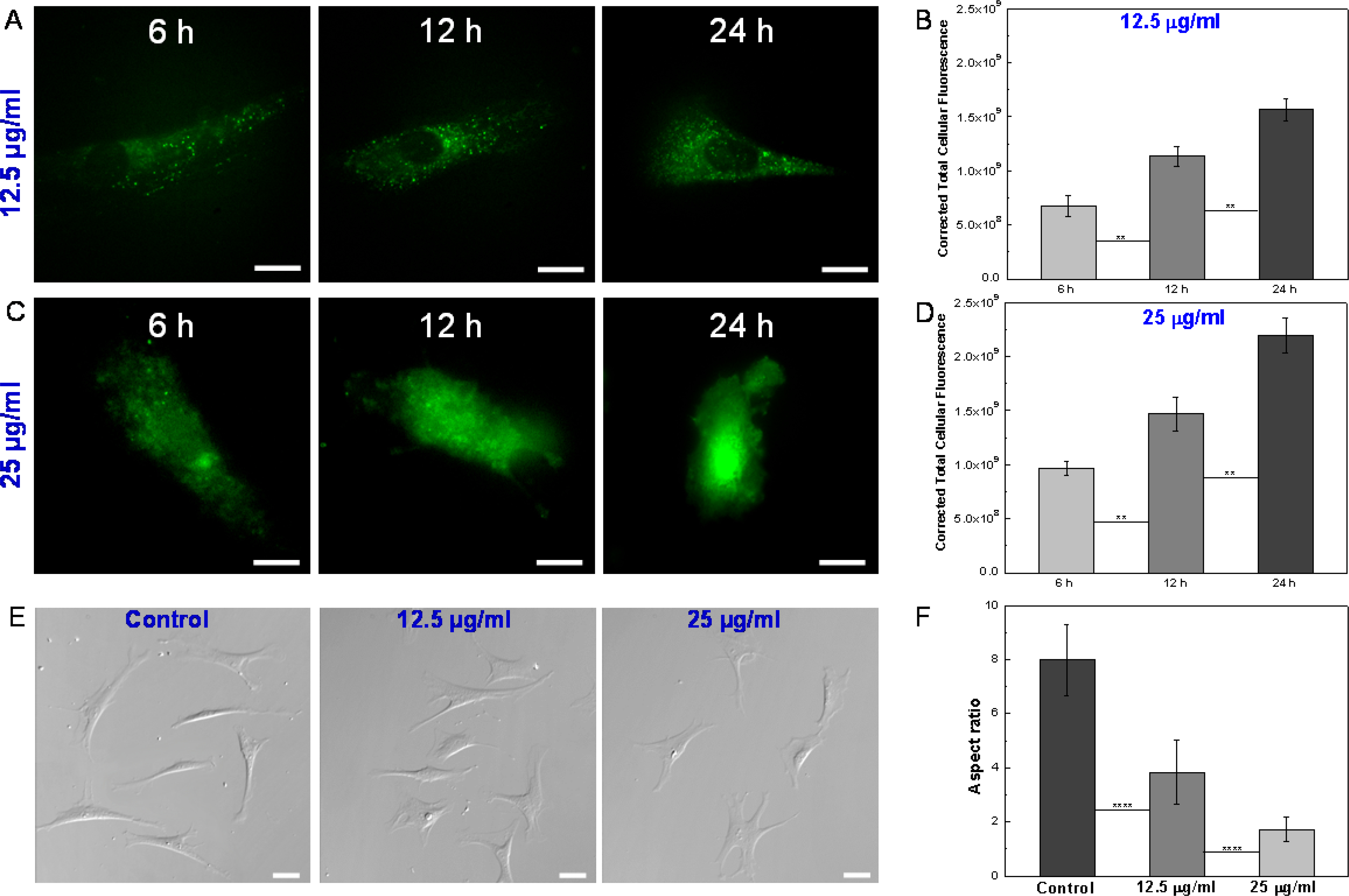
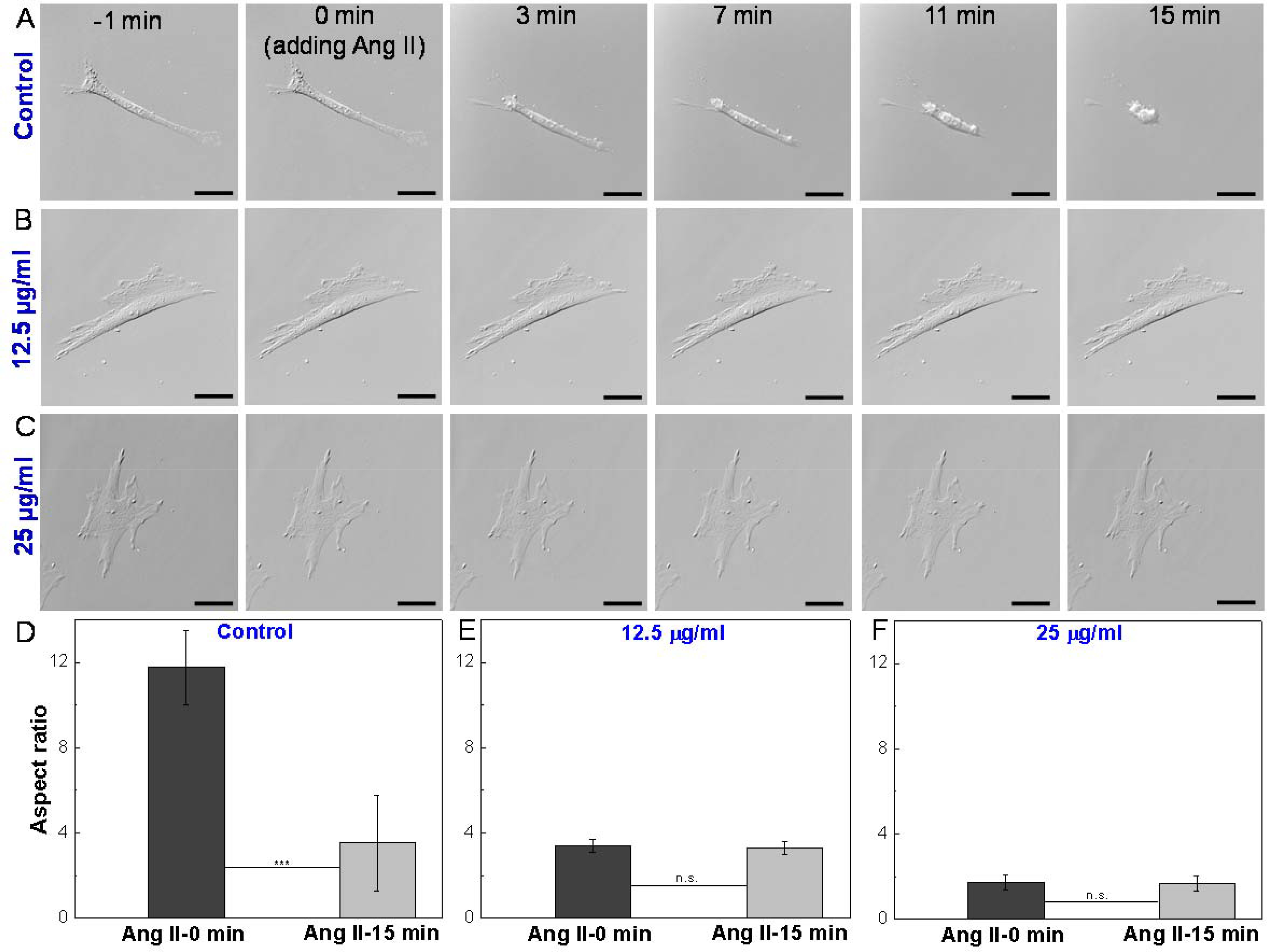
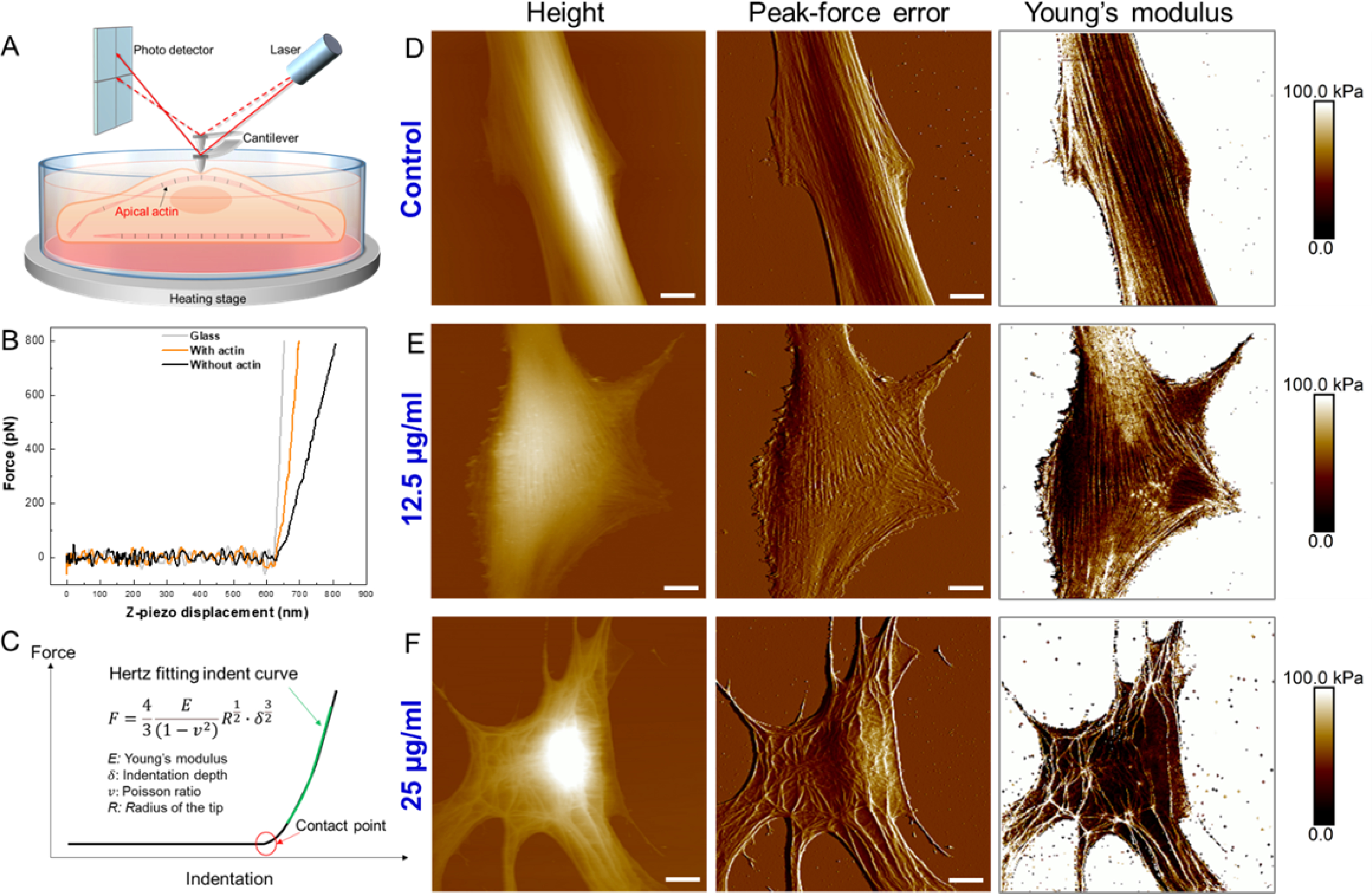
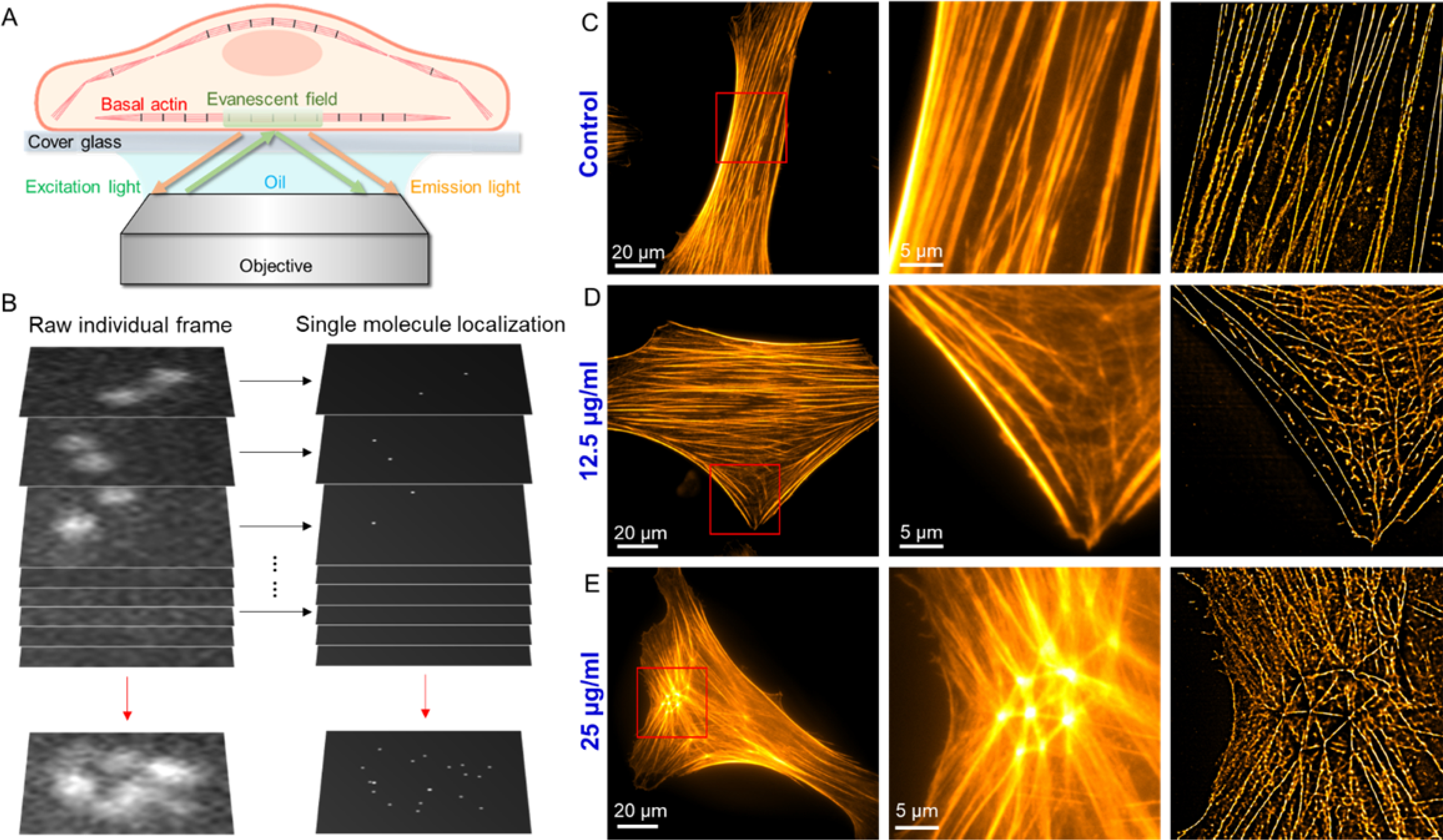
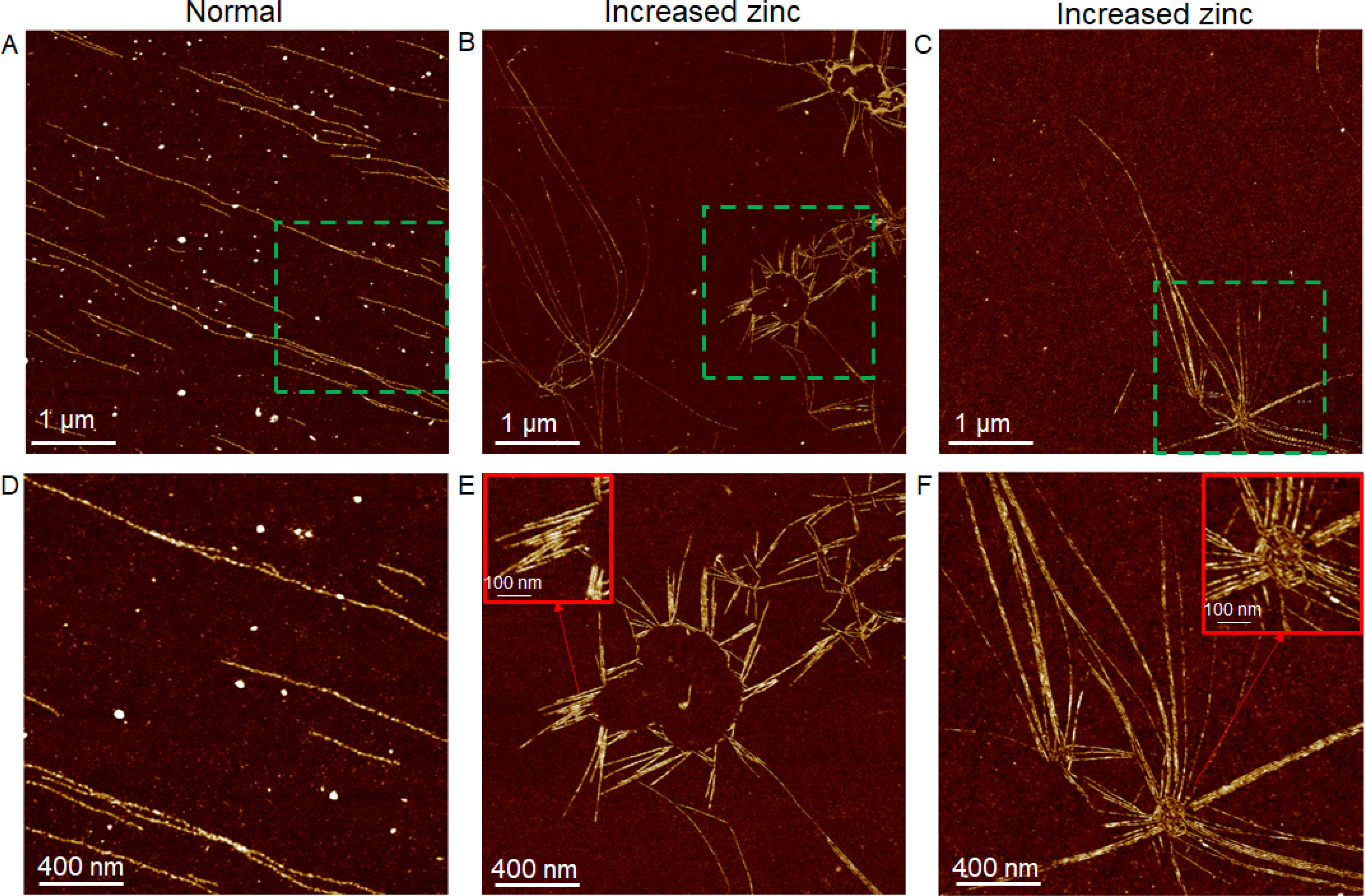
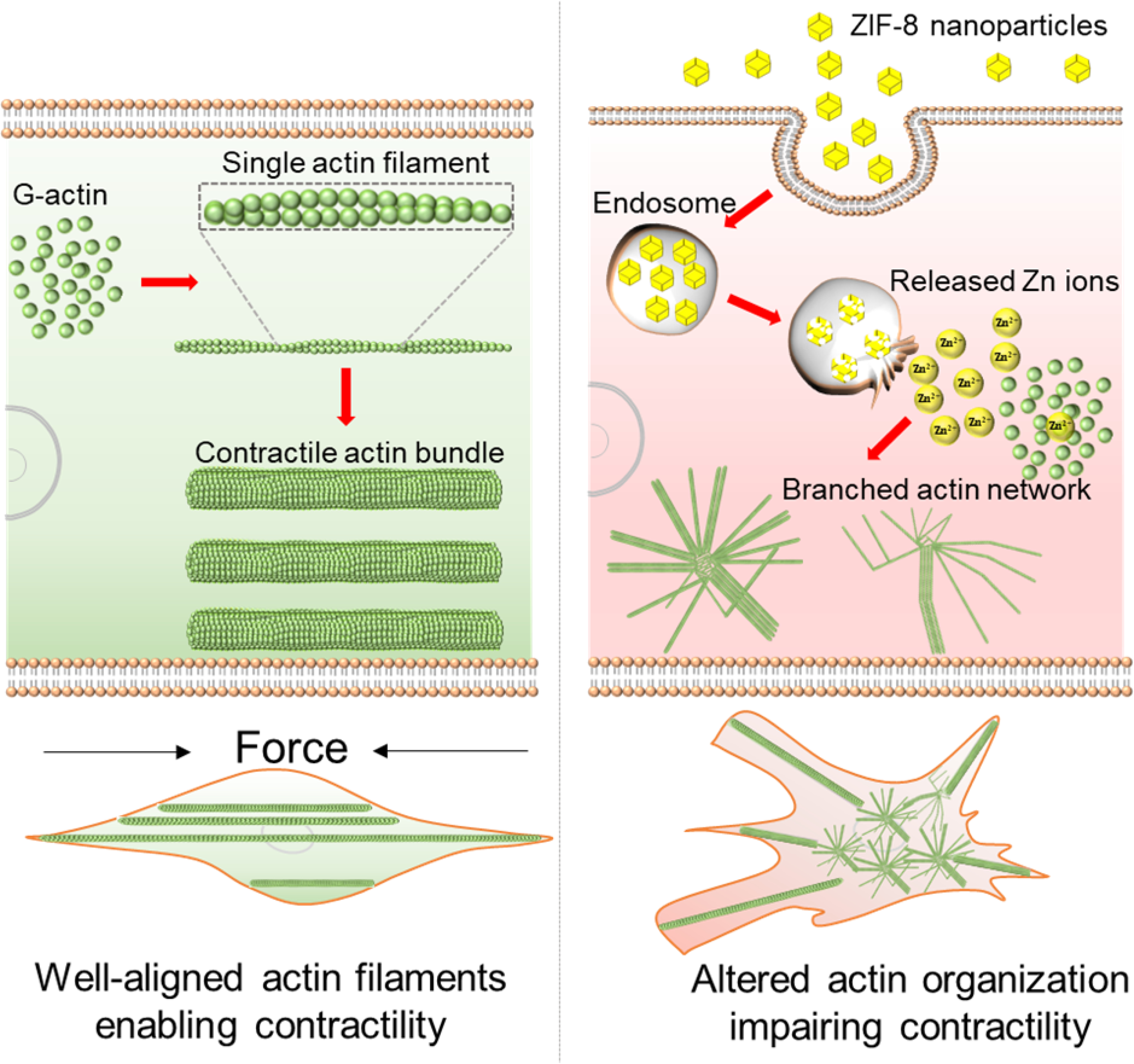
Zeolitic imidazolate framework-8 (ZIF-8) nanoparticles have emerged as a promising platform for drug delivery and controlled release. Considering most ZIF-8 nanoparticle drug carriers are designed to be administered intravenously, and thus would directly contact vascular smooth muscle cells (VSMCs) in many circumstances, the potential interactions of ZIF-8 nanoparticles with VSMCs require investigation. Here, the effects of low doses of ZIF-8 nanoparticles on VSMC morphology, actin organization, and contractility are investigated. Two nanoscale imaging tools, atomic force microscopy, and direct stochastic optical reconstruction microscopy, show that even at the concentrations (12.5 and 25 µg/ml) that were deemed "safe" by conventional biochemical cell assays (MTT and LDH assays), ZIF-8 nanoparticles can still cause changes in cell morphology and actin cytoskeleton organization at the cell apical and basal surfaces. These cytoskeletal structural changes impair the contractility function of VSMCs in response to Angiotensin II, a classic vasoconstrictor. Based on intracellular zinc and actin polymerization assays, we conclude that the increased intracellular Zn2+ concentration due to the uptake and dissociation of ZIF-8 nanoparticles could cause the actin cytoskeleton dis-organization, as the elevated Zn2+ directly disrupts the actin assembly process, leading to altered actin organization such as branches and networks. Since the VSMC phenotype change and loss of contractility are fundamental to the development of atherosclerosis and related cardiovascular diseases, it is worth noting that these low doses of ZIF-8 nanoparticles administered intravenously could still be a safety concern in terms of cardiovascular risks. Moving forward, it is imperative to re-consider the "safe" nanoparticle dosages determined by biochemical cell assays alone, and take into account the impact of these nanoparticles on the biophysical characteristics of VSMCs, including changes in the actin cytoskeleton and cell morphology.

Keywords: Actin cytoskeleton; Cell morphology; Contractility; Vascular smooth muscle cells; Zeolitic imidazolate framework-8 nanoparticles.


Conflict of interest statement

Conflicts of interest

There are no conflicts to declare.

Figures

Figure 1.
Figure 1.
(A) TEM image of synthesized ZIF-8 nanoparticles. (B) Powder XRD pattern of synthesized ZIF-8 nanoparticles and simulated powder XRD pattern of ZIF-8 (CCDC ID number: 864309). (C) MTT assay measuring VSMC viability after incubation with ZIF-8 nanoparticles for 24 h. (D) LDH assay measuring VSMC membrane integrity after incubation with ZIF-8 nanoparticles for 24 h. Data is mean ± standard deviation, n=6. Student’s t-test, **P < 0.01.
Figure 2.
Figure 2.
(A) Fluorescence images of single VSMCs after incubation with 12.5 μg/ml FITC-loaded ZIF-8 nanoparticles for 6 h, 12 h and 24 h. Scale bars: 20 μm. (B) Quantitative analysis of cell uptake of 12.5 μg/ml ZIF-8 nanoparticles based on correlated total cell fluorescence (CTCF). Data is mean ± standard deviation, n=50. Student’s t-test, **P < 0.01. (C) Fluorescence images of single VSMCs after incubation with 25 μg/ml FITC-loaded ZIF-8 nanoparticles for 6 h, 12 h and 24 h. Scale bars: 20 μm. (D) Quantitative analysis of cell uptake of 25 μg/ml ZIF-8 nanoparticles based on CTCF. Data is mean ± standard deviation, n=50. Student’s t-test, **P < 0.01. (E) DIC images of VSMCs after incubation with 0 (control), 12.5 and 25 μg/ml ZIF-8 nanoparticles for 24 h. Scale bars: 40 μm. (F) Aspect ratios of VSMCs after incubation with 0 (control), 12.5 and 25 μg/ml ZIF-8 nanoparticles for 24 h. Data is mean ± standard deviation, n=100. Student’s t-test, ****P < 0.0001. With the increase of ZIF-8 concentrations, the cells exhibited a less elongated, and more spread shape, leading to decreased aspect ratios.
Figure 3.
Figure 3.
Time-lapse DIC images of single VSMCs in response to Angiotensin II (Ang II) treatment. (A) Control: VSMC after incubation with standard medium for 24 h and subjected to Ang II treatment. (B) VSMC after incubation with 12.5 μg/ml ZIF-8 nanoparticles for 24 h and subjected to Ang II treatment. (C) VSMC after incubation with 25 μg/ml ZIF-8 nanoparticles for 24 h and subjected to Ang II treatment. Scale bars: 40 μm. (D-F) Aspect ratios of VSMCs before adding Ang II (0 min) and 15 min after adding Ang II. Data is mean ± standard deviation, n=10. Student’s t-test, ***P < 0.001. Un-treated VSMCs (control) showed contraction in response to Ang II treatment, whereas ZIF-8 nanoparticle treated VSMCs lost contractility.
Figure 4.
Figure 4.
(A) Schematic illustrating Peak-Force quantitative nanomechanical mapping of actin organization at the apical surfaces of live cells. (B) Raw force-displacement curves collected on glass, cell regions with apical actin filaments, and cell regions without apical actin filaments. (C) Hertz model fitting of force-indentation curve to obtain Young’s modulus at each pixel. (D) Height, peak-force error and Young’s modulus maps of single VSMC after incubation with 0 μg/ml (control) ZIF-8 nanoparticles for 24 h. (E) Height, peak-force error and Young’s modulus maps of single VSMC after incubation with 12.5 μg/ml ZIF-8 nanoparticles for 24 h. (F) Height, peak-force error and Young’s modulus maps of single VSMC after incubation with 25 μg/ml ZIF-8 nanoparticles for 24 h. Three types of maps showed excellent correlations to identify actin filaments at the apical surfaces of live cells. The ZIF-8 nanoparticles altered the VSMC morphology and actin organization in a concentration-dependent manner. Scale bars: 10 μm.
Figure 5.
Figure 5.
(A) Schematic illustrating total internal reflection fluorescence (TIRF) imaging of actin organization at the basal surfaces of phalloidin fluorescence stained cells. (B) The concept of dSTORM: The super resolution image is obtained by localizing single molecules within a stack of >10,000 frames, each capturing a different subset of individual molecules from the total ensemble. (C-E) Epi-fluorescence image of single VSMCs, red-boxed area of the epi-fluorescence image, and dSTORM image of the same area: (C) Control: VSMC after incubation with 0 μg/ml ZIF-8 nanoparticles for 24 h. (D) 12.5 μg/ml: VSMC after incubation with 12.5 μg/ml ZIF-8 nanoparticles for 24 h. (E) 25 μg/ml: VSMC after incubation with 12.5 μg/ml ZIF-8 nanoparticles for 24 h. ZIF-8 nanoparticle treatment led to the formation of actin branches and networks at the basal layers of the cells.
Figure 6.
Figure 6.
Actin polymerization assay investigating the effect of increased Zn2+ on actin assembly process. (A) and (D): AFM images of single actin filaments assembled from G-actin under normal assay conditions. (B-C) and (E-F): AFM images of actin bundles, branches and networks assembled from G-actin in the presence of increased Zn2+ concentrations. D-F are enlarged images of green-boxed areas in A-C, respectively.
Figure 7.
Figure 7.
Schematic illustrating how the ZIF-8 nanoparticles alter the actin organization in VSMCs. In the normal VSMCs (untreated cells), as shown in left column, G-actin assembles to single actin filaments, and then the filaments interact with actin-binding proteins such as myosin-II to form well-aligned actomyosin bundles, driving the contraction of cells. Upon uptake of ZIF-8 nanoparticles, as shown in right column, ZIF-8 nanoparticles dissolve in acidic endosomes/lysosomes and release Zn2+ into the cytoplasm, leading to increased intracellular Zn2+ levels. With increased intracellular Zn2+, the G-actin directly assembles to actin bundles and networks, instead of linear, single actin filaments, during the actin polymerization process (as demonstrated in Figure 6). Essentially, the actin cytoskeleton remodeling process is likely impacted by the increased intracellular Zn2+ levels, thus hindering the formation of well-aligned contractile filaments (i.e., actomyosin bundles) and the contraction of VSMCs.

Similar articles

Cited by

References

    1. Liu Y, Zhao Y, Chen X, Bioengineering of Metal-organic Frameworks for Nanomedicine, Theranostics, 9 (2019) 3122–3133. - PMC - PubMed
    1. Wu M-X, Yang Y-W, Metal–Organic Framework (MOF)-Based Drug/Cargo Delivery and Cancer Therapy, Advanced Materials, 29 (2017) 1606134. - PubMed
    1. Wang Y, Yan J, Wen N, Xiong H, Cai S, He Q, Hu Y, Peng D, Liu Z, Liu Y, Metal-organic frameworks for stimuli-responsive drug delivery, Biomaterials, 230 (2020) 119619. - PubMed
    1. Lu K, Aung T, Guo N, Weichselbaum R, Lin W, Nanoscale Metal–Organic Frameworks for Therapeutic, Imaging, and Sensing Applications, Advanced Materials, 30 (2018) 1707634. - PMC - PubMed
    1. Zhang W, Lu J, Gao X, Li P, Zhang W, Ma Y, Wang H, Tang B, Enhanced Photodynamic Therapy by Reduced Levels of Intracellular Glutathione Obtained By Employing a Nano-MOF with CuII as the Active Center, Angewandte Chemie International Edition, 57 (2018) 4891–4896. - PubMed

Publication types

LinkOut - more resources